Tetanus Prophylaxis and Soft Tissue Management in Orthopaedic Trauma
Key Takeaway
Tetanus remains a rare but life-threatening complication of open fractures. Proper management requires strict adherence to ATLS prophylaxis protocols, distinguishing between tetanus-prone and clean wounds. This guide details the administration of tetanus toxoid and human tetanus immune globulin, alongside essential principles of soft tissue debridement, fracture fixation, and thromboembolic prophylaxis in complex orthopaedic trauma patients.
Comprehensive Introduction and Patho-Epidemiology
The management of open fractures and severe soft tissue injuries in the acute trauma setting requires a comprehensive, strategic approach that extends far beyond the mechanical stabilization of osseous structures. Among the most critical, yet frequently underestimated, medical considerations in the acute polytrauma environment is the prevention of clinical tetanus. While widespread and aggressive vaccination programs have largely relegated tetanus to a rare complication in most developed nations, the catastrophic neurological consequences of Clostridium tetani infection demand unwavering, protocol-driven vigilance from the orthopaedic surgeon. The presence of high-energy trauma, characterized by devitalized tissue, profound ischemia, and gross contamination, creates the quintessential biological incubator for this deadly pathogen.
According to the Centers for Disease Control and Prevention (CDC), the incidence of tetanus in the United States remains exceptionally low, with an average of 29 cases reported annually, translating to an incidence of approximately 0.10 per million population. However, the clinical severity of the disease remains profound, with an overall mortality rate among reported cases hovering around 13%. Crucially, the mortality rate in patients aged 65 or older is nearly three times higher than in younger cohorts. This age-related vulnerability is directly correlated with waning immunity and inadequate longitudinal vaccination coverage. Epidemiological data indicates that tetanus vaccination coverage is only 57% in individuals between the ages of 18 and 64, and drops to a concerning 44% in those 65 years of age and older, highlighting a massive demographic at high risk during traumatic events.
Clostridium tetani is an obligate anaerobic, gram-positive, spore-forming bacillus that is ubiquitous in soil, dust, and animal feces. The spores are highly resilient, capable of surviving extreme environmental temperatures, desiccation, and standard chemical disinfectants. When introduced into an anaerobic environment—such as the devitalized, ischemic, or necrotic tissue typical of high-energy open fractures and crush injuries—the spores germinate into metabolically active vegetative bacilli. These bacilli produce tetanospasmin, an extraordinarily potent, plasmid-encoded neurotoxin. Tetanospasmin binds to peripheral motor neuron terminals, enters the axon, and retrogradely travels to the central nervous system. Upon reaching the spinal cord and brainstem, the toxin cleaves synaptobrevin, a SNARE protein critical for vesicle fusion, thereby irreversibly blocking the release of inhibitory neurotransmitters, specifically gamma-aminobutyric acid (GABA) and glycine. This unchecked excitatory motor neuron activity results in the pathognomonic clinical triad of severe spastic paralysis, intense muscle rigidity (trismus, risus sardonicus, opisthotonos), and profound autonomic instability.
A particularly insidious aspect of C. tetani in the realm of orthopaedic trauma is the "dormant spore" phenomenon. Spores introduced during the initial traumatic event may fail to germinate if the local oxygen tension is marginally sufficient to suppress vegetative growth, yet they remain viable. These spores can become encapsulated within dense avascular scar tissue or sclerotic, necrotic bone segments. Months or even years later, subsequent surgical interventions—such as nonunion takedown, hardware removal, or autogenous bone grafting—can disrupt this encapsulation, creating a renewed anaerobic microenvironment that triggers delayed germination and fulminant clinical tetanus in an under-immunized patient.
Detailed Surgical Anatomy and Biomechanics
A profound understanding of the soft tissue envelope's surgical anatomy is the cornerstone of preventing anaerobic microenvironments and ensuring successful fracture healing. The integumentary and muscular layers of the extremities are supplied by a complex, redundant network of perforating vessels arising from major axial arteries. These perforators form distinct three-dimensional vascular territories known as angiosomes. High-energy trauma, particularly crush injuries and high-velocity ballistic impacts, disrupts these angiosomes, leading to profound dermal ischemia, venous congestion, and subsequent tissue necrosis. The orthopaedic surgeon must respect these vascular boundaries during extensile exposures to prevent secondary iatrogenic necrosis of the skin flaps, which would inadvertently create a tetanus-prone wound bed.
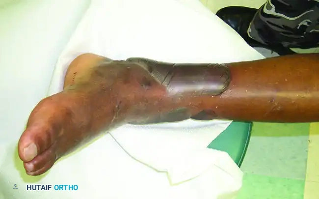
The condition of the soft tissue envelope dictates the timing, biomechanics, and strategy of surgical intervention. Severe dermal ischemia and venous congestion frequently manifest clinically as fracture blisters, which represent a traumatic cleavage at the dermo-epidermal junction secondary to unyielding interstitial edema. These blisters are broadly categorized into clear fluid-filled blisters and blood-filled blisters, each carrying distinct anatomical and surgical implications. Clear blisters indicate a more superficial separation where the underlying dermis remains largely intact, viable, and capable of re-epithelialization. Conversely, blood blisters represent a much deeper injury with complete disruption of the dermo-epidermal junction and significant dermal ischemia. Incising through a blood blister or its immediately adjacent compromised skin carries an unacceptably high risk of full-thickness skin necrosis, wound dehiscence, and deep periprosthetic infection.
Beneath the fascial layers, the skeletal muscle architecture is highly susceptible to the anaerobic conditions favored by C. tetani. Skeletal muscle relies on a dense capillary network to maintain oxygenation; however, the inelastic nature of the fascial compartments means that post-traumatic edema or hematoma rapidly elevates intracompartmental pressures. This phenomenon, if unchecked, leads to acute compartment syndrome, characterized by microvascular collapse, profound muscle ischemia, and eventual myonecrosis. The presence of necrotic muscle provides the ultimate substrate for tetanus spore germination. Therefore, prompt fasciotomy and radical excision of non-viable muscle are not merely limb-salvage procedures; they are critical biological interventions to eliminate the anaerobic environment.
From a biomechanical perspective, the management of fractures within a compromised soft tissue envelope requires a delicate balance between achieving rigid osseous stability and preserving the remaining precarious blood supply. In cases of severe open fractures, delayed unions and nonunions are frequent complications due to extensive periosteal stripping and endosteal ischemia. The primary biomechanical goal during reconstruction is to maximize the fatigue life of the fracture fixation construct. Because the biological healing potential is diminished, the implant must withstand cyclical loading for an extended duration. This necessitates the use of load-sharing devices (such as intramedullary nails) where biologically appropriate, or the strategic application of bridge plating techniques that bypass the zone of comminution, thereby preserving the fracture hematoma and the fragile periosteal blood supply critical for secondary bone healing.
Exhaustive Indications and Contraindications
The administration of tetanus prophylaxis and the timing of surgical intervention in orthopaedic trauma are governed by strict, evidence-based indications and contraindications. The fundamental principle of tetanus prevention is recognizing the wound environment that facilitates spore germination. The Subcommittee on Advanced Trauma Life Support (ATLS) of the American College of Surgeons has established definitive criteria for identifying tetanus-prone wounds, which dictate the subsequent pharmacological and surgical management.
A wound is classified as tetanus-prone if it exhibits any of the following characteristics: an age of more than 6 hours elapsed since the time of injury; a stellate, avulsion, or severe abrasion configuration; a depth of penetration greater than 1 cm; a mechanism of injury involving missiles (ballistics), crush, burns, or frostbite; the presence of infected, devitalized, denervated, or ischemic tissue; or visible contaminants including dirt, feces, soil, or saliva. The presence of any single criterion mandates an aggressive prophylactic approach. Conversely, a clean, minor wound is typically linear, less than 1 cm deep, less than 6 hours old, and devoid of devitalized tissue or gross contamination.
The pharmacological indications for prophylaxis depend entirely on the intersection of the wound classification and the patient's documented immunization history. Patients who have completed a primary tetanus toxoid active immunization series require only a booster dose of tetanus toxoid if their last dose was administered more than 5 years ago for tetanus-prone wounds, or more than 10 years ago for clean, minor wounds. For patients with an incomplete primary series, or those whose immunization history is unknown, aggressive prophylaxis is indicated. For clean wounds in this cohort, the tetanus toxoid vaccine is administered to initiate the series. For tetanus-prone wounds in unimmunized patients, the absolute indication is the simultaneous administration of Human Tetanus Immune Globulin (HTIG) to provide immediate passive immunity, alongside the tetanus toxoid to initiate active immunity.
Contraindications in this realm primarily revolve around outdated therapies and the management of concomitant systemic risks. Historically, equine tetanus antitoxin was utilized for severe open wounds; however, it is now strictly contraindicated due to the unacceptably high risk of severe anaphylaxis and serum sickness. HTIG is the absolute standard of care. Furthermore, in the realm of thromboembolic prophylaxis for these polytrauma patients, early chemical anticoagulation is contraindicated in the presence of active intracranial hemorrhage, severe solid organ injury, traumatic spinal epidural hematoma, or ongoing retroperitoneal hemorrhage from pelvic ring disruptions.
| Wound Classification | Patient Immunization Status | Tetanus Toxoid (Active) Indicated? | HTIG (Passive) Indicated? |
|---|---|---|---|
| Clean, Minor Wound | Unknown or < 3 doses | Yes | No |
| Clean, Minor Wound | ≥ 3 doses (Last dose < 10 yrs) | No | No |
| Clean, Minor Wound | ≥ 3 doses (Last dose > 10 yrs) | Yes | No |
| Tetanus-Prone Wound | Unknown or < 3 doses | Yes | Yes (250 Units IM) |
| Tetanus-Prone Wound | ≥ 3 doses (Last dose < 5 yrs) | No | No |
| Tetanus-Prone Wound | ≥ 3 doses (Last dose > 5 yrs) | Yes | No |
Pre-Operative Planning, Templating, and Patient Positioning
Pre-operative planning for the orthopaedic trauma patient with a severe open fracture begins in the trauma bay, strictly adhering to ATLS protocols. The initial assessment must prioritize the airway, breathing, and circulation before attention is directed to the extremity. The orthopaedic surgeon must recognize the "lethal triad" of trauma—coagulopathy, hypothermia, and acidosis—which significantly alters the surgical plan, often dictating a Damage Control Orthopaedics (DCO) approach rather than Early Total Care (ETC). Once life-threatening injuries are stabilized, the extremity is evaluated. The wound is photographed to prevent redundant examinations, grossly realigned, splinted, and covered with sterile, saline-soaked dressings. "Ward debridements" or probing of the wound in the emergency department are strictly forbidden, as they risk pushing superficial contaminants deeper into the soft tissue envelope.
Templating for these injuries requires a versatile approach. While standard orthogonal radiographs are mandatory, advanced imaging such as Computed Tomography (CT) with 3D reconstruction is frequently indicated for complex periarticular fractures or to assess bone loss. The surgeon must template for multiple eventualities, ensuring that spanning external fixator components, intramedullary nails of various diameters and lengths, and biologically friendly bridge plating systems are available. Furthermore, the pre-operative plan must include a definitive strategy for thromboembolic prophylaxis. Given the high risk of Deep Vein Thrombosis (DVT) and Pulmonary Embolism (PE) in immobilized polytrauma patients, the surgeon must decide between mechanical prophylaxis (intermittent pneumatic compression), chemical prophylaxis (Low-Molecular-Weight Heparin), or the placement of a retrievable Inferior Vena Cava (IVC) filter if chemical anticoagulation is absolutely contraindicated.
Patient positioning is critical to facilitate radical debridement, copious irrigation, and multi-planar fluoroscopic imaging. The patient is typically positioned supine on a radiolucent Jackson table or a standard operating table with a radiolucent extension. A bump may be placed under the ipsilateral hip to correct external rotation for lower extremity trauma. The entire limb must be prepped and draped free to allow for circumferential access, assessment of mechanical alignment, and extensile surgical approaches. In cases of massive bone loss or severe soft tissue degloving, the contralateral extremity should also be prepped and draped to allow for the harvesting of autogenous bone graft (e.g., iliac crest, RIA from the femur) or the execution of cross-leg soft tissue flaps.
A pneumatic tourniquet should be applied proximally but must be used judiciously. While a tourniquet provides a bloodless field for the initial exposure and identification of critical neurovascular structures, it must be deflated during the critical phase of muscle debridement. The assessment of muscle viability—the primary mechanical defense against tetanus—relies heavily on observing the tissue's capacity to bleed. Prolonged tourniquet times not only obscure this assessment but also exacerbate the ischemic injury to the already compromised soft tissue envelope, potentially precipitating the very anaerobic environment the surgeon is attempting to eliminate.
Step-by-Step Surgical Approach and Fixation Technique
The surgical approach to the tetanus-prone open fracture is centered on the principle of radical, aggressive debridement. This is not merely a procedure of infection control; it is the definitive mechanical eradication of the anaerobic microenvironment required for C. tetani spore germination. The traumatic wound must be systematically extended along standard, extensile surgical incisions, avoiding the creation of narrow skin bridges or compromising the vascularity of the flaps. The skin edges are excised back to healthy, bleeding dermis. Subcutaneous fat, which has a poor blood supply and is highly susceptible to necrosis, is aggressively resected if it appears contused or avulsed.
The critical phase of the operation is the evaluation and debridement of skeletal muscle. The surgeon must systematically evaluate all exposed muscle using the established "4 C's" criteria: Color, Consistency, Contractility, and Capacity to bleed. Healthy muscle is dark red, firm, contracts briskly when stimulated with electrocautery, and bleeds readily from its cut surface. Any muscle that is pale, dusky, friable, non-contractile, or avascular must be ruthlessly excised until healthy tissue is reached. This process may require the excision of entire muscle bellies. The presence of necrotic muscle or a severe crush component exponentially increases the risk of an anaerobic microenvironment, making this step the most vital surgical defense against tetanus.
Following radical debridement, the wound is subjected to copious, low-pressure irrigation. High-volume normal saline (typically 6 to 9 liters, depending on the Gustilo-Anderson classification) is utilized to mechanically wash away debris, hematoma, and residual bacterial load. The use of high-pressure pulsatile lavage is generally discouraged as it can drive contaminants deeper into the intramedullary canal and further damage fragile soft tissues. Furthermore, the addition of cytotoxic agents such as hydrogen peroxide, betadine, or chlorhexidine directly into the wound bed is avoided, as these agents cause profound cellular toxicity to the remaining viable host tissue, paradoxically increasing the risk of necrosis and subsequent infection.
Skeletal stabilization is the next critical step. Rigid fixation protects the fragile soft tissue envelope from ongoing mechanical trauma, reduces dead space, and restores length and alignment. In the setting of a highly contaminated, tetanus-prone wound, or a patient in extremis (DCO), a spanning external fixator is the implant of choice. Pins must be placed entirely outside the zone of injury. If the wound is adequately debrided and the soft tissue envelope allows for coverage, immediate intramedullary nailing may be performed for diaphyseal fractures. Following fixation, the wound is typically left open. Negative Pressure Wound Therapy (NPWT) or antibiotic-impregnated polymethylmethacrylate (PMMA) bead pouches are applied to seal the wound, obliterate dead space, and provide high local concentrations of antibiotics while preventing desiccation of the exposed tissues.
Simultaneous with the surgical intervention, pharmacological prophylaxis must be meticulously administered. For the unimmunized patient with a tetanus-prone wound, the "Separate Syringe" rule is a mandatory clinical pearl. Human Tetanus Immune Globulin (HTIG) provides immediate passive immunity, while the tetanus toxoid initiates active immunity. However, if administered in close proximity or with the same syringe, the HTIG will neutralize the toxoid in vivo, rendering the active vaccine useless. Therefore, separate syringes and separate anatomical sites of injection—typically the contralateral deltoids or anterolateral thighs—must be strictly utilized.
Complications, Incidence Rates, and Salvage Management
The complications associated with severe open fractures and tetanus-prone wounds are devastating, threatening both limb and life. The most feared complication, clinical tetanus, is fortunately rare due to modern prophylaxis, but carries a high mortality rate. If clinical tetanus develops, it constitutes a medical emergency requiring immediate ICU admission. Salvage management includes intubation and mechanical ventilation to secure the airway, aggressive administration of benzodiazepines and neuromuscular blocking agents to control spasticity and autonomic storms, and the administration of HTIG to neutralize unbound toxin. Intravenous metronidazole or penicillin is administered to eradicate the vegetative C. tetani bacilli, and the wound must undergo immediate, radical re-debridement to eliminate the biological source.
Deep periprosthetic infection and chronic osteomyelitis occur in up to 15-25% of severe Type III open fractures, despite appropriate initial management. These infections are typically polymicrobial, involving Staphylococcus aureus, Pseudomonas aeruginosa, and various anaerobes. Salvage management for deep infection requires the removal of all retained hardware, aggressive debridement of all necrotic bone (creating a critical-sized defect), and the placement of an antibiotic-loaded cement spacer. Once the infection is eradicated, reconstruction can be attempted using the Masquelet induced-membrane technique or distraction osteogenesis via an Ilizarov frame. If the soft tissue envelope is unsalvageable or the limb is insensate, early amputation must be considered as a definitive, life-saving salvage procedure.
Thromboembolic complications, including Deep Vein Thrombosis (DVT) and Pulmonary Embolism (PE), represent a massive systemic risk for the immobilized polytrauma patient. The incidence of DVT in major lower extremity and pelvic trauma can exceed 50% without prophylaxis, with fatal PE occurring in up to 2-5% of these patients. The "lethal triad" complicates the standard administration of chemical prophylaxis, forcing the surgeon to balance the risk of hemorrhagic complications against the embolic threat. Salvage management for a massive, hemodynamically unstable PE may require catheter-directed thrombolysis or open surgical embolectomy, while DVT is managed with therapeutic doses of low-molecular-weight heparin or direct oral anticoagulants once the bleeding risk has subsided.
| Complication | Estimated Incidence | Primary Etiology | Salvage Management Strategy |
|---|---|---|---|
| Clinical Tetanus | < 0.1% (in developed nations) | Failure of prophylaxis, missed anaerobic wound | ICU admission, airway control, massive HTIG, IV Metronidazole, radical re-debridement. |
| Deep Infection / Osteomyelitis | 10% - 25% (Type III open fractures) | Inadequate debridement, delayed soft tissue coverage | Hardware removal, radical bone resection, antibiotic spacers, delayed reconstruction (Masquelet/Ilizarov). |
| Aseptic Nonunion | 15% - 30% | Endosteal ischemia, periosteal stripping, construct fatigue | Optimization of mechanics (exchange nailing, rigid plating), autogenous bone grafting (RIA, iliac crest). |
| Deep Vein Thrombosis (DVT) | 20% - 50% (without prophylaxis) | Endothelial injury, venous stasis, hypercoagulability | Therapeutic anticoagulation (LMWH, DOACs), transition from mechanical prophylaxis. |
| Pulmonary Embolism (PE) | 2% - 5% (high-risk pelvic/LE trauma) | Embolization of massive DVT | Hemodynamic support, catheter-directed thrombolysis, surgical embolectomy, IVC filter placement. |
Phased Post-Operative Rehabilitation Protocols
The post-operative rehabilitation of a patient with a severe, tetanus-prone soft tissue injury and complex fracture is a protracted, phased process requiring meticulous coordination between the orthopaedic surgeon, plastic surgeon, infectious disease specialist, and physical therapist.
Phase 1: Acute Inpatient Management (Days 0 to 7)
The immediate post-operative phase is focused entirely on biological stabilization. The patient remains strictly non-weight-bearing on the affected extremity. The surgical wound, typically managed with Negative Pressure Wound Therapy (NPWT), is closely monitored for excessive drainage or foul odor. A mandatory "second-look" debridement is performed in the operating room at 48 to 72 hours post-injury to reassess muscle viability and ensure the absence of evolving necrosis. Soft tissue coverage (e.g., split-thickness skin grafting, local rotational flaps, or free tissue transfer) should ideally be achieved within 5 to 7 days, adhering to the principles established by Godina, to minimize the risk of nosocomial infection and seal the fracture environment. Thromboembolic prophylaxis is aggressively maintained, transitioning from mechanical to chemical modalities as the acute bleeding risk diminishes.
Phase 2: Subacute Sub-Acute and Transition (Weeks 1 to 6)
Once the soft tissue envelope has stabilized and fracture blisters have resolved, patients managed initially with damage control external fixation are transitioned to definitive internal fixation (e.g., intramedullary nailing or locked plating). Range of motion exercises for adjacent, uninjured joints are initiated immediately to prevent profound arthrofibrosis. For unimmunized patients who received their initial tetanus toxoid dose in the trauma bay, a critical milestone in this phase is the administration of the second dose of tetanus toxoid at 4 weeks post-injury. Failure to administer this second dose leaves the patient vulnerable to the "dormant spore" phenomenon during future reconstructive procedures.
Phase 3: Reconstruction and Progressive Loading (Months 2 to 12)
As radiographic evidence of early callus formation appears, the rehabilitation protocol shifts toward controlled, progressive weight-bearing. Axial loading stimulates secondary bone healing via callus formation, adhering to Wolff's Law, and prevents severe disuse osteopenia. This biological response is critical to protecting the orthopaedic implant from cyclical fatigue failure. In cases of oligotrophic or atrophic nonunion secondary to the initial severe soft tissue stripping, this phase may require return to the operating room for autogenous bone grafting. Crucially, the surgeon must ensure the patient receives their third and final dose of tetanus toxoid at 6 to 12 months to complete the primary active immunization series, ensuring durable, long-term protection.
Summary of Landmark Literature and Clinical Guidelines
The modern management of tetanus prophylaxis, soft tissue assessment, and open fracture care is built upon a foundation of landmark literature and rigorous clinical guidelines. The orthopaedic surgeon must be intimately familiar with these foundational texts to practice evidence-based medicine.
The Subcommittee on Advanced Trauma Life Support (ATLS) of the American College of Surgeons provides the definitive, globally recognized guidelines for the classification of tetanus-prone wounds and the subsequent algorithms for the administration of tetanus toxoid and Human Tetanus Immune Globulin (HTIG). These guidelines form the medicolegal standard of care in the acute trauma bay.
The classification and management of open fractures remain heavily influenced by the seminal work of Gustilo and Anderson (1976, 1984). Their classification system correlates the energy of the injury, the extent of soft tissue damage, and the degree of contamination with the subsequent risk of deep infection and amputation. Their work established the absolute necessity of radical surgical debridement and appropriate systemic antibiotic therapy as the primary defenses against both common pyogenic organisms and anaerobic pathogens like C. tetani.
Regarding soft tissue management, the landmark study by Marko Godina (1986) revolutionized the timing of microsurgical reconstruction. Godina demonstrated that early radical debridement followed by free tissue transfer within 72 hours of injury dramatically reduced infection rates, decreased nonunion rates, and shortened hospital stays compared to delayed reconstruction. While the exact timing remains a subject of ongoing debate, the principle of rapid, definitive soft tissue closure to seal the biological envelope is universally accepted.
Finally, the Eastern Association for the Surgery of Trauma (EAST) guidelines provide the most comprehensive, evidence-based recommendations for thromboembolic prophylaxis in the orthopaedic trauma patient. These guidelines delineate the critical balance between preventing fatal pulmonary emboli and avoiding catastrophic hemorrhage, establishing low-molecular-weight heparin as the pharmacological standard of care, and defining the strict, limited indications for the placement of prophylactic inferior vena cava (IVC) filters in the highest-risk polytrauma cohorts.
This academic synthesis is based on established protocols from Hutaifortho's Operative Orthopaedics and has been medically reviewed by Prof. Dr. Mohammed Hutaif, Consultant Orthopedic & Spine Surgeon. It is designed to assist orthopedic residents, fellows, and practicing surgeons in surgical preparation and board reviews (AAOS, FRCS, Arab Board).
