Mastering Preparation, Draping, and Tourniquet Protocols in Upper Extremity Surgery
Key Takeaway
Standardizing the preparation and draping of the upper extremity is critical for minimizing surgical site infections and optimizing operative workflow. This guide details evidence-based protocols for patient positioning, antiseptic selection, and safe pneumatic tourniquet application. Adherence to strict tourniquet time limits and proper skin preparation techniques ensures optimal surgical outcomes while mitigating the risk of neurovascular complications and chemical burns.
Comprehensive Introduction and Patho-Epidemiology
The meticulous preparation, sterile draping, and precise management of pneumatic tourniquets in upper extremity surgery represent the foundational pillars upon which all successful operative outcomes are built. While the surgical community often focuses intensely on the intricacies of osteosynthesis, microvascular anastomosis, or tendon repair, the absolute prerequisite for these advanced techniques is a flawlessly prepared and optimized surgical field. The methodology governing this preparatory phase must be strictly standardized, deeply rooted in evidence-based medicine, and executed with uncompromising discipline. A highly regimented routine not only streamlines the cognitive and physical workflow of the surgical team but also provides unencumbered spatial access to the operative field while profoundly minimizing the catastrophic risks of bacterial contamination, surgical site infections (SSIs), and iatrogenic neurovascular injuries. In the context of modern operative orthopedics, field preparation extends far beyond the rudimentary application of an antiseptic agent; it encompasses strategic patient positioning, the anticipation of autologous graft donor sites, the rigorous protection of vulnerable neurovascular structures, and the highly calculated biomechanical management of pneumatic tourniquets.
The patho-epidemiology of surgical site infections in upper extremity surgery underscores the critical nature of these protocols. The human skin, particularly in the axillary, cubital, and palmar regions, is heavily colonized by a diverse microbiome. The transient flora, which resides superficially on the stratum corneum, includes highly pathogenic organisms such as Staphylococcus aureus and various Gram-negative bacilli. More insidiously, the resident flora, predominantly coagulase-negative staphylococci (e.g., Staphylococcus epidermidis) and Cutibacterium acnes, resides deep within the sebaceous glands and hair follicles. These deeper reservoirs are notoriously difficult to eradicate with superficial mechanical scrubbing alone. When a surgical incision breaches the epidermal barrier, these endogenous organisms can rapidly inoculate the subcutaneous tissues and deeper fascial planes. The presence of orthopedic implants, such as titanium plates or Kirschner wires, dramatically lowers the threshold for clinical infection, as these foreign bodies provide an ideal scaffold for bacterial biofilm formation—a complex, extracellular polymeric matrix that renders bacteria highly recalcitrant to systemic antimicrobial therapy and host immune responses.
Concurrently, the patho-epidemiology of tourniquet-induced complications demands profound respect and understanding from the operating surgeon. The application of a pneumatic tourniquet induces a highly complex sequence of localized and systemic physiological alterations. Locally, the tissue distal to the cuff is subjected to profound ischemia, leading to a rapid depletion of intracellular adenosine triphosphate (ATP), the failure of sodium-potassium ATPase pumps, and subsequent cellular swelling. As the duration of ischemia progresses beyond the critical threshold of 90 to 120 minutes, irreversible microvascular endothelial damage occurs. Systemically, the eventual deflation of the tourniquet triggers ischemia-reperfusion injury, characterized by the massive release of reactive oxygen species (ROS), inflammatory cytokines, lactic acid, and potassium into the systemic circulation. This can lead to transient but significant hemodynamic instability, particularly in patients with compromised cardiopulmonary reserves.
Furthermore, mechanical compression from the tourniquet cuff itself poses a distinct pathological threat to the underlying neural architecture. Tourniquet paralysis, a devastating iatrogenic complication, is primarily a mechanical neurapraxia rather than a purely ischemic event. The pressure gradient is steepest at the edges of the tourniquet cuff, creating sheer forces that can cause invagination of the nodes of Ranvier and disruption of the myelin sheath. The radial nerve, as it spirals around the humerus, is particularly vulnerable to this compressive shear stress. Consequently, the operating surgeon must possess an exhaustive understanding of both the microbiological threats and the biomechanical hazards inherent in preparing the upper extremity for surgery, mitigating these risks through the rigorous application of standardized protocols.
Detailed Surgical Anatomy and Biomechanics
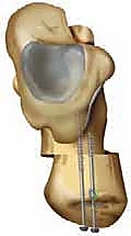
A profound understanding of the surgical anatomy of the upper extremity is non-negotiable when applying pneumatic tourniquets and executing sterile preparation. The proximal arm, the most common site for tourniquet application, is essentially a cylinder containing a single central bone surrounded by distinct muscular compartments. The brachial fascia encloses the anterior compartment (biceps brachii, brachialis, coracobrachialis) and the posterior compartment (triceps brachii). Crucially, the neurovascular bundles traverse these compartments in highly predictable, yet vulnerable, pathways. The brachial artery and the median and ulnar nerves course medially, relatively protected by the bulk of the biceps and triceps. However, the radial nerve exits the axilla, travels posteriorly, and lies directly against the periosteum of the humerus within the spiral groove before piercing the lateral intermuscular septum. This intimate bone-nerve relationship renders the radial nerve exceptionally susceptible to mechanical compression from an improperly placed or excessively pressurized tourniquet cuff. Forearm tourniquets, while useful for distal procedures, must similarly account for the superficial course of the ulnar nerve at the wrist and the superficial sensory branch of the radial nerve.
The cutaneous anatomy of the upper extremity dictates the pharmacological approach to antiseptic preparation. The epidermis consists of multiple stratified layers, with the outermost stratum corneum providing the primary physical barrier to microbial invasion. However, the presence of hair follicles, sebaceous glands, and sweat glands creates deep invaginations that harbor resident bacterial flora. Effective antiseptic agents must possess the chemical properties necessary to penetrate these lipid-rich environments. The palmar skin of the hand presents a unique anatomical challenge; it is thick, heavily keratinized, devoid of hair follicles, but rich in eccrine sweat glands. This thick keratin layer can trap dirt and organic debris, necessitating a vigorous mechanical scrub prior to the application of chemical antiseptics. Furthermore, the intricate anatomy of the nail folds and subungual spaces requires fastidious attention, as these areas are notorious reservoirs for fungal and bacterial pathogens that can easily contaminate the operative field during hand surgery.
The biomechanics of pneumatic tourniquet compression is a complex interplay of physics, tissue compliance, and vascular hemodynamics. The fundamental principle governing tourniquet efficacy is the transmission of pressure from the external cuff through the soft tissues to occlude the deep arterial vessels. This pressure transmission is not uniform; it diminishes as it moves centrally toward the bone. Therefore, the width of the tourniquet cuff is of paramount biomechanical importance. A wider cuff distributes the compressive force over a larger surface area, allowing for a more efficient transmission of pressure to the deeper tissues. Consequently, a wider cuff can achieve total arterial occlusion at a significantly lower inflation pressure compared to a narrow cuff. This reduction in absolute pressure dramatically decreases the mechanical shear stress exerted on the underlying nerves, thereby minimizing the risk of tourniquet-induced neurapraxia.
Modern tourniquet protocols increasingly rely on the concept of Limb Occlusion Pressure (LOP) rather than arbitrary, fixed pressure settings. LOP is defined as the minimum pressure required, at a specific time and in a specific patient, to completely occlude arterial blood flow distal to the cuff. LOP is influenced by the patient's systolic blood pressure, the circumference and tissue composition of the limb, and the design of the tourniquet cuff. Biomechanically, contoured cuffs that match the conical shape of the proximal arm provide superior pressure distribution compared to traditional cylindrical cuffs. By utilizing Doppler ultrasound or automated plethysmography to determine the patient's specific LOP, the surgeon can add a standard safety margin (typically 50 mmHg for LOP < 130 mmHg, or 75 mmHg for LOP > 130 mmHg) to determine the optimal, lowest effective inflation pressure. This personalized, biomechanically sound approach represents the gold standard in mitigating tourniquet-related morbidity.
Exhaustive Indications and Contraindications
The decision to utilize a pneumatic tourniquet, select a specific antiseptic agent, and employ mechanical exsanguination techniques must be based on a rigorous evaluation of the patient's physiological status and the specific requirements of the surgical procedure. While a bloodless field is highly advantageous for visualizing delicate structures such as digital nerves and flexor tendons, it is not without physiological cost. The indications for tourniquet use encompass nearly all major elective and traumatic interventions of the hand, wrist, and forearm, including complex intra-articular fracture fixation, microvascular nerve and vessel repair, tendon transfers, and extensive tenosynovectomies. In these scenarios, the obscuration of the surgical field by continuous hemorrhage poses a greater risk to the patient—through potential iatrogenic injury to vital structures—than the controlled ischemia of the tourniquet.
However, the absolute and relative contraindications to tourniquet application are numerous and must be meticulously respected. Absolute contraindications include the presence of severe peripheral vascular disease (PVD) or critical limb ischemia, where the temporary cessation of blood flow may precipitate irreversible gangrene. Sickle cell disease (and, in some cases, sickle cell trait) represents another absolute contraindication; the hypoxia, acidosis, and vascular stasis induced by the tourniquet will invariably trigger a massive, potentially limb-threatening sickling crisis. Furthermore, the presence of a deep vein thrombosis (DVT) in the operative limb absolutely precludes tourniquet use and mechanical exsanguination, as these maneuvers will likely dislodge the thrombus, resulting in a fatal pulmonary embolism. Severe crush injuries, where the soft tissues are already subjected to massive trauma and compartment swelling, are also contraindications, as the added ischemic insult of a tourniquet will exacerbate tissue necrosis and precipitate acute compartment syndrome.
The selection of antiseptic agents is similarly governed by strict indications and contraindications based on the agent's pharmacological profile and the patient's medical history. Chlorhexidine gluconate (CHG) in an alcohol base is the indicated gold standard for intact skin preparation in orthopedic surgery due to its rapid onset, broad spectrum, and superior residual bacteriostatic activity. However, CHG is absolutely contraindicated for use near the eyes, ears, and mucous membranes. It is highly keratotoxic and can cause permanent corneal opacification; it is also profoundly ototoxic and can cause irreversible sensorineural hearing loss if it enters the middle ear through a perforated tympanic membrane. Iodophors (e.g., povidone-iodine) are indicated when CHG is contraindicated, such as in facial trauma or when mucosal preparation is required. However, iodophors are contraindicated in patients with true, documented iodine allergies (anaphylaxis), though simple contact dermatitis to iodine is a relative contraindication that requires careful clinical judgment.
Mechanical exsanguination using an Esmarch bandage or elastic wrap is indicated to maximize the efficacy of the tourniquet and minimize the volume of blood trapped in the ischemic limb. However, mechanical exsanguination is strictly contraindicated in the presence of active, purulent infections (e.g., flexor tenosynovitis, deep space abscesses) and malignant neoplasms. In these pathological states, the intense proximal compression of the Esmarch bandage can force purulent exudate or malignant cells into the systemic venous and lymphatic circulation, leading to sepsis or metastatic dissemination. In such instances, the surgeon must rely on simple gravitational elevation of the limb for 3 to 5 minutes prior to tourniquet inflation.
Indications and Contraindications Table
| Modality / Agent | Primary Indications | Absolute Contraindications | Relative Contraindications |
|---|---|---|---|
| Pneumatic Tourniquet | Microvascular repair, tendon transfers, complex fracture fixation, extensive soft tissue dissection requiring a bloodless field. | Sickle cell disease, severe peripheral vascular disease, active DVT in the limb, severe crush injuries with impending compartment syndrome. | Poor skin integrity at cuff site, severe peripheral neuropathy, prolonged surgical duration (> 2 hours) without reperfusion capability. |
| Mechanical Exsanguination (Esmarch) | Routine elective upper extremity surgery to maximize bloodless field and minimize venous engorgement. | Purulent infections (tenosynovitis, abscess), malignant neoplasms in the extremity, active DVT. | Severe osteopenia/osteoporosis (risk of iatrogenic fracture during wrapping), recent delicate skin grafts. |
| Chlorhexidine Gluconate (CHG) | Standard preparation of intact skin for orthopedic procedures; superior residual bacteriostatic activity. | Application near eyes (keratotoxic), middle ear (ototoxic), mucous membranes, or meninges. Known CHG allergy. | Open wounds with exposed neurovascular structures (may cause chemical irritation), use on premature infants. |
| Povidone-Iodine (Iodophors) | Preparation of mucous membranes, facial/orbital surgery, open traumatic wounds, patients with CHG allergies. | True anaphylactic allergy to iodine. | Thyroid disease (systemic absorption risk in massive burns), prolonged use in neonates. |
Pre-Operative Planning, Templating, and Patient Positioning
The success of the surgical preparation phase is heavily dependent on exhaustive pre-operative planning and meticulous patient positioning. This process begins long before the patient enters the operating theater. The surgeon must cognitively "template" the procedure, anticipating the exact anatomical exposure required, the potential need for proximal extension of the incision, and the possible requirement for autologous tissue grafts. This foresight dictates the boundaries of the sterile field and the precise location of the tourniquet. If an iliac crest bone graft (ICBG) is anticipated, the patient must be positioned supine with a radiolucent bump placed under the ipsilateral hip to elevate the crest. The sterile prep must then seamlessly incorporate the upper extremity, the chest wall, the abdomen, and the hemipelvis. Similarly, if a palmaris longus, plantaris, or sural nerve harvest is planned, the respective donor extremities must be strategically positioned, prepped, and draped into the primary operative field to allow simultaneous access by a two-team surgical approach.
Patient positioning for upper extremity surgery typically involves the use of a specialized, radiolucent hand table. The patient is positioned supine, with the operative limb extended onto the table. The critical biomechanical principle during this phase is the absolute protection of the brachial plexus and peripheral nerves. The arm must never be abducted greater than 90 degrees from the torso, nor should it be subjected to extreme external rotation or hyperextension. Such non-physiological positioning places immense traction on the roots and trunks of the brachial plexus, potentially resulting in devastating, long-lasting postoperative neuropraxia. The head and neck should be maintained in a neutral position to prevent contralateral stretch on the plexus. Furthermore, all superficial bony prominences, including the ulnar nerve at the cubital tunnel and the common peroneal nerve at the fibular head (if lower extremity donor sites are used), must be heavily padded with gel or foam protectors to prevent pressure-induced ischemia during prolonged procedures.
Hair removal, a critical component of pre-operative planning, must be executed according to strict, evidence-based protocols. The historical practice of shaving the operative site with a razor the night before surgery has been universally abandoned. Razors create microscopic lacerations in the epidermis, which serve as highly fertile niduses for bacterial colonization and multiplication, leading to exponentially higher SSI rates. Modern protocols mandate that if hair removal is necessary to facilitate incision planning, wound closure, or the adherence of sterile adhesive drapes, it must be performed using electric clippers immediately prior to the surgical prep, ideally in the preoperative holding area. The clipping should be minimal, removing only the hair that directly interferes with the surgical field.
Electrosurgical safety is another paramount consideration during the setup phase. The grounding pad (dispersive electrode) must be applied to a well-vascularized, muscular area, typically the ipsilateral or contralateral anterior thigh. The pad must have complete, uniform contact with clean, dry, hairless skin. Improper placement over bony prominences, excessive hair, or scar tissue significantly increases the electrical resistance at the pad-skin interface. This can lead to capacitive coupling and the concentration of radiofrequency energy, resulting in severe, full-thickness electrical burns. The surgeon must visually verify the correct placement and secure adherence of the grounding pad before the sterile draping sequence commences.
Step-by-Step Surgical Approach and Fixation Technique
In the context of field preparation, the "surgical approach and fixation technique" refers to the highly regimented, sequential execution of tourniquet application, skin antisepsis, and the mechanical fixation of sterile draping barriers. The initial step is the secure application of the pneumatic tourniquet. The selected site, typically the proximal third of the arm, must be meticulously inspected for skin integrity. Two layers of smooth, wrinkle-free cast padding (Webril) or a specialized, seamless stockinette are applied to the limb. Wrinkles in the padding are strictly forbidden, as they will translate into deep cutaneous blistering and pressure necrosis under the inflated cuff. The appropriately sized tourniquet cuff—selected to be the widest possible that fits the limb anatomy—is then wrapped snugly over the padding. The cuff must overlap itself sufficiently to ensure the Velcro fasteners engage securely, preventing catastrophic sudden deflation or "popping" during the procedure. The pneumatic tubing is directed proximally and secured to the drapes to prevent traction on the cuff.
Following the mechanical fixation of the tourniquet, the limb is subjected to a preliminary mechanical scrub. A non-sterile assistant elevates the limb by the fingertips, ensuring the arm remains suspended. The assistant performs a vigorous, mechanical wash using an antiseptic soap (e.g., 4% chlorhexidine gluconate scrub) and sterile water, starting from the fingertips and working proximally to the distal edge of the tourniquet. This step is designed to remove gross organic debris, desquamated skin cells, and transient flora. Crucially, during this process, a sterile towel or an adhesive U-drape must be tightly wrapped circumferentially around the limb immediately distal to the tourniquet. This physical barrier is absolutely essential to prevent any liquid from wicking proximally and pooling beneath the tourniquet cuff, which is the primary mechanism for devastating chemical burns.
Once the preliminary scrub is complete and the limb is dried with a sterile towel, the formal skin preparation is executed by the operating surgeon or a designated sterile team member. When utilizing a chlorhexidine-alcohol applicator, the technique requires friction. The applicator is activated, and the solution is applied with a vigorous, back-and-forth scrubbing motion for a minimum of 30 seconds on dry skin (or 2 minutes on moist areas). This mechanical friction is necessary to drive the antiseptic into the epidermal crevices and sebaceous glands. The prep proceeds from the planned incision site outward to the periphery, never returning a contaminated applicator to a previously prepped area. The digits are prepped individually, ensuring the web spaces and subungual regions are thoroughly saturated. Following application, the absolute most critical step is allowing the alcohol-based solution to dry completely. This typically requires 3 to 5 minutes. The desiccation process is required for the full bactericidal effect to manifest and, vitally, to allow all flammable alcohol vapors to dissipate, thereby eliminating the risk of surgical fires upon the activation of electrocautery.
The final phase is the fixation of the sterile drapes. An impervious, sterile stockinette is rolled down the limb from the fingertips to the proximal edge of the prepped field. The limb is then passed through the fenestration of a specialized upper extremity drape. The drape is secured circumferentially around the proximal arm, tightly sealing the sterile field from the unsterile tourniquet and axilla. The stockinette is then sharply incised over the planned surgical site, and an iodine-impregnated or clear adhesive incise drape (Ioban) may be applied directly to the skin. This adhesive drape mechanically immobilizes the surrounding skin flora, preventing bacteria from migrating into the wound during prolonged retraction and manipulation. With the field isolated, mechanical exsanguination is performed using an Esmarch bandage, and the tourniquet is rapidly inflated to the predetermined LOP, finalizing the preparation sequence.
Complications, Incidence Rates, and Salvage Management
Despite the most rigorous adherence to established protocols, complications related to tourniquet use, patient positioning, and chemical preparation can and do occur. The operating surgeon must be intimately familiar with the pathophysiology of these complications to execute rapid salvage management and mitigate long-term morbidity. Tourniquet paralysis is perhaps the most feared complication. It is characterized by a spectrum of neurological deficits ranging from transient sensory paresthesias to profound, irreversible motor paralysis. The mechanism is primarily mechanical compression leading to microvascular ischemia of the vasa nervorum and subsequent demyelination at the nodes of Ranvier. The incidence of clinically significant tourniquet palsy is estimated at 1 in 8,000 cases, though subclinical neuropraxia is undoubtedly much higher. Salvage management for tourniquet paralysis is largely expectant; the vast majority of neuropraxic injuries will resolve spontaneously within 3 to 6 months. However, if clinical recovery plateaus, serial electromyography (EMG) and nerve conduction studies (NCS) are mandated to differentiate neuropraxia from more severe axonotmesis, which may ultimately require surgical neurolysis or nerve grafting.
Chemical burns represent a severe, yet entirely iatrogenic and preventable complication. These burns occur when alcohol-based or iodine-based antiseptic solutions are permitted to pool beneath the tourniquet cuff or the patient's dependent body parts. The combination of prolonged chemical contact, the pressure of the inflated cuff, and the localized ischemia results in deep, full-thickness skin necrosis. The incidence is low, but the medicolegal implications are immense. Salvage management of a chemical burn requires immediate recognition postoperatively. The area must be copiously irrigated. Superficial burns may be managed with topical silver sulfadiazine and meticulous wound care. However, deep, full-thickness burns often require formal surgical debridement and split-thickness skin grafting, significantly complicating the patient's recovery from the primary orthopedic procedure.
Systemic physiological reactions to tourniquet inflation and deflation are common, particularly in cases with prolonged ischemia times. Tourniquet pain, characterized by a dull, aching, burning sensation that becomes increasingly intolerable, is mediated by unmyelinated C-fibers that are resistant to the local anesthetic effects of regional blocks. This pain often manifests clinically as unexplained hypertension and tachycardia in the anesthetized patient, typically occurring 45 to 60 minutes after inflation. Management requires deepening the plane of general anesthesia or the administration of supplemental intravenous analgesics. Conversely, tourniquet deflation results in the sudden release of acidic, hyperkalemic, and hypoxic blood into the systemic circulation. This can cause transient hypotension and arrhythmias. The anesthesia team must be acutely aware of the impending deflation to manage these hemodynamic shifts proactively with fluid boluses or vasopressors if necessary.
Post-ischemic compartment syndrome is a rare but catastrophic complication associated with prolonged tourniquet times, particularly in the setting of severe trauma or massive fluid resuscitation. The reperfusion of ischemic muscle tissue leads to a profound increase in capillary permeability, resulting in massive intracellular and interstitial edema. If this edema occurs within the rigid fascial compartments of the forearm or hand, tissue pressures can rapidly exceed capillary perfusion pressure, leading to secondary ischemic necrosis of the musculature. The surgeon must maintain a high index of suspicion in the immediate postoperative period. If compartment syndrome is suspected based on clinical signs (pain out of proportion, pain with passive stretch, tense compartments), emergent salvage management via extensive fasciotomies of all involved compartments is absolutely mandatory to prevent irreversible Volkmann's ischemic contracture.
Complications Table
| Complication | Estimated Incidence | Primary Mechanism | Salvage Management / Mitigation |
|---|---|---|---|
| Tourniquet Paralysis / Neuropraxia | ~1 in 8,000 to 1 in 10,000 | Mechanical shear stress at cuff edges; microvascular ischemia of vasa nervorum. | Expectant observation; physical therapy. Serial EMG/NCS at 6-12 weeks if no recovery. Surgical neurolysis for refractory cases. |
| Chemical Burns | Rare, highly variable | Pooling of alcohol/iodine under the tourniquet cuff under pressure. | Immediate copious irrigation. Topical antimicrobials for superficial burns. Debridement and skin grafting for full-thickness necrosis. |
| Tourniquet Pain / Systemic Hypertension | Common (>50% after 60 mins under regional) | C-fiber activation bypassing regional blocks; sympathetic nervous system surge. | Deepen general anesthesia; administer IV opioids/dexmedetomidine. Deflate tourniquet if time limits are exceeded. |
| Reperfusion Edema / Compartment Syndrome | < 1% (higher in major trauma) | Increased capillary permeability post-ischemia; massive fluid shifts into rigid fascial compartments. | Immediate removal of all constrictive dressings. Keep limb at heart level. Emergent surgical fasciotomy if pressures > 30 mmHg. |
Phased Post-Operative Rehabilitation Protocols
The postoperative rehabilitation of the upper extremity following surgery under a pneumatic tourniquet must account not only for the healing of the surgically repaired tissues (bone, tendon, nerve) but also for the physiological recovery of the entire limb from the profound ischemic insult. The rehabilitation protocol is phased, reflecting the biological timeline of cellular recovery and tissue repair. The immediate postoperative phase (0 to 72 hours) is dominated by the management of reperfusion edema. Following tourniquet deflation, the limb experiences a hyperemic response and significant third-spacing of fluid. The strict elevation of the extremity above the level of the heart is mandatory to facilitate venous and lymphatic drainage. Bulky, mildly compressive dressings are applied to provide a counter-pressure to interstitial swelling, but care must be taken to ensure these dressings do not act as secondary, unyielding tourniquets. Active range of motion (AROM) of the uninvolved digits is initiated immediately to utilize the muscle pump mechanism, further aiding in edema reduction and preventing early joint stiffness.
The early mobilization phase (Days 3 to 14) focuses on overcoming the transient "muscle stunning" induced by the tourniquet. Prolonged ischemia depletes muscle glycogen stores and disrupts the sarcoplasmic reticulum, leading to temporary weakness and a sluggish contractile response, even in the absence of frank nerve injury. Physical therapy during this phase emphasizes gentle, progressive tendon gliding exercises and isometric contractions. The therapist must carefully monitor the limb for signs of excessive inflammation or the insidious onset of Complex Regional Pain Syndrome (CRPS). The intense nociceptive stimulus of tourniquet ischemia, combined with surgical trauma
