Mastering Operative Orthopaedics: Advanced Surgical Approaches, Arthroplasty, and Salvage Techniques
Key Takeaway
This comprehensive academic guide outlines fundamental operative orthopaedic techniques, drawing upon established principles of surgical approaches, arthroplasty, and limb salvage. Designed for orthopedic residents and practicing consultants, it provides an evidence-based review of indications, biomechanics, patient positioning, and step-by-step surgical execution. Mastery of these foundational procedures—ranging from joint reconstruction to complex osteomyelitis management—is essential for achieving optimal patient outcomes and minimizing postoperative complications in advanced orthopedic practice.
Comprehensive Introduction and Patho-Epidemiology
The foundation of modern operative orthopaedics rests upon a profound understanding of surgical anatomy, biomechanics, and meticulous tissue handling. As codified in seminal texts such as Campbell’s Operative Orthopaedics and the contemporary Hutaifortho's Operative Orthopaedics, the evolution of surgical techniques—from extensile anatomical approaches to complex joint arthroplasty, joint preservation, and limb salvage—demands rigorous academic study and precise technical execution. This masterclass synthesizes the critical operative techniques required for advanced orthopedic practice. Designed for postgraduate residents, fellows, and practicing consultants, this comprehensive guide provides an in-depth, evidence-based exploration of essential surgical approaches, joint reconstruction, arthrodesis, amputation principles, and the aggressive management of deep musculoskeletal infections.
The patho-epidemiology of musculoskeletal disease dictates the vast array of surgical interventions required in modern practice. Osteoarthritis (OA) remains the leading cause of global musculoskeletal disability, driven by a complex interplay of mechanical wear, cartilage senescence, and low-grade synovial inflammation. The rising life expectancy and increasing prevalence of obesity have exponentially expanded the volume of primary and revision total joint arthroplasties. Concurrently, high-energy trauma, often exhibiting a bimodal demographic distribution affecting young males and osteoporotic elderly individuals, necessitates mastery of complex pelvic and appendicular exposures to restore anatomical alignment and biomechanical stability.
Furthermore, the burden of prosthetic joint infections (PJI) and chronic osteomyelitis presents a formidable challenge to the orthopedic surgeon. The formation of bacterial biofilms on inert implant surfaces and necrotic bone (sequestrum) renders systemic antimicrobial therapy largely ineffective without concomitant radical surgical debridement. Understanding the epidemiology of these infections, often involving virulent pathogens such as methicillin-resistant Staphylococcus aureus (MRSA) and complex polymicrobial flora, underscores the necessity for aggressive dead-space management and soft-tissue reconstruction.
Ultimately, the orthopedic surgeon must be equipped with a versatile armamentarium. The transition from joint-preserving osteotomies in the dysplastic young adult to definitive arthroplasty in the degenerative joint, and finally to salvage arthrodesis or amputation in the face of catastrophic failure, represents the continuum of orthopedic care. Mastery of this continuum requires not only technical dexterity but also a profound appreciation for the underlying patho-epidemiology that drives surgical decision-making, ensuring that each intervention is tailored to the patient's specific physiological and functional demands.
Detailed Surgical Anatomy and Biomechanics
The Hip and Pelvis
The surgical anatomy of the hip and pelvis is unforgiving, demanding absolute precision to navigate complex neurovascular networks and muscular envelopes. The posterior approach to the hip relies on understanding the relationship between the short external rotators and the sciatic nerve. The sciatic nerve exits the greater sciatic foramen inferior to the piriformis in the vast majority of patients, though anatomical variants (such as the nerve piercing the piriformis) must be anticipated. The medial femoral circumflex artery (MFCA), the primary blood supply to the femoral head, courses anterior to the superior gemellus and posterior to the obturator externus; its deep branch must be meticulously protected during joint preservation procedures like surgical dislocation.
Biomechanically, the hip functions as a class-one lever. The center of rotation, femoral offset, and leg length are the critical parameters that must be restored during total hip arthroplasty (THA). The abductor moment arm, defined by the distance from the center of the femoral head to the insertion of the gluteus medius on the greater trochanter, must be optimized to minimize the joint reactive force. Failure to restore femoral offset results in abductor weakness, a persistent Trendelenburg gait, and increased risk of posterior dislocation due to bony or soft-tissue impingement.
In pelvic trauma, the ilioinguinal approach dictates a thorough mastery of the neurovascular structures crossing the pelvic brim. The approach utilizes three distinct anatomical windows: medial to the external iliac vessels, between the vessels and the iliopsoas/femoral nerve, and lateral to the iliopsoas. The surgeon must be acutely aware of the "Corona Mortis," an anastomotic vascular ring connecting the external iliac (or deep inferior epigastric) and obturator systems, present in up to 80% of individuals. This structure crosses the superior pubic ramus and, if inadvertently avulsed during dissection, can retract into the true pelvis, leading to catastrophic and potentially fatal hemorrhage.
The Knee Joint
The knee is a complex, modified hinge joint whose stability relies entirely on the intricate interplay of its ligamentous constraints and the dynamic forces of the crossing musculature. In the coronal plane, the mechanical axis of the lower extremity passes from the center of the femoral head through the center of the knee to the center of the ankle. During Total Knee Arthroplasty (TKA), the distal femoral resection is typically executed at 5° to 7° of valgus relative to the intramedullary anatomical axis to recreate this neutral mechanical alignment. In the sagittal plane, the posterior slope of the proximal tibia (typically 3° to 7°) must be reproduced to optimize knee flexion and patellofemoral tracking.
The biomechanics of the posterior cruciate ligament (PCL) are paramount in TKA design and execution. The PCL dictates femoral rollback—the posterior translation of the femoral condyles on the tibial plateau during deep flexion, which clears the posterior femur from impinging on the posterior tibia. In a PCL-retaining TKA, failure to appropriately balance the flexion gap and the PCL will lead to paradoxical anterior femoral translation, resulting in accelerated polyethylene wear, anterior knee pain, and severely restricted range of motion.
Soft tissue balancing in the knee requires a precise understanding of collateral ligament isometry. The medial collateral ligament (MCL) consists of superficial and deep components, with the superficial MCL serving as the primary restraint to valgus stress. In the varus knee, sequential release of the deep MCL, posteromedial capsule, and occasionally the semimembranosus is required. Conversely, the valgus knee demands a careful "pie-crusting" or sequential release of the lateral structures, including the iliotibial band (ITB), lateral collateral ligament (LCL), and the popliteus tendon, taking extreme care to avoid iatrogenic injury to the common peroneal nerve as it courses around the fibular neck.
Lower Extremity Neurovascular Topography
In the context of limb salvage and amputation, the neurovascular topography of the lower extremity dictates flap design and healing potential. The Burgess long posterior flap technique for transtibial amputations relies on the robust vascularity of the posterior calf musculature. The gastrocnemius and soleus muscles are supplied by the sural arteries (branches of the popliteal artery) and the posterior tibial artery, which are typically less affected by atherosclerotic occlusive disease than the anterior tibial artery supplying the anterior compartment.
Nerve management during amputation requires an intimate knowledge of peripheral nerve anatomy to prevent disabling neuroma formation. The tibial nerve, deep peroneal nerve, superficial peroneal nerve, and saphenous nerve must be systematically identified. The physiological principle of traction neurectomy involves applying gentle distal traction, ligating the nerve to prevent bleeding from the vasa nervorum, and transecting it sharply. This allows the proximal stump to retract deep into a well-vascularized, unscarred muscle bed, isolating it from the mechanical pressure of the future prosthetic socket.
Furthermore, the management of chronic osteomyelitis using vascularized tissue transfer necessitates an understanding of angiosomes and microvascular pedicles. The use of a free latissimus dorsi or rectus abdominis flap to cover massive tibial defects relies on the anastomosis of the thoracodorsal or deep inferior epigastric vessels, respectively, to the recipient vessels in the leg (often the posterior tibial or anterior tibial vessels). This influx of highly vascularized tissue is critical for delivering systemic antibiotics to the site of infection and promoting the incorporation of subsequent bone grafts.
Exhaustive Indications and Contraindications
The decision-making process in advanced operative orthopaedics requires a meticulous balancing of patient-specific factors, local tissue conditions, and the biomechanical demands of the proposed intervention.
Arthroplasty and Joint Preservation
Primary total joint arthroplasty (THA/TKA) is indicated for end-stage degenerative joint disease, inflammatory arthropathies, post-traumatic osteoarthrosis, and certain acute fractures (e.g., displaced femoral neck fractures in the elderly). The primary goal is the eradication of pain and the restoration of functional mobility. Absolute contraindications include active local or systemic infection, profound neurological deficits precluding joint control (e.g., severe Charcot arthropathy), and critically compromised soft tissue envelopes that would inevitably lead to wound breakdown and secondary prosthetic joint infection. Relative contraindications encompass severe medical comorbidities, extreme morbid obesity, and active intravenous drug abuse.
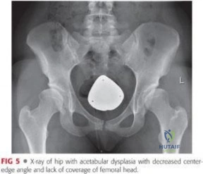
Conversely, joint preservation techniques are strictly indicated for young, active patients with symptomatic structural abnormalities but preserved articular cartilage. The Bernese Periacetabular Osteotomy (PAO) is the gold standard for symptomatic developmental dysplasia of the hip (DDH) in adults with closed triradiate cartilages and minimal osteoarthritic changes (Tönnis grade 0 or 1). Surgical dislocation of the hip is indicated for complex femoroacetabular impingement (FAI), slipped capital femoral epiphysis (SCFE) deformities, and the excision of osteochondral lesions. These procedures are contraindicated in the presence of advanced joint space narrowing, diffuse cartilage delamination, or severe osteopenia that would compromise osteotomy fixation.
Salvage Procedures and Amputation
Arthrodesis (joint fusion) serves as a highly durable salvage procedure when arthroplasty is contraindicated or has catastrophically failed. Ankle arthrodesis is the treatment of choice for end-stage post-traumatic tibiotalar arthrosis, particularly in young laborers who would rapidly wear out a total ankle replacement. Knee arthrodesis is primarily indicated as a salvage for an infected, multiply-revised TKA where the extensor mechanism has been destroyed or bone stock is insufficient for reimplantation. Contraindications to arthrodesis include bilateral adjacent joint disease (which would severely limit overall mobility) and an inability to achieve a biologically viable bone bed.
Amputation must never be viewed as a failure of care, but rather as a definitive reconstructive procedure aimed at restoring mobility via a prosthesis. Indications include unsalvageable severe extremity trauma (e.g., Mangled Extremity Severity Score > 7), overwhelming necrotizing infections, malignant bone or soft tissue tumors where limb salvage would compromise oncological margins, and end-stage peripheral vascular disease with intractable rest pain or gangrene. Contraindications are relative and primarily involve the patient's physiological inability to survive the surgical insult or the lack of proximal vascular inflow to heal the chosen amputation level.
| Procedure Category | Primary Indications | Absolute Contraindications | Relative Contraindications |
|---|---|---|---|
| Total Joint Arthroplasty | End-stage OA, Inflammatory arthritis, Displaced femoral neck fractures (elderly) | Active local/systemic infection, Neuropathic joint (Charcot), Absent extensor mechanism (TKA) | Morbid obesity, Severe medical comorbidities, Poor dentition |
| Joint Preservation (PAO/Dislocation) | Symptomatic DDH, Complex FAI, SCFE deformities, Pipkin fractures | Advanced OA (Tönnis > 1), Open triradiate cartilage (for PAO), Severe osteopenia | Age > 45 years, Obesity, Significant joint stiffness |
| Arthrodesis (Fusion) | Post-traumatic arthrosis (young laborers), Failed infected arthroplasty, Paralytic deformities | Adjacent joint severe arthrosis, Active untreated infection (for internal fixation) | Significant limb length discrepancy, Poor bone stock |
| Amputation | Unsalvageable trauma, Ischemic gangrene, Invasive malignancy, Chronic refractory infection | Lack of proximal vascular inflow to heal the stump level | Severe cardiopulmonary instability, Non-ambulatory status (may opt for higher level for healing) |
Pre-Operative Planning, Templating, and Patient Positioning
Digital Templating and Deficit Management
The success of any reconstructive orthopedic procedure is predicated on exhaustive preoperative planning. In total hip and knee arthroplasty, digital templating on calibrated radiographs is mandatory. For THA, the surgeon must identify the center of rotation, calculate the required femoral offset, and assess the leg length discrepancy. The acetabular component size is templated to achieve a line-to-line fit or slight press-fit with the native subchondral bone, ensuring the teardrop and the ilioischial line are respected. The femoral stem is templated to determine the level of the neck cut and the required intramedullary sizing to achieve rigid diaphyseal or metaphyseal fixation.
In revision scenarios, preoperative planning must account for massive bone loss. The Paprosky Classification is utilized to grade acetabular and femoral defects and dictate the surgical strategy. For acetabular cavitary defects (Paprosky Type I and IIA), particulate cancellous impaction bone grafting is planned. Segmental defects (Type IIB and IIC) require structural allografts or highly porous trabecular metal augments. In the setting of pelvic discontinuity (Type III), where the superior and inferior hemipelvis are mechanically separated, the surgeon must prepare for the utilization of an antiprotrusio cage, a custom triflange component, or a cup-cage construct combined with massive structural allografting.
For TKA, templating involves determining the mechanical axis and the degree of deformity. The surgeon plans the distal femoral and proximal tibial resection levels to balance the flexion and extension gaps. In cases of severe extra-articular deformity or retained hardware, conventional intramedullary guides may be contraindicated, necessitating the use of computer-assisted navigation, robotic-arm assistance, or custom patient-specific instrumentation (PSI) based on preoperative advanced imaging (CT or MRI).
Patient Positioning and Fluoroscopic Setup
Patient positioning is the critical first step in the operating theater; improper positioning inevitably leads to compromised exposure and malpositioned implants. For the posterior approach to the hip, the patient is placed in the lateral decubitus position. Rigid pelvic fixation using anterior and posterior supports is absolutely mandatory. If the pelvis rolls anteriorly or posteriorly during the procedure, the surgeon's spatial orientation is lost, leading to inaccurate assessment of acetabular component version and inclination, which dramatically increases the risk of postoperative dislocation.
The ilioinguinal approach requires the patient to be positioned supine on a radiolucent Jackson or OSI table to allow for unrestricted intraoperative fluoroscopy. The ipsilateral lower extremity must be draped freely to allow for manipulation, specifically flexion of the hip and knee, which relaxes the iliopsoas muscle and the femoral nerve, facilitating their retraction and opening the middle anatomical window. A Foley catheter is placed to decompress the bladder, and the abdomen is prepped widely to allow for proximal extension of the incision if necessary.
For complex trauma and joint preservation surgeries, the fluoroscopic setup must be confirmed prior to incision. The C-arm must be able to obtain perfect orthogonal views (e.g., AP, Judet oblique views for the pelvis; precise lateral views for the knee or femur) without being obstructed by the operating table or patient positioning devices. In procedures like the Bernese PAO, continuous fluoroscopic feedback is essential to ensure the ischial and pubic osteotomies are complete and that the posterior column remains structurally intact.
Step-by-Step Surgical Approach and Fixation Technique
The Posterior Approach to the Hip (Moore/Gibson)
The posterior approach remains the workhorse for primary and revision THA, hemiarthroplasty, and the dependent drainage of septic hips. It provides unparalleled, extensile exposure of the acetabulum and proximal femur without violating the critical hip abductor mechanism (gluteus medius and minimus).
- Incision and Superficial Dissection: A 10 to 15 cm curved incision is centered over the posterior aspect of the greater trochanter, extending proximally toward the posterior superior iliac spine (PSIS) and distally along the axis of the femoral shaft. The subcutaneous tissue is divided, and the fascia lata is incised distally. This incision is carried proximally to split the coarse fibers of the gluteus maximus bluntly, utilizing the internervous plane between the superior gluteal nerve (supplying the proximal half) and the inferior gluteal nerve (supplying the distal half).
- Deep Dissection and Nerve Protection: A Charnley or self-retaining retractor is placed to maintain the split in the gluteus maximus. The bursa overlying the short external rotators is excised. The piriformis, superior gemellus, obturator internus, and inferior gemellus are identified. The sciatic nerve must be visually or palpably identified exiting the greater sciatic notch inferior to the piriformis; it is protected throughout the procedure, strictly avoiding excessive medial retraction which induces neuropraxia.
- Tendon Release and Capsulotomy: The short external rotators and the conjoint tendon are tagged with heavy non-absorbable sutures and transected directly at their femoral insertion on the greater trochanter. The quadratus femoris is partially released at its superior border if additional exposure is required, taking extreme care to isolate and electrocoagulate the terminal branches of the medial femoral circumflex artery (MFCA) to prevent postoperative hematoma. A T-shaped or crucial capsulotomy is then performed, preserving the capsule for later meticulous repair, which significantly reduces the risk of posterior dislocation.
The Ilioinguinal Approach to the Acetabulum (Letournel and Judet)
The ilioinguinal approach is the definitive exposure for anterior column, anterior wall, and associated both-column acetabular fractures. It allows access to the entire internal iliac fossa, the pelvic brim, and the superior pubic ramus.
- Incision and Superficial Dissection: The incision begins at the pubic symphysis, extends laterally along the inguinal ligament to the anterior superior iliac spine (ASIS), and continues proximally along the anterior two-thirds of the iliac crest. The external oblique aponeurosis is incised in line with the inguinal ligament, opening the inguinal canal. The spermatic cord or round ligament is mobilized and protected with a Penrose drain.
- Creation of the Three Windows: The approach relies on working through three distinct anatomical windows.
- The Lateral Window: Created by elevating the iliacus muscle from the internal iliac fossa, providing access to the sacroiliac joint and the proximal anterior column.
- The Middle Window: Accessed by mobilizing the iliopsoas muscle and the femoral nerve laterally, and the external iliac vessels medially. The iliopectineal fascia, which separates these structures, must be sharply divided down to the pelvic brim.
- The Medial Window: Accessed medial to the external iliac vessels and lateral to the rectus abdominis.
- Vascular Management and Fixation: During the development of the medial window, the posterior aspect of the superior pubic ramus is exposed. The surgeon must meticulously search for the Corona Mortis. If present, it must be doubly ligated and divided to prevent retraction and exsanguination. Fracture reduction is achieved using specialized pelvic reduction forceps (e.g., Jungbluth clamps) applied across the pelvic brim, and fixation is secured with reconstruction plates contoured precisely to the complex three-dimensional anatomy of the innominate bone.
Total Hip and Knee Arthroplasty Execution
Cementless THA Execution: Following dislocation and femoral neck resection, the acetabulum is exposed using specialized retractors (e.g., anterior, posterior, and inferior Hohmann retractors). The labrum and pulvinar are excised. The acetabulum is reamed sequentially to bleeding subchondral bone, preserving the subchondral plate for structural support. The cementless cup is impacted with a press-fit technique, aiming strictly for the safe zone of 40° of inclination and 15° to 20° of anteversion. The femoral canal is then sequentially broached. The cementless stem relies on initial mechanical stability via diaphyseal or metaphyseal press-fit, which allows for secondary biological fixation (osteointegration) via the titanium porous coating.
Primary TKA Execution: A medial parapatellar arthrotomy is the standard approach, though subvastus or midvastus approaches may be utilized in select patients to preserve the extensor mechanism. The distal femoral resection is executed based on the preoperative templating (typically 5° to 7° valgus). Femoral sizing and external rotation (usually 3°) are established using the posterior condylar axis, Whiteside’s line (the anteroposterior axis), and the surgical transepicondylar axis. The proximal tibia is resected perpendicular to its mechanical axis in the coronal plane, incorporating the native posterior slope. Soft tissue balancing is then meticulously performed; varus deformities require medial releases (deep MCL, posteromedial corner), while valgus deformities require lateral pie-crusting (ITB, LCL, popliteus). The goal is perfectly symmetric rectangular flexion and extension gaps.
Hip Preservation: Surgical Dislocation and Periacetabular Osteotomy
Surgical Dislocation (Ganz): This revolutionary technique allows full 360-degree visualization of the femoral head. A lateral approach is utilized, and a trochanteric flip osteotomy is performed, leaving the gluteus medius and vastus lateralis attached to the mobile greater trochanter fragment. The capsule is incised via a Z-capsulotomy. The hip is dislocated anteriorly with external rotation and flexion. The deep branch of the MFCA is protected by keeping the external rotators intact and avoiding any tension on the posterior capsule. Following the treatment of the intra-articular pathology (e.g., osteochondroplasty for FAI), the hip is reduced, and the trochanter is rigidly fixed with cortical screws.
Bernese Periacetabular Osteotomy (PAO): The PAO involves a series of extra-articular osteotomies to reorient the dysplastic acetabulum. Through a modified Smith-Petersen or bikini incision, the surgeon sequentially performs an incomplete ischial osteotomy (using a curved osteotome directed infracotyloid), a complete superior pubic ramus osteotomy, and a complex biplanar iliac osteotomy. The final retroacetabular cut connects the iliac and ischial osteotomies, completely mobilizing the acetabular fragment while leaving the posterior column intact for pelvic stability. The fragment is then rotated laterally and anteriorly to optimize femoral head coverage and fixed with multiple cortical screws.
Arthrodesis, Amputation, and Infection Management
Ankle Arthrodesis (Transfibular Approach): The distal fibula is resected approximately 5 cm proximal to the joint line and bisected longitudinally to serve as a biological onlay strut graft. The articular cartilage of the tibiotalar joint is meticulously debrided using curettes and osteotomes down to bleeding subchondral bone. The joint must be positioned perfectly: neutral dorsiflexion, 0° to 5° of valgus, and 5° of external rotation to match the contralateral limb. Rigid internal fixation is achieved using two or three large-fragment (6.5mm or 7.3mm) crossed cannulated lag screws, or a dedicated anterior locking plate, ensuring massive compression across the fusion interface.
Transtibial Amputation (Burgess Technique): The tibia is transected 12 to 15 cm distal to the joint line. The anterior cortex of the tibia is beveled at a 45° angle and smoothed with a rasp to prevent skin breakdown over a sharp bony prominence. The fibula is transected 1 to 2 cm shorter than the tibia. The tibial and peroneal nerves are managed via traction neurectomy. The critical step is the myodesis: the robust posterior muscle fascia of the gastrocnemius-soleus complex is brought anteriorly and sutured directly to the anterior periosteum and deep fascia via drill holes in the distal tibia. This provides a dynamic, robust soft-tissue envelope capable of withstanding the sheer forces of a prosthetic socket.
Management of Chronic Osteomyelitis: The eradication of deep bone infection requires principles akin to oncological surgery. Radical sequestrectomy is performed; all necrotic, avascular bone and fibrotic soft tissue must be excised until punctate bleeding—the "paprika sign"—is uniformly observed on the remaining bone surfaces. The resulting massive dead space must be managed. An antibiotic bead pouch is often utilized: Polymethylmethacrylate (PMMA) beads are mixed with heat-stable antibiotics (e.g., Vancomycin, Tobramycin), placed into the bony defect, and sealed with an artificial membrane to generate local antibiotic concentrations hundreds of times higher than systemic levels without nephrotoxicity. Once the infection is cleared, the defect is reconstructed via the Papineau technique (open cancellous grafting), bone transport (Ilizarov), or vascularized fibular grafting.
Complications, Incidence Rates, and Salvage Management
Surgical intervention in advanced orthopaedics carries inherent risks. The surgeon must be intimately familiar with the incidence, prevention, and salvage management of catastrophic complications.
Intraoperative and Early Postoperative Complications
Neurovascular injury is a devastating intraoperative complication. Sciatic nerve palsy occurs in approximately 1% to 3% of primary THAs and up to 8% in complex revisions or PAOs. It typically presents as a peroneal division deficit (foot drop) due to the superficial and lateral anatomical position of the peroneal fibers within the sciatic sheath. Management includes immediate removal of any compressive hematoma, ensuring the limb is not lengthened excessively, and utilizing an ankle-foot orthosis (AFO) while awaiting nerve recovery, which can take up to 18 months.
Vascular injuries, though rare (incidence < 0.2% in THA/TKA), are limb-threatening emergencies. The Corona Mortis hemorrhage during the ilioinguinal approach or popliteal artery injury during the posterior capsular release in TKA requires immediate recognition. The surgeon must pack the wound, obtain proximal and distal control, and consult vascular surgery immediately for primary repair or interposition grafting. Tourniquet use in TKA must be judicious to minimize the risk of reperfusion injury and deep vein thrombosis (DVT).
Prosthetic Joint Infection (PJI) is the most dreaded early to intermediate complication, occurring in 1% to 2% of primary arthroplasties. Acute postoperative infections (within 4 weeks) or acute hematogenous infections can sometimes be managed with a DAIR procedure (Debridement, Antibiotics, and Implant Retention) combined with modular polyethylene exchange. However, chronic PJI requires a definitive two-stage exchange arthroplasty: complete removal of all hardware and cement, placement of an articulating or static antibiotic-loaded PMMA spacer, 6 weeks of targeted intravenous antibiotics, and subsequent reimplantation once serum inflammatory markers (CRP, ESR) normalize and joint aspirations are sterile.
Late Complications and Revision Strategies
Aseptic loosening is the most common cause of late failure in joint arthroplasty, driven by macrophage-mediated osteolysis in response to particulate wear debris (polyethylene, metal, or cement). This insidious process leads to massive cavitary and segmental bone loss. Salvage management requires complex revision surgery utilizing the principles of bypass fixation. In the femur, a loose primary stem is bypassed using a long, diaphyseal-engaging, fluted, tapered titanium stem to achieve rigid fixation distal to the osteolytic defect.
In amputation surgery, late complications include symptomatic neuroma formation, heterotopic ossification, and socket-related skin breakdown. A symptomatic neuroma that fails conservative management (gabapentinoids, targeted steroid injections) requires surgical excision. The salvage technique involves resecting the neuroma and burying the proximal nerve stump deep within a muscle belly or into a drilled hole in the adjacent bone (TMR - Targeted Muscle Reinnervation is an advanced alternative) to shield it from mechanical irritation.
| Complication | Estimated Incidence | Primary Prevention Strategy | Salvage / Management Protocol |
|---|---|---|---|
| Sciatic Nerve Palsy | 1% - 3% (Primary THA) | Direct visualization, avoid excessive lengthening, careful retraction | AFO bracing, hematoma evacuation, neurolysis if entrapped |
| Corona Mortis Hemorrhage | Up to 80% prevalence | Meticulous dissection of superior pubic ramus, double ligation | Immediate packing, proximal/distal control, vascular repair |
| Prosthetic Joint Infection (PJI) | 1% - 2% (Primary) | Strict asepsis, prophylactic antibiotics, optimized host factors |
