Masterclass in Fundamental Orthopaedic Surgical Techniques and Approaches
Key Takeaway
Mastering fundamental orthopaedic surgical techniques and anatomical approaches is paramount for achieving optimal patient outcomes. This comprehensive guide details essential perioperative protocols, including tourniquet application, patient positioning, and bone grafting principles. Furthermore, it provides a step-by-step analysis of major surgical approaches to the hip, knee, and shoulder, emphasizing biomechanics, internervous planes, and evidence-based postoperative care for orthopaedic surgeons and residents.
Comprehensive Introduction and Patho-Epidemiology
The field of orthopaedic surgery is defined by a continuous, dynamic evolution of innovative techniques, advanced instrumentation, and a profoundly deepening understanding of musculoskeletal biomechanics. For the orthopaedic resident, fellow, and practicing consultant, absolute mastery of surgical approaches and fundamental operative techniques forms the unequivocal bedrock of successful patient outcomes. This masterclass synthesizes the core principles of operative orthopaedics, transitioning from essential perioperative management—such as precise tourniquet application, physiological patient positioning, and strict infection control—to the intricate, step-by-step execution of foundational surgical approaches. The modern orthopaedic surgeon must transcend the role of a mere technician, operating instead as an applied anatomist and biomechanical engineer who respects the delicate interplay between the soft tissue envelope and osseous structures.
The epidemiological burden of musculoskeletal disease continues to rise exponentially, driven by an aging global population, the increasing prevalence of obesity, and the high-energy trauma associated with modern transportation and extreme sports. Degenerative joint diseases, particularly osteoarthritis of the hip and knee, now represent one of the leading causes of global disability, necessitating millions of arthroplasty procedures annually. Simultaneously, the incidence of fragility fractures in the osteoporotic population demands surgical interventions that not only restore anatomical alignment but also respect the diminished biological healing capacity of the host. Consequently, the operative techniques employed must be meticulously selected to optimize biomechanical stability while minimizing iatrogenic trauma to the surrounding vascular and neurological networks.
Strict adherence to anatomical internervous planes, meticulous soft tissue handling, and evidence-based postoperative protocols are paramount in mitigating the inherent risks of surgical intervention. An internervous plane—a dissection path between two muscles supplied by different peripheral nerves—allows for extensile exposure without denervating the musculature, thereby preserving dynamic joint stability and accelerating functional recovery. Furthermore, the biological imperatives of fracture healing and soft tissue integration dictate that surgical approaches must preserve the periosteal blood supply and minimize disruption to the local hematoma when applicable.
This comprehensive text provides a postgraduate-level analysis of these critical components, designed to serve as an authoritative reference for FRCS, EBOT, and AAOS board candidates. By deconstructing the patho-epidemiology of musculoskeletal disorders and correlating it with advanced surgical anatomy, the surgeon is equipped to navigate complex primary and revision scenarios. The subsequent sections will systematically detail the anatomical, technical, and rehabilitative pillars required to execute the most pivotal surgical approaches in the orthopaedic armamentarium, ensuring the highest standard of evidence-based patient care.
Detailed Surgical Anatomy and Biomechanics
A profound understanding of surgical anatomy and regional biomechanics is the quintessential element that separates the master surgeon from the surgical mechanic. In the hip, the posterior approach (Moore or Southern approach) relies on a deep understanding of the pelvitrochanteric musculature and the sciatic nerve's trajectory. The gluteus maximus, innervated solely by the inferior gluteal nerve, is split in line with its fibers, representing a safe, non-internervous dissection that avoids denervation. Deep to this, the short external rotators (piriformis, superior gemellus, obturator internus, inferior gemellus, and quadratus femoris) function as dynamic stabilizers of the hip. The critical vascular anatomy here involves the medial circumflex femoral artery (MCFA), the primary blood supply to the femoral head, which courses superior to the upper border of the quadratus femoris. Biomechanically, the hip is a constrained ball-and-socket joint where stability is dictated by bony offset, abductor tension, and the integrity of the posterior capsule, all of which must be meticulously managed during reconstruction.
In the knee, the medial parapatellar approach requires navigation of the extensor mechanism, a complex biomechanical pulley system governed by the quadriceps tendon, patella, and patellar ligament. The dissection violates the medial retinaculum and the vastus medialis obliquus (VMO) expansion, which are critical for dynamic patellar tracking. The knee's biomechanics are defined by a complex "roll-and-glide" mechanism dictated by the cruciate ligaments and the meniscofemoral geometry. Surgical exposure must respect the blood supply to the patella, predominantly derived from the superior and inferior genicular arteries, forming an anastomotic ring. Overzealous lateral release combined with a medial arthrotomy can critically compromise this vascularity, leading to devastating avascular necrosis of the patella.
The shoulder's deltopectoral approach is a masterclass in utilizing a true internervous plane. The interval lies between the deltoid, innervated by the axillary nerve, and the pectoralis major, innervated by the medial and lateral pectoral nerves. The cephalic vein serves as the anatomical lighthouse for this interval. Deep dissection exposes the conjoined tendon (coracobrachialis and short head of the biceps), which must be retracted medially with extreme caution to avoid traction neurapraxia to the musculocutaneous nerve, which typically enters the muscle belly 5 to 8 centimeters distal to the coracoid process. Biomechanically, the glenohumeral joint is inherently unstable, relying on the rotator cuff force couples for dynamic concavity compression. Management of the subscapularis during this approach is therefore critical; whether via tenotomy, peel, or lesser tuberosity osteotomy, its anatomical restoration is non-negotiable for postoperative anterior stability and internal rotation strength.
In the ankle, the lateral approach to the fibula navigates the delicate subcutaneous tissue of the lateral leg, demanding strict attention to the superficial peroneal nerve proximally and the sural nerve distally. The internervous plane proximally lies between the peroneus tertius (deep peroneal nerve) and the peroneus brevis (superficial peroneal nerve). The lateral malleolus serves as the primary lateral buttress of the ankle mortise, resisting talar shift. Biomechanically, even a 1-millimeter lateral shift of the talus can decrease the tibiotalar contact area by up to 42%, exponentially increasing joint contact stresses and precipitating rapid post-traumatic arthrosis. Therefore, the surgical anatomy must be exposed with minimal periosteal stripping to facilitate an anatomical, rigid reduction of the fibula and restoration of the syndesmotic ligamentous complex.
Exhaustive Indications and Contraindications
The decision to proceed with operative intervention requires a rigorous synthesis of clinical pathology, patient-specific physiological reserves, and a deep understanding of the capabilities and limitations of specific surgical approaches. Absolute indications for the major orthopaedic approaches typically revolve around catastrophic structural failure, such as displaced intra-articular fractures, severe degenerative joint disease refractory to exhaustive conservative management, and acute neurovascular compromise secondary to fracture-dislocations. For instance, the posterior approach to the hip is absolutely indicated for the treatment of displaced femoral neck fractures in the elderly requiring hemiarthroplasty, as well as for primary total hip arthroplasty (THA) in the setting of end-stage osteoarthritis where posterior acetabular exposure is required.
Relative indications encompass scenarios where surgery offers significant functional improvement but must be weighed against host factors. A deltopectoral approach for a proximal humerus fracture may be relatively indicated in a physiologically robust 70-year-old with a three-part fracture, whereas conservative management might be chosen for a frail patient with the identical fracture pattern. Similarly, the use of structural allograft versus autologous iliac crest bone graft (ICBG) is a relative decision based on the required volume of graft, the need for immediate structural support (osteoconduction), versus the biological necessity for osteoprogenitor cells (osteogenesis) in the setting of a recalcitrant nonunion.
Contraindications are equally critical in surgical decision-making. Absolute contraindications universally include active, untreated local or systemic infection (unless the procedure is explicitly for source control, such as an irrigation and debridement), severe medical comorbidities precluding safe anesthesia, and an uncooperative patient incapable of adhering to postoperative rehabilitation protocols. For specific approaches, severe peripheral vascular disease may absolutely contraindicate a tourniquet-assisted medial parapatellar approach due to the risk of critical limb ischemia. Relative contraindications often involve compromised soft tissue envelopes, such as severe venous stasis disease or prior irradiated skin over the lateral fibula, which exponentially increases the risk of wound dehiscence and deep hardware infection.
| Surgical Approach / Technique | Absolute Indications | Relative Indications | Absolute Contraindications | Relative Contraindications |
|---|---|---|---|---|
| Posterior Hip Approach | Displaced femoral neck fracture (hemiarthroplasty); End-stage hip OA; Posterior acetabular fractures. | Revision THA with posterior bone loss; Removal of posterior intra-articular loose bodies. | Active joint infection (for primary arthroplasty); Severe patient non-compliance. | History of recurrent posterior dislocations; Severe hip flexion contracture. |
| Medial Parapatellar Knee | End-stage knee OA (TKA); Complex intra-articular distal femur/proximal tibia fractures. | Extensive synovectomy; Complex ligamentous multiligament reconstruction. | Active joint infection (for primary TKA); Charcot arthropathy (relative to absolute). | Prior compromised lateral soft tissue envelope; Severe patella baja. |
| Deltopectoral Shoulder | End-stage glenohumeral OA (TSA); Displaced 3- or 4-part proximal humerus fractures. | Recurrent anterior instability (open Bankart); Chronic retracted subscapularis tears. | Active joint infection; Deltoid paralysis (for anatomic TSA). | Prior extensive anterior surgical scarring; Severe glenoid retroversion requiring posterior approach. |
| Lateral Fibula Approach | Displaced lateral malleolus fractures; Unstable syndesmotic injuries. | Chronic lateral ligamentous instability (Brostrom); Fibular osteotomies. | Active cellulitis over the lateral incision site; Critical limb ischemia. | Severe venous stasis dermatitis; Poorly controlled diabetes mellitus with neuropathy. |
| Iliac Crest Bone Grafting | Atrophic nonunions requiring osteoprogenitor cells; Major cavitary bone defects. | Arthrodesis procedures (spine, foot/ankle); Augmentation of hardware fixation. | Donor site active infection; Previous catastrophic harvest site morbidity. | Severe osteoporosis (poor graft quality); Chronic pain syndromes at the pelvis. |
Pre-Operative Planning, Templating, and Patient Positioning
Meticulous preoperative planning is the invisible foundation upon which all successful orthopaedic procedures are built. This begins with high-quality, orthogonally acquired radiographs and the rigorous application of digital templating software. Templating is not merely an exercise in implant sizing; it is a critical predictive tool for restoring joint biomechanics, calculating necessary bony resections, and anticipating intraoperative challenges. In total hip arthroplasty, templating dictates the center of rotation, femoral offset, and leg length restoration. The surgeon must utilize a known magnification marker (typically a 25mm radiopaque sphere positioned at the level of the greater trochanter) to calibrate the digital images accurately. Failure to template accurately can result in catastrophic leg length discrepancies, global joint instability, and premature catastrophic wear of the bearing surfaces.
The pneumatic tourniquet is an indispensable tool in extremity surgery, providing a bloodless operative field that facilitates meticulous dissection and precise implant placement. However, its use requires strict adherence to physiological principles. Tourniquet pressure must be individualized based on the patient's Limb Occlusion Pressure (LOP). Modern practice dictates adding a safety margin to the LOP (e.g., 40 mm Hg for LOP <130 mm Hg; 60 mm Hg for LOP 131–190 mm Hg). Time limits are equally critical; ischemic time should ideally not exceed 120 minutes. If prolonged surgery is anticipated, the tourniquet must be deflated for 15 to 20 minutes to allow for reperfusion and clearance of metabolic byproducts before reinflation. The limb must be exsanguinated using an Esmarch bandage prior to inflation, except in cases of infection or malignancy, where simple elevation for 3 to 5 minutes is preferred to prevent systemic dissemination. Tourniquet paralysis is a devastating complication typically resulting from excessive mechanical pressure rather than ischemia; therefore, adequate padding with cast padding, completely devoid of wrinkles, is mandatory.
Optimal patient positioning is critical for surgical exposure, anesthesia access, and the prevention of perioperative complications such as pressure ulcers and peripheral nerve injuries. The supine position is standard for anterior approaches to the hip, knee, and ankle. The surgeon must ensure the heels are floated and the ulnar nerve at the cubital tunnel is meticulously padded. The lateral decubitus position, frequently utilized for posterior approaches to the hip, requires the patient to be rigidly secured with a pelvic positioner (peg board or bean bag). An axillary roll is absolutely mandatory, placed just caudal to the axilla of the dependent arm, to prevent catastrophic brachial plexus traction injuries. The beach-chair position for shoulder surgery requires careful head and neck neutral alignment to prevent cerebral hypoperfusion and cervical nerve root traction.
Surgical site infections (SSIs) remain a catastrophic complication, particularly in the presence of orthopaedic hardware. Skin preparation with alcohol-based chlorhexidine solutions is the gold standard, demonstrating superior efficacy in reducing skin flora compared to aqueous povidone-iodine. The solution must be allowed to dry completely to maximize its bactericidal effect and prevent catastrophic operating room fires. Impervious, sterile drapes must isolate the operative field, and iodine-impregnated incise drapes (Ioban) are frequently utilized to sequester skin flora. Copious irrigation with normal saline is fundamental throughout the procedure, and the addition of dilute betadine (0.35%) to irrigation solutions prior to closure has been definitively shown in recent literature to significantly reduce SSI rates across multiple orthopaedic subspecialties.
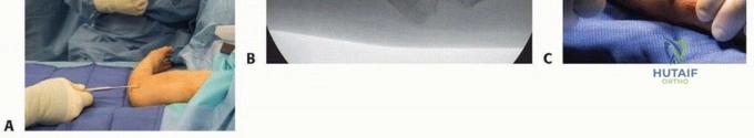
Step-by-Step Surgical Approach and Fixation Technique
The execution of a major orthopaedic approach requires absolute precision, respect for tissue planes, and a systematic progression from skin incision to deep articular exposure.
The Posterior Approach to the Hip
The patient is positioned in the lateral decubitus position. A 10-15 cm curved incision is centered over the posterior third of the greater trochanter, extending proximally toward the posterior superior iliac spine (PSIS) and distally along the femoral shaft. Superficial dissection involves incising the fascia lata distally and bluntly splitting the gluteus maximus proximally in line with its fibers. Deep dissection requires immediate identification and protection of the sciatic nerve, which is often palpable posterior to the short external rotators. The short external rotators (piriformis, superior gemellus, obturator internus, and inferior gemellus) are tagged with heavy non-absorbable suture, released at their tendinous insertion on the greater trochanter, and reflected posteriorly to act as a soft-tissue buffer protecting the sciatic nerve. A T-shaped or crucial capsulotomy is then performed, allowing for dislocation of the femoral head via internal rotation, flexion, and adduction.
The Medial Parapatellar Approach to the Knee
With the patient supine and a tourniquet applied, a longitudinal midline incision is made from 5 cm proximal to the superior pole of the patella, extending distally to the medial border of the tibial tubercle. Superficial dissection elevates full-thickness medial and lateral fasciocutaneous flaps to expose the entire extensor mechanism. The deep arthrotomy begins by incising the quadriceps tendon longitudinally, leaving a 3-4 mm cuff of tendon on the vastus medialis to facilitate robust closure. The incision continues distally around the medial border of the patella and along the medial border of the patellar tendon down to the tibial tubercle. The patella is then carefully everted or subluxated laterally. The retropatellar fat pad is partially excised, and the anterior horns of the menisci are released to fully expose the tibiofemoral joint, taking care to protect the collateral ligaments during retractor placement.
The Deltopectoral Approach to the Shoulder
The patient is positioned in the beach-chair position. A linear incision is made from the tip of the coracoid process, extending distally and laterally along the deltopectoral groove. The cephalic vein is identified, marking the internervous plane. The vein is typically retracted laterally with the deltoid to preserve its major venous tributaries, minimizing bleeding. Deep dissection involves retracting the conjoined tendon medially, taking extreme care not to place vigorous traction that could injure the musculocutaneous nerve. The clavipectoral fascia is incised, exposing the subscapularis tendon. Depending on the procedure, the subscapularis is managed via a tenotomy, a peel off the lesser tuberosity, or a lesser tuberosity osteotomy. The underlying capsule is then incised, providing direct access to the humeral head and glenoid.
Principles of Fixation and Bone Grafting
Once exposure is achieved, reconstructive techniques such as tendon-to-bone fixation and bone grafting are frequently employed. The reattachment of tendon to bone (e.g., rotator cuff repair, subscapularis repair) relies on achieving rigid initial fixation to allow for the biological formation of Sharpey's fibers. Locking suture configurations, such as the Krackow or whipstitch, are mandatory as they provide superior pull-out strength by gripping the longitudinal collagen fibers of the tendon. These are secured to a decorticated bony bed using modern titanium, PEEK, or biocomposite suture anchors. When addressing bone defects or nonunions, the Iliac Crest Bone Graft (ICBG) remains the gold standard. A successful graft must provide osteogenesis (viable osteoprogenitor cells), osteoinduction (Bone Morphogenetic Proteins), and osteoconduction (a structural scaffold). When harvesting from the anterior iliac crest, the incision is made 2 cm posterior to the Anterior Superior Iliac Spine (ASIS) to avoid iatrogenic injury to the Lateral Femoral Cutaneous Nerve (LFCN). The inner table of the ilium should be preserved to maintain structural integrity and prevent catastrophic pelvic herniation.
Complications, Incidence Rates, and Salvage Management
Despite meticulous surgical technique and exhaustive preoperative planning, perioperative complications remain an inherent risk of orthopaedic surgery. The surgeon must be intimately familiar with the incidence, etiology, and immediate salvage management of these adverse events. Surgical Site Infection (SSI) is perhaps the most devastating complication, particularly in the presence of metallic implants. Periprosthetic joint infection (PJI) occurs in approximately 1-2% of primary arthroplasties and up to 5% of revision cases. The etiology is typically intraoperative contamination by skin flora (Staphylococcus aureus, Coagulase-negative staphylococci) or hematogenous seeding. Salvage management depends on the chronicity of the infection; acute postoperative infections (<4 weeks) may be managed with a thorough Debridement, Antibiotics, and Implant Retention (DAIR) with modular part exchange, whereas chronic infections mandate a two-stage revision arthroplasty utilizing an antibiotic-impregnated cement spacer.
Neurological injuries, while less frequent, carry profound medicolegal and functional implications. In the posterior approach to the hip, the sciatic nerve is at risk (incidence <1% in primary THA, up to 3% in revisions). Injury typically results from excessive retractor pressure, direct laceration, or excessive lengthening of the operative limb. Management includes immediate intraoperative exploration if a laceration is suspected, removal of compressive hematomas, and postoperative use of an Ankle-Foot Orthosis (AFO) to manage the resultant foot drop while awaiting potential nerve recovery, which can take up to 18-24 months. Similarly, the axillary nerve is at risk during the deltopectoral approach if dissection strays inferior to the subscapularis, leading to deltoid paralysis and catastrophic shoulder dysfunction.
Hardware failure and periprosthetic fractures represent significant biomechanical complications. Aseptic loosening is the most common long-term complication of joint replacement, driven by particulate wear debris leading to macrophage-induced osteolysis. Periprosthetic fractures can occur intraoperatively (e.g., broaching a femur) or postoperatively due to trauma. Intraoperative fractures must be recognized immediately and bypassed with longer stems or fixed with cerclage wires and locking plates. Postoperative fractures are managed based on the Vancouver (hip) or Felix (knee) classification systems, which dictate whether the implant is stable (amenable to ORIF) or loose (mandating revision arthroplasty).
| Complication | Estimated Incidence Rate | Primary Etiology | Salvage / Management Strategy |
|---|---|---|---|
| Periprosthetic Joint Infection (PJI) | 1.0% - 2.0% (Primary) | Intraoperative contamination; Hematogenous seeding. | Acute: DAIR with modular exchange. Chronic: Two-stage revision with antibiotic spacer. |
| Sciatic Nerve Palsy | 0.5% - 1.0% (Primary THA) | Retractor compression; Excessive limb lengthening; Hematoma. | Remove compression; AFO for foot drop; EMG at 6 weeks; Tendon transfers if no recovery at 1 year. |
| Tourniquet Paralysis | < 0.1% | Excessive mechanical pressure; Inadequate padding; Wrinkled drapes. | Supportive care; Physical therapy; Usually resolves spontaneously over weeks to months. |
| Intraoperative Periprosthetic Fracture | 1.0% - 3.0% (Uncemented THA) | Over-broaching; Poor bone quality; Excessive impaction force. | Immediate recognition; Cerclage wiring; Bypass fracture by 2 cortical diameters with a longer stem. |
| Aseptic Loosening / Osteolysis | 5.0% - 10.0% (at 15 years) | Particulate wear debris leading to macrophage-mediated bone resorption. | Revision arthroplasty with bone grafting of cavitary defects and use of highly cross-linked polyethylene. |
Phased Post-Operative Rehabilitation Protocols
The ultimate success of any orthopaedic procedure is inextricably linked to the execution of a highly structured, biologically phased postoperative rehabilitation protocol. The rehabilitation timeline must respect the physiological phases of tissue healing: the inflammatory phase (days 1-7), the reparative/proliferative phase (weeks 2-6), and the remodeling phase (months 2-12). Phase I (Acute/Tissue Healing) focuses on protecting the surgical repair, managing edema, and preventing catastrophic complications such as deep vein thrombosis (DVT) and arthrofibrosis. For procedures involving rigid internal fixation of fractures or primary arthroplasty (e.g., TKA, THA), early mobilization is paramount. Immediate weight-bearing as tolerated and aggressive early range of motion (ROM) exercises are initiated on postoperative day zero to stimulate synovial fluid diffusion, nourish the articular cartilage, and prevent capsular contracture. Conversely, procedures involving tendon-to-bone repairs (e.g., rotator cuff repair, subscapularis repair in TSA) require strict immobilization to protect the fragile Sharpey's fibers forming at the interface.
Phase II (Intermediate/Mobility) typically commences around weeks 4 to 6, coinciding with the transition from soft callus to hard callus in bone healing, and the maturation of collagen in soft tissue repairs. In the posterior approach to the hip, strict adherence to posterior hip precautions (avoiding flexion past 90 degrees, adduction across the midline, and internal rotation) is maintained for 6 weeks to allow the posterior capsule and short external rotators to heal, thereby preventing dislocation. Once this milestone is reached, precautions are gradually weaned, and therapy focuses on active ROM, concentric muscle activation, and gait normalization. For the deltopectoral approach, active internal rotation and passive external rotation are strictly restricted for the first 6 weeks to protect the subscapularis repair. In Phase II, active-assisted and active ROM are initiated to restore the glenohumeral force couples.
Phase III (Advanced/Strengthening and Proprioception) begins at months 3 to 4 and extends up to a year postoperatively. This phase is characterized by the application of progressive resistance, closed kinetic chain exercises, and plyometrics for the athletic patient. Bone remodeling continues in response to mechanical stress according to Wolff's Law. For the lateral malleolus fracture patient, therapy advances from simple weight-bearing in a boot to dynamic proprioceptive training on a balance board to restore the neuromuscular control of the peroneal musculature. Return to sport or heavy manual labor is dictated by objective criteria, including the restoration of 90% of contralateral limb strength, absence of effusion, and psychological readiness. The surgeon and physical therapist must maintain open, continuous communication to tailor these protocols to the patient's specific physiological responses and functional goals.
Summary of Landmark Literature and Clinical Guidelines
The practice of modern operative orthopaedics is deeply rooted in a robust foundation of landmark literature and evidence-based clinical guidelines. The American Academy of Orthopaedic Surgeons (AAOS) and the Centers for Disease Control and Prevention (CDC) provide definitive, peer-reviewed guidelines that dictate standard of care. Regarding Venous Thromboembolism (VTE) prophylaxis in total joint arthroplasty, the AAOS guidelines advocate for a risk-stratified approach. For standard-risk patients, the use of aspirin (typically 81mg twice daily for 4 weeks) combined with mechanical prophylaxis (sequential compression devices) has been definitively shown to provide equivalent efficacy to aggressive anticoagulants (e.g., low molecular weight heparin, direct oral anticoagulants) while significantly reducing the risk of devastating postoperative hematomas and subsequent periprosthetic joint infections.
The prevention of Surgical Site Infections (SSIs) has been heavily influenced by the work of Parvizi et al. and the International Consensus Meeting on Periprosthetic Joint Infection. Landmark consensus statements mandate the optimization of preoperative host factors (HbA1c < 7.0%, smoking cessation for 4-6 weeks, malnutrition correction), the use of weight-based prophylactic antibiotics (e.g., Cefazolin) administered within 60 minutes of incision, and the use of dilute povidone-iodine lavage prior to wound closure. These evidence-based interventions have collectively reduced the incidence of PJI from historical highs to the current benchmark of 1-2%.
Surgical approaches themselves are built upon the foundational anatomical treatises of pioneers such as Hoppenfeld and deBoer, whose texts on surgical exposures remain the definitive blueprint for internervous plane dissection. The posterior approach to the hip was popularized by Austin Moore, whose original descriptions of the pelvitrochanteric anatomy paved the way for modern, tissue-sparing modifications. In the shoulder, the legacy of Charles Neer dictates the management of proximal humerus fractures and shoulder arthroplasty via the deltopectoral interval. Neer's classification system and his emphasis on the preservation of the anterior humeral circumflex artery remain central to preventing avascular necrosis of the humeral head. Mastery of these historical texts, combined with a continuous appraisal of contemporary randomized controlled trials, ensures that the orthopaedic surgeon operates not merely on experience, but on the absolute forefront of scientific evidence.
