Febrile Foot Ulcers: A Deep Dive into Diagnosis, Surgical Anatomy, and Indications for Orthopedic Management
Key Takeaway
Febrile foot ulcers signal systemic infection, often indicating deep space infection or osteomyelitis, particularly in diabetic patients. Orthopedic management involves prompt diagnosis using clinical signs, elevated ESR/CRP, and advanced imaging. Surgical intervention is crucial for source control, especially with abscesses, confirmed osteomyelitis, or necrotizing fasciitis, requiring a thorough understanding of foot anatomy and biomechanics.
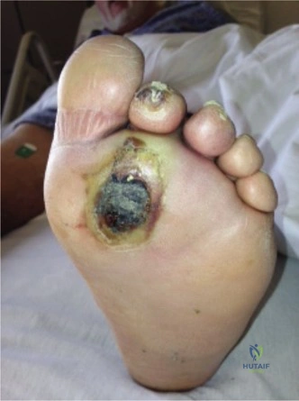
A 62-year-old patient with long-standing Type 2 diabetes presents to the emergency department with a 4-day history of a worsening, foul-smelling ulcer on the plantar aspect of the first metatarsal head. They are febrile (38.4°C) and tachycardic. On examination, there is surrounding erythema extending to the mid-foot and a positive probe-to-bone test. How would you systematically manage this patient?
Candidate: I would immediately stabilize the patient, start broad-spectrum IV antibiotics, and order inflammatory markers (ESR/CRP) and blood cultures. I would get plain radiographs, then admit the patient for surgical debridement and bone biopsy, involving a multidisciplinary team including endocrinology, ID, and vascular surgery.
Candidates often jump straight to "take them to theatre" without mentioning initial resuscitation, metabolic optimization (glucose control), or the critical need for a multidisciplinary team. Failing to emphasize the bone biopsy as the definitive diagnostic step for osteomyelitis is a significant oversight.
A high-scoring answer follows the Resuscitation-Diagnosis-Multidisciplinary-Debridement framework: 1. Stabilize: ABCDE approach, glycemic control, and systemic sepsis management. 2. Investigations: Bloods (CBC, ESR, CRP, cultures), imaging (X-rays, followed by MRI if osteomyelitis suspected). 3. MDT Approach: Immediate referral to Vascular (perfusion assessment), Endocrinology (glycemic optimization), and ID (antibiotic stewardship). 4. Surgical Goal: Urgent debridement of necrotic tissue and abscess drainage, with mandatory intraoperative deep bone biopsy (histopathology and cultures) to confirm osteomyelitis before finalizing antibiotic duration.
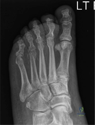
You have decided to proceed to the operating theatre for debridement. The radiograph shows evidence of cortical erosion at the first metatarsal head. Please describe your surgical technique for the debridement and how you would address the bone.
Candidate: I would perform a wide excision of the ulcer and all necrotic soft tissues. Regarding the bone, I would use a rongeur or saw to resect the infected metatarsal head until I reach bleeding, healthy bone (the 'paprika sign'), ensuring I take adequate samples for histology and culture.
Failing to mention the preservation of neurovascular structures or not discussing the "aftercare" of the bone (e.g., whether to leave the wound open for secondary intention or use NPWT). Also, failing to highlight that separate samples are needed for aerobic, anaerobic, and fungal cultures.
The perfect response focuses on "Source Control": 1. Incision: Incorporate the ulcer, avoid tension, and plan for potential flap coverage. 2. Debridement: Excising all non-viable soft tissue/tendon. 3. Bone Management: Resection to bleeding, viable margins. Using the term "saucerization" for cortical lesions or "ray amputation" if the metatarsal head is non-salvageable. 4. Specimen Handling: Explicitly state: "I will take deep bone samples for histology and separate culture media (aerobic/anaerobic/fungal)." 5. Closure Strategy: Wound left open, packing with non-adherent dressings or initiating Negative Pressure Wound Therapy (NPWT) if the base is healthy.
The patient recovers post-operatively, but 3 weeks later, they present with a recurrent ulcer near the site of the previous surgery. The patient is no longer febrile. What is your differential diagnosis and how do you investigate this recurrence?
Candidate: I would be concerned about residual or recurrent osteomyelitis, inadequate offloading, or vascular insufficiency. I would repeat inflammatory markers, repeat imaging (MRI is the gold standard here), and check their vascular status with ABIs/TBI or formal duplex imaging.
Focusing only on infection and forgetting biomechanics. Candidates often forget that recurrent ulceration in a neuropathic foot is frequently caused by offloading failure or secondary Charcot arthropathy resulting from altered gait mechanics.
Structure the answer into three pillars: 1. Infectious: Residual osteomyelitis vs. new infection. (Check ESR/CRP trend vs. baseline). 2. Vascular: Assess for progression of peripheral artery disease (Is there enough inflow for healing?). 3. Mechanical: Address the "offloading failure." Did the patient comply with TCC/specialized footwear? Evaluate for occult structural deformity/Charcot arthropathy (MRI/CT). Conclusion: If these are ruled out, it indicates a failure of the initial surgical correction, warranting a revisit to the MDT for potential structural reconstruction or higher-level amputation.
