
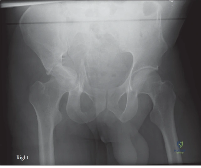
A 35-year-old male arrives in the Emergency Department following a high-energy motorcycle collision. He is hemodynamically stable. An AP pelvis radiograph is obtained.
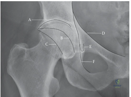
Describe your initial management and the specific radiographic findings you are looking for in this patient.
Candidate: I would follow ATLS protocols to ensure systemic stability. I'd perform a focused neurovascular examination, particularly looking for sciatic nerve palsy. Radiographically, I need Judet views—iliac and obturator obliques—and a high-resolution CT scan with 2D and 3D reconstructions to classify the fracture using the Judet and Letournel system, specifically assessing the articular congruity and displacement of the columns.
Candidates often jump straight to "I would plan for a Kocher-Langenbeck approach." This ignores the trauma context (ATLS/systemic safety) and lacks a structured approach to imaging. Furthermore, failing to mention the specific importance of the femoral head's concentric reduction on CT is a major oversight.
Start with a "Patient First" approach: ATLS, secondary survey, and neurovascular assessment (sciatic/femoral). In terms of imaging, define the goal: Anatomical assessment. Mention: 1. Plain film analysis (AP and Judet views) to identify column involvement. 2. CT with 2D/3D reconstruction as the definitive planning tool. 3. Specifically mention looking for: articular step-off (>2mm), marginal impaction of the posterior wall, quadrilateral surface displacement, and ensuring the femoral head remains concentrically reduced within the acetabulum.
Following your assessment, you identify a displaced posterior wall fracture with associated posterior hip subluxation. What are the absolute indications for surgical intervention, and how would you surgically approach this?
Candidate: Surgery is indicated for instability, incongruity, or significant displacement. For a posterior wall fracture, if the articular surface is significantly involved—typically >40%—or if the hip remains unstable on stress examination, I would proceed with a Kocher-Langenbeck approach to allow for anatomical reduction and buttress plate fixation.
Ignoring the "soft tissue" component. Candidates often forget to mention the risk to the sciatic nerve during the Kocher-Langenbeck approach or fail to address marginal impaction, which is common in posterior wall injuries and must be elevated and bone-grafted to prevent late failure.
Structure the answer clearly: Indications: Instability, intra-articular step-off >2mm, or incarceration of fragments. Surgical Approach: Kocher-Langenbeck. Key Technical Pearls: (1) Meticulous protection of the sciatic nerve. (2) Need to perform an arthrotomy to inspect the joint for loose bodies and articular impaction. (3) Elevate any impacted marginal cartilage and use bone graft. (4) Apply a buttress plate along the posterior rim to restore the posterior wall and provide rotational stability to the femoral head.
Detailed Chapters & Topics
Dive deeper into specialized chapters regarding acetabulum-fracture