
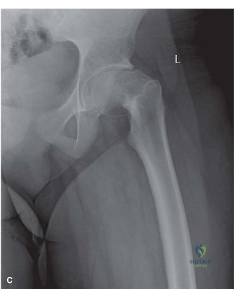
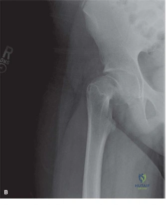
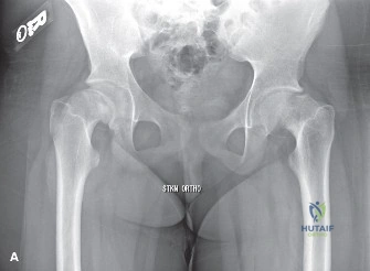
You are presented with a 13-year-old male who presents to the emergency department with acute onset of severe right hip pain following a minor fall. He is unable to bear weight. On examination, the hip is held in external rotation and he has a limited range of motion. AP and frog-leg lateral radiographs are ordered.
How do you classify this patient's condition, and what is your immediate priority in management?
Candidate: I would classify this as an unstable Slipped Capital Femoral Epiphysis (SCFE) because the patient is unable to bear weight. My immediate priority is to make the patient NPO, obtain urgent orthopaedic consultation, and plan for emergent surgical stabilization, likely with an in-situ pinning.
Candidates often fail to explicitly mention the "emergency" nature of an unstable slip. A poor answer focuses solely on the X-ray diagnosis without emphasizing that the inability to weight-bear classifies this as a surgical emergency to prevent AVN.
The candidate must define this as an Unstable SCFE (Loder's classification). Key elements: 1) Clinical emergency due to 50% risk of AVN. 2) Need for immediate NPO status and fluid resuscitation. 3) Discussion of the need for urgent in situ fixation. 4) Acknowledging that aggressive reduction is contraindicated to protect the medial femoral circumflex artery.
You are in the operating theatre preparing for the stabilization of this unstable SCFE. Describe the specific radiographic landmarks and the trajectory you will use to achieve optimal epiphyseal engagement while avoiding iatrogenic injury.
Candidate: I would make a small incision over the lateral proximal femur. I will place the guide wire in the anterior-superior quadrant of the epiphysis on the lateral view to counteract the posterior displacement. On the AP view, I will aim for the center of the head. I will ensure the tip is 2/3 to 3/4 into the epiphysis without penetrating the joint.
Failing to mention the specific danger of the posterior-inferior segment (where the retinacular vessels reside) or neglecting the importance of using multiple fluoroscopic views (AP, lateral, and oblique) to definitively rule out joint penetration.
A high-scoring answer focuses on "Anti-slip Trajectory." The candidate explains: 1) Entry point at the distal metaphysis to protect the trochanteric physis. 2) Aiming for the anterior-superior quadrant on the lateral view to provide mechanical resistance against further posterior translation. 3) Using multiple views (AP and true lateral) to verify that threads are entirely epiphyseal, with 3-4 threads of purchase, ensuring zero articular penetration.
The surgery for the right hip was successful. The parents are now asking about the long-term risks for the right hip, and they are concerned about the left hip as well. What do you tell them?
Candidate: For the right hip, the main risks are AVN, chondrolysis, and potential Femoroacetabular Impingement (FAI) from the residual cam deformity. Regarding the left hip, I would discuss the 20-60% risk of a contralateral slip and recommend prophylactic pinning, especially given the patient's age and potential underlying endocrine factors.
Dismissing the risk of FAI or failing to offer a structured plan for the contralateral hip. Borderline candidates fail to mention the need for metabolic/endocrine screening in younger or atypical patients.
The candidate demonstrates maturity by: 1) Categorizing risks (Short-term: AVN/Chondrolysis; Long-term: FAI/OA). 2) Addressing the "Cam deformity" as the cause of FAI. 3) Presenting the debate on prophylactic pinning objectively: acknowledging the 20-60% risk and the benefit of preventing a second emergency. 4) Suggesting an endocrine workup (thyroid, renal, etc.) given the patient's potential predisposition.
Detailed Chapters & Topics
Dive deeper into specialized chapters regarding 6-pediatrics-cases