Traumatic Hip Dislocations: Epidemiology, Anatomy, and Management Essentials
Key Takeaway
Traumatic hip dislocations (THD) are orthopedic emergencies caused by high-energy trauma, often involving motor vehicle collisions. They involve the femoral head displacing from the acetabulum, predominantly posteriorly. Prompt recognition and reduction are crucial due to the significant risk of complications like avascular necrosis of the femoral head, sciatic nerve injury, and post-traumatic arthritis if treatment is delayed.
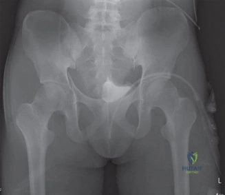
A 32-year-old male arrives in the Emergency Department following a high-speed motorcycle accident. He complains of severe left hip pain and is unable to bear weight. On examination, the left lower limb is held in a position of flexion, adduction, and internal rotation. There is no distal neurovascular deficit. Your resident has ordered an AP pelvis radiograph. What is your immediate management plan?
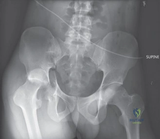
Candidate: I would immediately secure the airway and stabilize the patient according to ATLS protocols. Once stable, I would confirm the diagnosis of a posterior hip dislocation on the radiograph. The priority is urgent closed reduction under sedation/general anesthesia, ideally within 6 hours. Post-reduction, I would obtain a CT scan to rule out incarcerated fragments and associated fractures.
Candidates often jump straight to "take them to theater for closed reduction." They fail to mention the systemic trauma workup (ATLS), neglect to perform and document a dedicated neurovascular examination (specifically the sciatic nerve), or fail to emphasize that a CT scan is mandatory post-reduction, not just a plain film.
Start with ATLS principles and patient stability. State clearly: "This is an orthopedic emergency." 1. Clinical Exam: Document neurovascular status (sciatic nerve). 2. Immediate Goal: Urgent closed reduction (aiming for < 6 hours, ideally < 2 hours) to minimize AVN risk. 3. Procedural: Reduction under adequate anesthesia, followed by clinical assessment. 4. Post-procedure: Must obtain a CT scan to assess for femoral head fractures (Pipkin), acetabular rim fractures, and intra-articular debris. Do not clear the patient until the CT is reviewed for concentric joint reduction.
The patient has undergone a successful closed reduction. You are now reviewing the post-reduction CT scan. You identify a Pipkin Type II fracture of the femoral head. Discuss your management strategy.
Candidate: A Pipkin Type II fracture involves the femoral head superior to the fovea, which is the weight-bearing zone. Because this is an articular fracture, I would recommend Open Reduction Internal Fixation (ORIF). I would likely use a posterior approach (Kocher-Langenbeck) to visualize the fracture, anatomical reduction, and fix it with headless compression screws, ensuring they are buried beneath the cartilage to prevent chondral damage.
Candidates often fail to classify the fracture correctly or suggest conservative management. A Pipkin II is inherently unstable and articular; treating it non-operatively leads to rapid post-traumatic arthritis. Furthermore, forgetting to mention the protection of the sciatic nerve during the approach is a critical oversight.
Systematically justify the surgical intervention: "Pipkin Type II involves the weight-bearing dome, requiring ORIF to prevent secondary arthritis." 1. Approach: Kocher-Langenbeck for posterior exposure. 2. Key steps: Careful identification of the sciatic nerve. Perform capsulotomy. 3. Fixation: Use small-diameter, headless variable-pitch screws (e.g., Herbert screws) placed from the articular side or retrograde, ensuring the construct is stable and hardware is submerged. 4. Rehab: Emphasize early protected range of motion, but strict weight-bearing restrictions (non-weight bearing or touch-down) for 6–12 weeks to protect the femoral head vascularity and fracture union.
