Solving Unstable SCFE: Subcapital Realignment with Extended Retinacular Flap
Key Takeaway
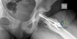
This topic focuses on Solving Unstable SCFE: Subcapital Realignment with Extended Retinacular Flap, The extended retinacular flap is a vital part of the subcapital realignment with surgical dislocation technique, chosen to treat unstable Slipped Capital Femoral Epiphysis (SCFE). This advanced surgical method facilitates gentle reduction and stabilization of severe hip slips, aiming to restore proper femoral head alignment while carefully preserving vascularity for optimal patient outcomes.
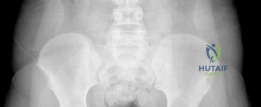
A 13-year-old male presents to the Emergency Department with an acute onset of severe left hip pain following a minor fall. He is unable to bear any weight on the limb. He is obese and reports 3 months of intermittent aching in the same hip. On examination, the hip is held in external rotation, and any attempt at internal rotation causes severe pain. An AP pelvis radiograph is provided. What is your diagnosis and immediate management plan?
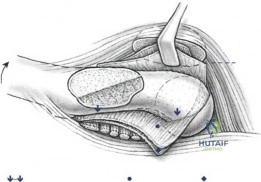
Candidate: The diagnosis is an unstable Slipped Capital Femoral Epiphysis (SCFE). I would confirm this with a cross-table lateral view—avoiding the frog-leg view to prevent further displacement. Management involves admission, strict non-weight bearing, and urgent surgical stabilization, likely a modified Dunn procedure given it is unstable.
Candidates often suggest obtaining a "frog-leg lateral" view to confirm the slip. This is dangerous in an unstable SCFE as it risks further displacement and vascular compromise of the femoral head. Additionally, failing to explicitly mention "unstable" (Loder criteria) shows a lack of clinical nuance.
Classify as an Unstable SCFE (Loder criteria). State clearly that the frog-leg view is contraindicated due to risk of vascular injury. Propose a management plan: 1) Immediate admission and strict non-weight bearing. 2) Urgent MRI to assess femoral head perfusion. 3) Plan for anatomical reduction via the modified Dunn procedure (surgical hip dislocation) to prevent AVN and long-term FAI, noting this must be performed by a surgeon with specific fellowship expertise.
You have decided to perform a modified Dunn procedure. Explain the vascular anatomy at risk during this surgery and how your technique specifically protects it.
Candidate: The primary blood supply is from the medial femoral circumflex artery (MFCA). The terminal branches are in the posterior retinaculum. I would protect these by elevating an extended retinacular flap and shortening the femoral neck to allow reduction without tension.
Failing to mention the "extended retinacular flap" by name or failing to link the need for metaphyseal shortening to the reduction of tension on the posterior retinacular vessels. Examiners want to hear the mechanical rationale for the shortening.
The blood supply is dominated by the deep branch of the MFCA, which travels in the posterior retinaculum. In SCFE, the anterior vessels are often already disrupted, making the head reliant on the posterior supply. The extended retinacular flap technique allows for subperiosteal dissection of the posterior retinaculum as a continuous sleeve with the epiphysis. Crucially, I must resect the metaphyseal prominence (the cam lesion); this shortening is not just for impingement, but to create the necessary slack to prevent iatrogenic tension on the retinacular vessels during anatomical reduction.
During the surgery, you have completed the capsulotomy and dislocation. What specific technique is shown here, and what is the primary risk if this step is performed incorrectly?
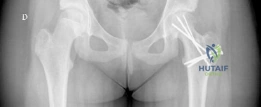
Candidate: This is a surgical hip dislocation using a trochanteric flip osteotomy. The primary risk is damaging the abductors or the blood supply to the femoral head if the retinaculum is not protected.
Describing it simply as "an open approach." The examiner is looking for the specific terminology: "Digastric Trochanteric Flip Osteotomy". Failing to highlight why this is used (preserving the abductor muscle mass while getting 360-degree joint access) is a missed opportunity.
This is a digastric trochanteric flip osteotomy. It preserves the abductor origin on the proximal fragment and the vastus lateralis on the distal fragment, protecting the blood supply to the gluteus medius. The risk of an improper osteotomy includes trochanteric non-union, which leads to abductor weakness and a permanent Trendelenburg gait. Furthermore, if the dislocation is performed without the "extended retinacular flap" protection, the risk of iatrogenic avascular necrosis is significantly elevated.
Post-operatively, the patient develops profound joint stiffness and radiographic narrowing of the joint space. What is your differential diagnosis, and how would you manage this?
Candidate: This sounds like chondrolysis. I would check for hardware penetration, encourage physiotherapy, and potentially remove the hardware if it is causing irritation.
Treating it as a benign finding or only focusing on hardware. Chondrolysis is a catastrophic complication. A poor candidate forgets to mention the differential—specifically septic arthritis or AVN—which must be ruled out before assuming it is purely chondrolysis.
My top differential is chondrolysis, but I must exclude septic arthritis (infection) and AVN (vascular collapse) first. I would perform an urgent inflammatory workup (CRP/ESR), consider aspiration, and review the imaging for hardware penetration. If it is established chondrolysis, management is challenging: aggressive physical therapy for ROM, NSAIDs for anti-inflammatory effects, and hardware removal. I would warn the patient/family about the high risk of poor long-term outcomes, potentially leading to total hip arthroplasty in early adulthood.
