Comprehensive Diagnosis of Developmental Dysplasia of the Hip (DDH) in a Young Adult
Key Takeaway
Diagnosing Developmental Dysplasia of the Hip (DDH) in young adults involves a comprehensive approach. This includes patient history of chronic hip pain, clinical examination (ROM, provocative tests), and advanced imaging. Key diagnostics: X-rays showing acetabular undercoverage, MRI for soft tissues, and 3D CT scans for precise anatomical confirmation.
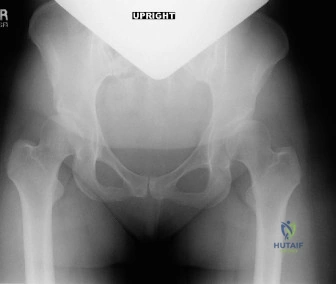
A 28-year-old female presents with a 3-year history of progressive right hip pain. She has a history of DDH managed with a Pavlik harness as an infant. On examination, she has a positive FADIR test and restricted internal rotation. Below is her AP Pelvis radiograph. Describe the radiographic findings and what specific measurements would you perform to guide your management?
Candidate: The radiograph shows evidence of hip dysplasia. Specifically, I see reduced acetabular coverage of the femoral head, a disrupted Shenton’s line, and a steep acetabular slope. To assess this, I would measure the Lateral Center-Edge Angle (LCEA), the Tönnis angle (Acetabular Index), and the femoral head extrusion index. I would also check for signs of early osteoarthritis, such as subchondral sclerosis or cysts.
Failing to quantify the findings (e.g., just saying "the hip looks shallow") or forgetting the Tönnis classification for osteoarthritis. Candidates often forget to mention the False Profile view, which is essential to assess anterior coverage, or they focus only on the bone and miss the joint space changes.
Identify the key parameters: 1) LCEA (Wiberg): < 20° is diagnostic of dysplasia. 2) Tönnis Angle (Acetabular Index): > 10° indicates acetabular steepness. 3) Extrusion Index: > 25% suggests subluxation. 4) Tönnis Grade: Essential for surgical candidacy (Grade 0-1 is ideal for PAO). Always mention that you would also use the False Profile view to evaluate the Anterior Center-Edge Angle (ACEA) to determine the degree of anterior undercoverage.
The patient is confirmed to have symptomatic residual hip dysplasia with early degenerative changes. She is requesting surgery. What is your surgical strategy, and what are the critical anatomical structures you must protect during a Periacetabular Osteotomy (PAO)?
Candidate: My surgical strategy is a Bernese PAO to reorient the acetabulum, improve coverage, and delay the need for THA. Regarding structures, I need to protect the sciatic nerve posteriorly, the femoral neurovascular bundle medially, and the lateral femoral cutaneous nerve (LFCN) during the approach.
Forgetting to discuss the importance of the posterior column. A common mistake is focusing only on the soft tissue structures while failing to mention the preservation of the posterior hinge, which is critical for the stability and vascularity of the mobile acetabular fragment.
Structure the answer by anatomy and procedure: 1) Vascularity: Maintaining the medial soft tissue attachments to the acetabular fragment is vital to prevent AVN. 2) Neurology: Highlight protection of the Sciatic nerve (posteriorly), Femoral bundle (medially), and LFCN. 3) Procedure: Emphasize the four-cut osteotomy technique (ischial, pubic, iliac, and posterior column) and the use of the "greenstick" technique at the posterior column to ensure controlled fragment mobilization while maintaining essential vascular supply.
