العربية
Part of the Master Guide

AAOS & ABOS Foot & Ankle Board Review MCQs (Set 2): Ankle Fractures, Lisfranc, Diabetic Foot

AAOS & ABOS Orthopedic MCQs: Foot & Ankle Set 3 | Board Prep Questions

23 Apr 2026 66 min read 103 Views
Foot & Ankle 2009 MCQs - Part 3

Key Takeaway

This high-yield Foot & Ankle MCQ set (Set 3) is designed for AAOS and ABOS board preparation. It covers crucial topics like acute ankle injuries, complex foot trauma, degenerative conditions, and common hindfoot deformities, ensuring comprehensive review for orthopedic residents and practitioners.

AAOS & ABOS Orthopedic MCQs: Foot & Ankle Set 3 | Board Prep Questions

Comprehensive 100-Question Exam


00:00

Start Quiz

Question 1

What is the most common malignant tumor of the foot?


Explanation

Whereas chondrosarcoma is the most frequently occurring malignant bone tumor of the foot and synovial sarcoma is the most common soft-tissue foot malignancy, the most common malignant tumor overall is melanoma. It constitutes approximately 25% of lesions found on the lower extremity. Furthermore, 31% of all melanomas arise in the foot. Mizel MS, Miller RA, Scioli MW (eds): Orthopaedic Knowledge Update: Foot and Ankle 2. Rosemont, IL, American Academy of Orthopaedic Surgeons, 1998, pp 11-26.

Question 2

A 40-year-old man underwent an ankle arthroscopy 6 months ago for a talus osteochondral defect. He continues to have pain and burning on the lateral portal but states that the pain is now more superficial than his original pain. Examination reveals that he has shooting pain to his medial foot and ankle when his lateral portal is tapped. A previous injection around the lateral portal gave him relief for about 2 weeks. What treatment will best eliminate his pain?


Explanation

The patient clearly has entrapment of the superficial peroneal nerve in the lateral portal. It is most likely only the medial branch by examination. If the nerve is in good condition, it can simply be released. If the nerve is cut or severely thinned, it is better excised and buried. The sural nerve most likely would be caught in a posterior-lateral portal. Jobe MT, Wright PE: Peripheral nerve injuries, in Canale ST (ed): Campbell's Operative Orthopaedics. St Louis, MO, Mosby, 1998, pp 3839-3844.

Question 3

When performing a Weil osteotomy of a lesser metatarsal, the desired angle of the saw cut should be approximately


Explanation

Appropriate orientation of the saw cut when performing a Weil osteotomy is approximately parallel with the plantar surface of the foot. This is done in an effort to minimize plantar displacement of the capital fragment. The removal of additional bone from the osteotomy site either by removing a separate wafer of bone or using a thicker saw blade has also been described to minimize plantar displacement of the distal fragment. Trnka H, Nyska M, Parks BG, et al: Dorsiflexion contracture after the Weil osteotomy: Results of cadaver study and three-dimensional analysis. Foot Ankle Int 2001;22:47-50.

Question 4

A patient with diabetic peripheral neuropathy undergoes a partial first ray amputation for a chronic ulcer beneath the first metatarsal head. The insertion of the anterior tibialis is preserved. The patient has 10 degrees of passive dorsiflexion at the ankle and no other foot deformities or ulcers. Which of the following is considered appropriate shoe wear for this patient?


Explanation

The steel shank is a flat 1-inch steel strip placed between layers of the shoe to extend the foot lever and prevent deformity at the toe break seen following a partial first ray amputation. A rocker sole may be added as well to facilitate transition from foot flat to the toe-off phase of gait. Proper shoe fit is important, but "snug" fitting shoes in a patient with peripheral neuropathy and likely fluctuations in volume from intermittent swelling are to be avoided. A custom shoe is an unnecessary expense. The patient has at least 10 degrees of dorsiflexion at the ankle with an intact anterior tibialis muscle; therefore, catching the sole on carpeting should not be a problem. Philbin TM, Leyes M, Sferra JJ, et al: Orthotic and prosthetic devices in partial foot amputations. Foot Ankle Clin 2001;6:215-228.

Question 5

A 32-year-old laborer reports left ankle pain and deformity. History reveals that he sustained a left ankle fracture 2 years ago and was treated with closed reduction and casting. Radiographs are shown in Figures 25a through 25c. What is the most appropriate management?


Explanation

Corrective osteotomy of fibular malunions, with appropriate lengthening, even in the presence of early arthritis, has been shown to decrease ankle pain and increase stability. Reduction and bone grafting of the medial malleolar nonunion is also needed. There is no evidence supporting the use of intra-articular steroids or hyaluronic acid in the ankle joint. Lateral talar displacement of even 1 mm has been reproducibly shown to decrease tibiotalar contact by 40% to 42%, causing a predisposition to arthritis. Weber D, Friederich NF, Muller W: Lengthening osteotomy of the fibula for post-traumatic malunion: Indication, technique and results. Int Orthop 1998;22:149-152. Lloyd J, Elsayed S, Hariharan K, et al: Revisiting the concept of talar shift in ankle fractures. Foot Ankle Int 2006;27:793-796. Offierski CM, Graham JD, Hall JH, et al: Later revision of fibular malunion in ankle fractures. Clin Orthop Relat Res 1982;171:145-149.


Question 6

Preservation or reconstruction of which of the following structures is essential to minimize the risk of hallux valgus developing after removal of part or all of the medial sesamoid?


Explanation

Complications of medial sesamoidectomy include stiffness, claw toe, and hallux valgus. Each sesamoid sits within its respective head of the flexor hallucis brevis tendon. Excision of one sesamoid can result in slack in its flexor hallucis brevis tendon; therefore, it is imperative to preserve or repair the flexor hallucis brevis tendon when removing the medial sesamoid. Dedmond BT, Cory JW, McBryde A Jr: The hallucal sesamoid complex. J Am Acad Orthop Surg 2006;14:745-753.

Question 7

In the nonsurgical management of posterior tibial tendon dysfunction with flexible deformity, a common strategy is to prescribe an ankle-foot orthosis or a University of California Biomechanics Laboratory (UCBL) orthosis with medial posting. A high patient satisfaction rating and favorable outcome with this nonsurgical management is most likely in which of the following situations?


Explanation

Most authors recommend an initial trial of nonsurgical management in the treatment of adult-acquired flatfoot deformity such as posterior tibial tendon dysfunction. Chao and associates found that there is high patient satisfaction with ankle-foot orthoses and UCBL-type inserts in elderly patients with a relatively sedentary lifestyle. Alternatively, there was a higher dissatisfaction rate in young active patients, those with balance and ambulation difficulties (Parkinson's, severe arthritis of the hip or knee), and patients with inflammatory systemic disorders. Chao W, Wapner KL, Lee TH, et al: Nonoperative management of posterior tibial tendon dysfunction. Foot Ankle Int 1996;17:736-741.

Question 8

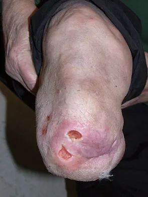
Figure 26 shows the clinical photograph of a patient who has developed a residual limb ulcer following a traumatic transtibial amputation 2 years ago. What is the preferred treatment to resolve the ulcer?


Explanation

The first step in the treatment of an amputation residual limb (stump) ulcer is local wound care and adjustment of the residual limb-prosthetic interface, as well as adjusting prosthetic alignment. Surgical revision should be undertaken only when prosthetic modification is unsuccessful. Murnaghan JJ, Bowker JH: Musculoskeletal complications, in Smith DG, Michael JW, Bowker JH (eds): Atlas of Amputations and Limb Deficiencies, ed 3. Rosemont, IL, American Academy of Orthopaedic Surgeons, 2004, pp 683-700.


Question 9

The spring ligament of the foot connects what two bones?


Explanation

The spring ligament is also known as the calcaneonavicular ligament and connects the calcaneus to the navicular. This ligament supports the talar head and is an important anatomic supporting structure of the medial longitudinal arch of the foot. Choi K, Lee S, Otis JC, et al: Anatomical reconstruction of the spring ligament using peroneus longus tendon graft. Foot Ankle Int 2003;24:430-436.

Question 10

An obese 62-year-old man reports a 10-year history of progressive flatfoot deformity and a 3-month history of a painful callus along the plantar medial midfoot that has not improved with custom shoe wear, pedorthics, and callus care. There is no hindfoot motion, but functional ankle motion remains. He does not have diabetes mellitus. Radiographs are shown in Figures 27a and 27b. What is the best surgical option at this point?


Explanation

The deformity is long-standing, the hindfoot is immobile, and the radiographs reveal severe degenerative arthritis involving the entire hindfoot, severe deformity, and talonavicular dislocation. The "exostosis" responsible for the callus is the talar head; resection would severely destabilize the foot. Degenerative arthritis and fixed deformity preclude lateral column lengthening, medial slide calcaneal osteotomy, and talonavicular arthrodesis. Triple arthrodesis is the only viable option. Johnson JE, Yu JR: Arthrodesis techniques in the management of Stage II and III acquired adult flatfoot deformity. Instr Course Lect 2006;55:531-542.


Question 11

A 20-year-old collegiate football player sustains an injury to his left foot 3 weeks before the start of the fall season. Examination reveals localized tenderness over the lateral midfoot and normal foot alignment. Radiographs are shown in Figures 28a through 28c. What is the treatment of choice?


Explanation

Due to the relatively high incidence of delayed union and nonunion associated with this mildly displaced Jones-type fracture, and the temporal proximity to his playing season, intramedullary screw fixation is the treatment of choice in this collegiate athlete to best ensure healing and expedite his return to football. If nonsurgical management were elected, application of a non-weight-bearing short leg cast would be appropriate since a higher likelihood of healing is expected with it versus a short leg walking cast. The risk of recurrent fracture of fractures that heal with nonsurgical management has reportedly been high (approximately 30%). Quill GE: Fractures of the proximal fifth metatarsal. Orthop Clin North Am 1995;26:353-361. Torg JS, Balduini FC, Zelko RR, et al: Fractures of the base of the fifth metatarsal distal to the tuberosity: Classification and guidelines for nonsurgical and surgical management. J Bone Joint Surg Am 1984;66:209-214.


Question 12

When the great toe deviates into a valgus position, the action of the abductor hallucis muscle becomes one of


Explanation

The abductor hallucis muscle inserts together with the medial tendon of the flexor hallucis brevis into the medial base of the proximal phalanx of the great toe. When the hallux assumes a valgus position, the action of the abductor becomes one of flexion and pronation of the first metatarsal. Resch S: Functional anatomy and topography of the foot and ankle, in Myerson M (ed): Foot and Ankle Disorders. Philadelphia, PA, WB Saunders, 2000, vol 1, pp 25-49.

Question 13

When performing a bunionectomy with a release of the lateral soft-tissue structures, the surgeon is cautioned against releasing the conjoined tendon that inserts along the lateral base of the proximal phalanx of the great toe. This conjoined tendon is made up of what two muscles?


Explanation

Owens and Thordardson cautioned surgeons not to release the conjoined tendon from the base of the proximal phalanx of the great toe because of an increased risk of iatrogenic hallux varus. Release of the transverse and oblique heads of the adductor hallucis is largely accomplished by releasing the soft tissue adjacent to the lateral sesamoid, without releasing tissue from the base of the proximal phalanx. The conjoined tendon is made up of the flexor hallucis brevis and the adductor hallucis. Owens S, Thordardson DB: The adductor hallucis revisited. Foot Ankle Int 2001;22:186-191.

Question 14

Figures 29a and 29b show a clinical photograph and radiographs of a patient who sustained an open calcaneus fracture in a motor vehicle accident. The patient received immediate IV antibiotics and an emergent irrigation and debridement. The swelling has subsided by 3 weeks and the medial wound is clean. What do you tell the patient about the likelihood of infection if a formal open reduction and internal fixation via a lateral approach is performed?


Explanation

Multiple authors have shown similar infection rates for grade 1 and 2 open medial fractures and closed fractures that have been treated with an extensile lateral approach and open reduction and internal fixation. Patients only need IV antibiotics for 2 to 3 days after surgery. Formal open reduction and internal fixation is not recommended for grade 3 medial wounds and most lateral wounds. Heier KA, Infante AF, Walling AK, et al: Open fractures of the calcaneus: Soft-tissue injury determines outcome. J Bone Joint Surg Am 2003;85:2276-2282.


Question 15

When compared to traditional open repair through a posterior incision, percutaneous Achilles tendon repair clearly results in a reduction of what complication?


Explanation

Prospective studies, including randomized and randomized multicenter reports, have shown that percutaneous or mini-open acute Achilles tendon repair has comparable functional results when compared to traditional open techniques. Calder and Saxby reported one superficial infection out of 46 patients with a mini-open repair; Assal and associates and Cretnik and associates had no wound complications or infections. The other complications have not proved to be less likely with the mini-open or percutaneous technique. Assal M, Jung M, Stern R, et al: Limited open repair of Achilles tendon ruptures: A technique with a new instrument and findings of a prospective multicenter study. J Bone Joint Surg Am 2002;84:161-170. Calder JD, Saxby TS: Early, active rehabilitation following mini-open repair of Achilles tendon rupture: A prospective study. Br J Sports Med 2005;39:857-859.

Question 16

A 24-year-old woman was struck by a mini van in a parking lot and sustained a closed segmental tibia fracture that was treated with an intramedullary nail the following morning. Follow-up examinations reveal a slowly progressive clawing of all five toes, a progressive equinocavovarus contracture, and the patient is unable to perform a single heel rise on the affected limb. At 1 year after surgery, the patient now has a 10-degree equinus contracture that is not relieved with knee flexion. Treatment should now consist of


Explanation

This is an example of a missed deep posterior compartment syndrome that typically presents 6 months after the injury with progressive clawing due to necrosis, scarring, and contracture of the posterior tibial tendon, flexor digitorum longus, and flexor hallucis longus. Treatment consists of debridement of necrotic muscle and scar tissue with corresponding tendon excision. After debridement and posterior capsule release, if the equinus is relieved with knee flexion, a gastrocnemius slide may be performed. Otherwise, the lengthening should be at the level of the Achilles tendon. Bracing will not address the claw toes. Hansen ST Jr: Functional Reconstruction of the Foot and Ankle. Philadelphia, PA, Lippincott Williams & Wilkins, 2000, pp 212-213. Manoli A II, Smith DG, Hansen ST Jr: Scarred muscle excision for the treatment of established ischemic contracture of the lower extremity. Clin Orthop Relat Res 1993;292:309-314.

Question 17

A 26-year-old man with chronic lateral ankle instability underwent a modified Broström procedure 8 months ago. He reports persistent pain and swelling of the lateral ankle. Examination reveals lateral ankle tenderness and swelling and a negative anterior drawer test. Laboratory studies show a WBC count of 6,500/mm3 and an erythrocyte sedimentation rate of 15 mm/h. Radiographs of the ankle are normal. What is the most likely cause of this problem?


Explanation

Chronic lateral instability is commonly associated with a longitudinal split tear of the peroneus brevis tendon. The interrelationship of lateral ankle instability with superior retinacular laxity and resultant peroneus brevis split can account for persistent lateral ankle pain in this patient. Surgical treatment must identify and correct the underlying tendon pathology and should attempt to repair or debride the peroneus brevis tendon, reconstruct the superior peroneal retinaculum, flatten the posterior edge of the fibula by removing the sharp bony prominence, or deepening the fibular groove, along with addressing lateral ankle ligamentous instability. The laboratory values are not consistent with infection. A negative anterior drawer test confirms stability of the repair. Ankle arthritis is not seen on radiographs and usually takes longer than 3 months to develop. Bonnin M, Tavernier T, Bouysset M: Split lesions of the peroneus brevis tendon in chronic ankle laxity. Am J Sports Med 1997;25:699-703.

Question 18

A 52-year-old woman with a 2-year history of a flexible (stage II) adult-acquired flatfoot deformity has failed to respond to nonsurgical management consisting of immobilization, custom orthotics, nonsteroidal anti-inflammatory drugs, and physical therapy. The patient is unable to perform a single limb heel rise. Weight-bearing radiographs are shown in Figures 30a through 30c. What is the most appropriate surgical correction?


Explanation

The patient has an atypical adult flatfoot deformity. The radiographs reveal forefoot abduction, mild loss of calcaneal pitch, and marked plantar flexion sag through the naviculocuneiform joint. The inability to perform a single limb heel rise indicates that the posterior tibial tendon is nonfunctional; however, the deformity remains flexible. In this patient, surgical treatment should include a tendon transfer, lateral column lengthening, medial column arthrodesis, and heel cord lengthening. Because a substantial portion of the deformity stems from the naviculocuneiform joint in this instance, tendon transfer and lateral column lengthening alone provide insufficient deformity correction. Triple arthrodesis and heel cord lengthening is best reserved for fixed flatfoot deformities. Soft-tissue procedures alone are associated with a high failure rate, as are attempted tendon repairs. Greisberg J, Assal M, Hansen ST Jr, et al: Isolated medial column stabilization improves alignment in adult-acquired flatfoot. Clin Orthop Relat Res 2005;435:197-202.


Question 19

Optimal management of the injury shown in Figure 31 should include which of the following?


Explanation

The radiograph shows a displaced calcaneal beak fracture, a tongue-type fracture variant. The fracture fragment typically includes the insertion point of the Achilles tendon, which places marked tension on the thin overlying soft-tissue envelope and can lead to full-thickness necrosis if not acutely addressed. Cast immobilization does not adequately address the increased soft-tissue tension, as the fragment will be difficult to control. Arthroscopic-assisted techniques or primary arthrodesis are not indicated because calcaneal beak fractures are typically extra-articular. Sanders RW, Clare MP: Fractures of the calcaneus, in Coughlin MJ, Mann RA, Saltzman CL (eds): Surgery of the Foot and Ankle, ed 8. Philadelphia, PA, Mosby-Elsevier, 2007, vol 2, pp 2017-2073.


Question 20

A 23-year-old man who was the restrained driver in a car involved in a high-speed motor vehicle accident sustained the closed injury shown in Figures 32a through 32c. Which of the following factors has the greatest impact on the risk of osteonecrosis?


Explanation

The incidence of osteonecrosis following displaced talar neck fractures is most related to the extent of initial fracture displacement. With increasing fracture displacement, the tenuous vascular supply to the talar body is more at risk for damage, thereby increasing the risk of osteonecrosis. Although displaced talar neck fractures have historically been considered a surgical emergency, recent studies have shown that the timing of surgical intervention bears no impact on the development of osteonecrosis. While nicotine use has an influence on fracture healing, it has never been shown to be a factor in osteonecrosis, nor has posterior-to-anterior screw fixation or the quality of fracture reduction. Lindvall E, Haidukewych G, Dipasquale T, et al: Open reduction and stable fixation of isolated, displaced talar neck and body fractures. J Bone Joint Surg Am 2004;86:2229-2234.


Question 21

A 30-year-old woman injured her ankle playing soccer 3 months ago. She now reports popping and pain over the lateral side of her ankle. An MRI scan is shown in Figure 33. What structure needs to be repaired to alleviate the popping?


Explanation

The symptoms and MRI scan indicate dislocated peroneal tendons. In this patient, the structure that needs to be repaired is the superior peroneal retinaculum. If the popping was coming from a torn peroneal tendon, repair would involve the peroneal longus or brevis tendon, but this is not shown in the MRI scan. The anterior talofibular ligament or the calcaneofibular ligament would need to be repaired if the patient had ankle instability due to an ankle sprain. Jones DC: Tendon disorders of the foot and ankle. J Am Acad Orthop Surg 1993;1:87-94.


Question 22

A 35-year-old woman with type 1 diabetes mellitus has been treated for the past 2 years at a wound care center for persistent bilateral fifth metatarsal head ulcers. Management has consisted of shoe wear modifications, treatment with multiple enzymatic ointments, and a fifth metatarsal head resection on the left side. Physical examination reveals intact pulses, minimal ankle dorsiflexion, neutral hindfoot, and a persistent ulcer under the fifth metatarsal heads. What treatment will best help heal the ulcers?


Explanation

The patient likely has a significant Achilles contracture that causes her to always bear more weight on her forefoot. A gastrocnemius recession takes the ankle out of plantar flexion and she will be able to return to a normal gait and reduce the pressures on her forefoot. A forefoot amputation is a salvage option. The other choices are appropriate; however, the patient has had this problem for 2 years and she has already had multiple attempts at shoe wear modification. Laughlin RT, Calhoun JH, Mader JT: The diabetic foot. J Am Acad Orthop Surg 1995;3:218-225.

Question 23

The hallucal sesamoids are held together by which of the following structures?


Explanation

The two sesamoids of the metatarsophalangeal joint are embedded in the tendons of the short flexor of the great toe. They are held together by the intersesamoid ligament and the plantar plate, which inserts on the base of the proximal phalanx of the hallux. The flexor hallucis longus tendon inserts onto the distal phalanx of the great toe. The plantar calcaneonavicular (spring) ligament, by supporting the head of the talus, principally maintains the arch of the foot. The plantar fascia inserts distally onto the skin and to the flexor tendons and transverse metatarsal ligaments at each metatarsophalangeal joint. The intermetatarsal ligament attaches to the base of the second through fifth metatarsals. Lewis WH (ed): Gray's Anatomy of the Human Body, ed 20. Philadelphia, PA, Lea & Febiger, 2000.

Question 24

Figures 34a and 34b show the clinical photograph and a weight-bearing radiograph of a patient with diabetes mellitus who has had recurrent ulcers under the head of the talus that have previously resolved with a series of non-weight-bearing total contact casts. The deformity does not correct passively. Dorsalis pedis and posterior tibial pulses are palpable. The patient is insensate to the Semmes-Weinstein 5.07 (10 gm) monofilament. The ulcer is currently healed. What is the best option to prevent recurrent ulceration and infection?


Explanation

This is a nonplantigrade deformity in a patient with a Charcot foot deformity. Longitudinal studies have shown that recurrent ulceration/infection is likely unless the deformity is corrected. Achilles tendon lengthening is advised for simple forefoot ulcers. The current approach to this problem is best managed with surgical correction of the deformity, Achilles tendon lengthening, and therapeutic footwear. Bevan WP, Tomlinson MP: Radiographic measures as a predictor of ulcer formation in diabetic charcot midfoot. Foot Ank Int 2008;29:568-573. Simon SR, Tejwani SG, Wilson DL, et al: Arthrodesis as an early alternative to nonoperative management of Charcot arthropathy of the diabetic foot. J Bone Joint Surg Am 2000;82:939-950.


Question 25

Which of the following conditions precludes performing a tendon transfer?


Explanation

Several conditions must be met before a tendon transfer has the potential to correct a dynamic deformity. If the target joint cannot be passively corrected to neutral, it indicates that a static joint contracture or bony deformity exists that cannot be corrected with a dynamic tendon transfer. While in-phase muscles are best, out-of-phase muscles are often the only muscles available for transfer. Tendon transfer should pull in a straight line to avoid tethering and late failure. Canale ST (ed): Campbell's Operative Orthopaedics, ed 10. St Louis, MO, Mosby, 2003, pp 1283-1287.

Question 26

A 55-year-old woman presents with a painful, flexible flatfoot deformity. Standing radiographs reveal greater than 40% talonavicular uncoverage on the AP view. She is diagnosed with Stage IIb posterior tibial tendon dysfunction. What is the most appropriate surgical management?


Explanation

Stage IIb posterior tibial tendon dysfunction is characterized by a flexible deformity with forefoot abduction (>40% talonavicular uncoverage). A lateral column lengthening must be added to the FDL transfer and calcaneal osteotomy to correct the abduction.

Question 27

A 22-year-old collegiate running back sustains a purely ligamentous Lisfranc injury. There are no fractures identified on CT scan, but weight-bearing radiographs demonstrate 4 mm of diastasis between the medial and middle cuneiforms. What is the most appropriate definitive treatment?


Explanation

Primary arthrodesis of the medial column (1st-3rd TMT joints) is recommended for purely ligamentous Lisfranc injuries. This approach yields better functional outcomes and lower reoperation rates compared to ORIF in ligamentous variants.

Question 28

A 30-year-old male is involved in a high-speed motor vehicle collision and sustains a Hawkins II talar neck fracture.

Which of the following blood vessels provides the primary blood supply to the talar body and is at greatest risk in this injury?


Explanation

The artery of the tarsal canal, a branch of the posterior tibial artery, provides the majority of the blood supply to the talar body. It enters inferiorly and is highly susceptible to disruption in displaced talar neck fractures.

Question 29

When comparing operative repair to nonoperative management utilizing early functional bracing for acute Achilles tendon ruptures, which of the following statements is supported by current literature?


Explanation

Recent randomized controlled trials show that nonoperative management using early functional rehabilitation protocols has rerupture rates comparable to surgical repair. Furthermore, nonoperative management avoids surgical wound complications and infections.

Question 30

A 45-year-old female presents with severe bunion pain. Radiographs reveal a hallux valgus angle (HVA) of 45 degrees, an intermetatarsal angle (IMA) of 18 degrees, and clinical hypermobility of the 1st tarsometatarsal (TMT) joint. Which of the following procedures is most appropriate?


Explanation

The Lapidus procedure (1st TMT arthrodesis) is indicated for severe hallux valgus (IMA >15 degrees) combined with 1st TMT hypermobility. It provides powerful correction of the intermetatarsal angle and stabilizes the medial column.

Question 31

A 40-year-old male is evaluated for persistent lateral hindfoot pain 8 months after nonoperative management of a joint-depressed calcaneus fracture. Examination reveals tenderness inferior to the lateral malleolus and pain with active eversion against resistance. What is the most likely etiology of his pain?


Explanation

Lateral wall blowout is a classic complication of nonoperative management in displaced calcaneus fractures. This leads to subfibular impingement of the peroneal tendons, causing lateral hindfoot pain and pain with resisted eversion.

Question 32

An elite football player sustains a hyperdorsiflexion injury to his first MTP joint (turf toe). MRI confirms a complete tear of the plantar plate. Which of the following is an absolute indication for surgical repair?


Explanation

Surgical intervention for turf toe is indicated in Grade III injuries demonstrating gross instability, intra-articular loose bodies, or significant proximal retraction of the sesamoids. Operative repair restores the anatomy of the sesamoid complex and plantar plate.

Question 33

A 60-year-old patient with long-standing, poorly controlled diabetes presents with a unilaterally warm, swollen, and erythematous foot without ulceration. Radiographs show periarticular debris, fragmentation, and joint subluxation at the midfoot. What is the most appropriate initial management?


Explanation

This presentation is classic for Eichenholtz Stage I (fragmentation stage) acute Charcot arthropathy. The gold standard for initial treatment is immobilization and offloading via a total contact cast until the acute inflammatory phase resolves.

Question 34

A patient undergoes excision of a symptomatic mass in the 3rd intermetatarsal space after failing shoe modifications and injections. A Mulder's click was present preoperatively. What histopathologic finding is expected in the excised specimen?


Explanation

Morton's neuroma is not a true neoplastic neuroma but rather a compressive neuropathy. Histology classically shows perineural fibrosis, endoneurial edema, and axonal degeneration of the common digital nerve.

Question 35

A 65-year-old male with end-stage post-traumatic ankle arthritis is undergoing an isolated tibiotalar arthrodesis. To optimize postoperative gait, what is the ideal position for ankle fusion?


Explanation

Optimal position for ankle arthrodesis is neutral dorsiflexion (0 degrees), 0 to 5 degrees of hindfoot valgus, and external rotation equal to the contralateral limb (typically 5 to 10 degrees). This alignment minimizes adjacent joint stress.

Question 36

A 21-year-old collegiate track athlete complains of vague, chronic midfoot pain. CT scan reveals a non-displaced stress fracture of the tarsal navicular. Why is this specific fracture at high risk for delayed union or nonunion?


Explanation

The central third of the tarsal navicular is a vascular watershed zone. Stress fractures typically occur in this avascular region, making them prone to delayed union and nonunion, often requiring strict non-weight-bearing cast immobilization or surgical fixation.

Question 37

A 14-year-old boy presents with frequent ankle sprains and a rigid, painful flatfoot. Lateral radiographs demonstrate a prominent 'C-sign.'

This finding is most indicative of which pathology?


Explanation

The 'C-sign' on a lateral radiograph represents a continuous outline formed by the medial border of the talar dome and the posterior border of the sustentaculum tali. It is highly indicative of a talocalcaneal coalition.

Question 38

A 28-year-old male complains of deep ankle pain. MRI reveals a 1.8 cm^2 osteochondral lesion of the posteromedial talar dome. Nonoperative management has failed. What is the most appropriate surgical intervention?


Explanation

For osteochondral lesions of the talus larger than 1.5 cm^2, microfracture has a high failure rate. Osteochondral autograft transfer (OATS) or autologous chondrocyte implantation (ACI) is indicated for lesions of this size.

Question 39

A 22-year-old elite basketball player sustains a fifth metatarsal base fracture located at the metaphyseal-diaphyseal junction (Zone 2). What is the recommended treatment to ensure the fastest safe return to play?


Explanation

Zone 2 (Jones) fractures occur in a vascular watershed area and have a high risk of nonunion. In elite athletes, early intramedullary screw fixation is recommended to decrease nonunion rates and expedite return to play.

Question 40

During surgical treatment for insertional Achilles tendinopathy and a prominent Haglund deformity, severe tendon degeneration requires detachment of 60% of the Achilles insertion to adequately debride the calcification and retrocalcaneal bursa. What is the recommended next step in managing the tendon?


Explanation

If 50% or more of the Achilles tendon insertion is detached during debridement for insertional tendinopathy, it must be formally reattached to the calcaneal footprint using suture anchors to prevent postoperative rupture and weakness.

Question 41

A 58-year-old male presents with dorsal foot pain and inability to wear dress shoes. Examination shows less than 10 degrees of 1st MTP dorsiflexion and pain throughout the midrange of motion. Radiographs show severe joint space narrowing and large dorsal osteophytes (Grade 3 hallux rigidus). What is the most reliable definitive treatment?


Explanation

For advanced (Grade 3 or 4) hallux rigidus with pain in the midrange of motion and diffuse joint space loss, 1st MTP arthrodesis provides the most reliable pain relief and functional improvement. Cheilectomy is reserved for earlier stages with pain only at terminal dorsiflexion.

Question 42

A patient undergoes open reduction and internal fixation of a pronation-external rotation ankle fracture, which includes placement of two quadricortical syndesmotic screws. According to the current orthopedic consensus, when should these screws be routinely removed?


Explanation

Current evidence suggests that routine removal of syndesmotic screws is unnecessary. Retained, broken, or loose screws do not significantly worsen functional outcomes, and removal should be reserved for symptomatic patients to avoid unnecessary secondary surgery.

Question 43

A 55-year-old woman presents with medial ankle pain, a progressive flatfoot deformity, and the inability to perform a single-leg heel raise. Radiographs demonstrate >40% talonavicular uncoverage and significant forefoot abduction. Which of the following surgical interventions is most appropriate for this stage of deformity?


Explanation

This patient has Stage IIb posterior tibial tendon dysfunction, characterized by forefoot abduction (>40% talonavicular uncoverage). Treatment requires FDL transfer and medial calcaneal osteotomy, plus a lateral column lengthening to correct the significant abduction.

Question 44

In a patient with Charcot-Marie-Tooth disease presenting with a classic cavovarus foot deformity, the plantarflexed first ray is primarily driven by the relative overpull of which of the following muscles?


Explanation

In Charcot-Marie-Tooth, the peroneus longus retains its strength longer than the tibialis anterior, leading to a strong plantarflexing force on the first ray. This creates a forefoot-driven cavovarus deformity.

Question 45

The major blood supply to the talar body, which is most at risk in a Hawkins Type III talar neck fracture, originates from which of the following arteries?


Explanation

The artery of the tarsal canal, a branch of the posterior tibial artery, provides the dominant blood supply to the talar body. It is highly susceptible to injury in displaced talar neck fractures.

Question 46

The Lisfranc ligament is best described as a stout band that originates on the medial cuneiform and inserts on the base of the second metatarsal. On which aspect of these bones does this ligament primarily attach?


Explanation

The Lisfranc ligament is an interosseous ligament that connects the lateral aspect of the medial cuneiform to the medial aspect of the base of the second metatarsal, primarily providing plantar and interosseous stability.

Question 47

Which of the following represents an absolute contraindication to performing a primary total ankle arthroplasty?


Explanation

Neuropathic (Charcot) arthropathy with loss of protective sensation is an absolute contraindication to total ankle arthroplasty due to unacceptably high rates of failure, bone loss, and component subsidence.

Question 48

A 42-year-old female presents with severe bunion pain. Weight-bearing radiographs reveal a hallux valgus angle of 45 degrees, an intermetatarsal angle of 18 degrees, and obvious widening and hypermobility of the first tarsometatarsal (TMT) joint. Which of the following is the most appropriate surgical treatment?


Explanation

First tarsometatarsal (TMT) arthrodesis, or the Lapidus procedure, is indicated for severe hallux valgus with an intermetatarsal angle >15 degrees and associated first TMT hypermobility.

Question 49

During an extensile lateral approach to the calcaneus for open reduction and internal fixation of a highly comminuted intra-articular fracture, careful placement of the horizontal limb of the incision is required to protect which of the following structures?


Explanation

The sural nerve crosses the lateral aspect of the hindfoot and is at highest risk of injury during the horizontal limb incision of the extensile lateral approach to the calcaneus.

Question 50

A 24-year-old elite track athlete complains of vague, aching dorsal midfoot pain. A CT scan confirms a non-displaced stress fracture of the tarsal navicular. The relative avascularity of the central third of the navicular predisposes this area to nonunion. Which arteries supply the margins of this avascular zone?


Explanation

The navicular is supplied radially by branches of the dorsalis pedis and medial plantar arteries. This leaves a central avascular zone that is highly prone to stress fractures and delayed unions.

Question 51

A 28-year-old skier presents with acute lateral ankle pain and swelling after a fall. Radiographs demonstrate a small cortical avulsion fracture off the posterolateral aspect of the distal fibula (a "fleck sign"). This radiographic finding is pathognomonic for an injury to which of the following structures?


Explanation

The "fleck sign" on an ankle radiograph represents an avulsion of the superior peroneal retinaculum from the posterolateral fibula, strongly indicating peroneal tendon subluxation or dislocation.

Question 52

A 30-year-old male sustains a high-energy motor vehicle collision resulting in a displaced talar neck fracture with both subtalar and tibiotalar dislocation (Hawkins Type III). Which of the following best represents his risk of developing avascular necrosis (AVN) of the talar body?


Explanation

Hawkins Type III fractures involve dislocation of the subtalar and ankle joints, disrupting all three major blood supplies to the talar body. This results in a historical AVN risk exceeding 80%, though modern prompt reduction may marginally lower this.

Question 53

In a patient with Charcot-Marie-Tooth disease, the classic cavovarus foot deformity is primarily driven by specific muscle imbalances. Which of the following accurately describes the primary deforming forces?


Explanation

In Charcot-Marie-Tooth, the peroneus longus outpowers the weaker tibialis anterior, causing plantarflexion of the first ray and forefoot pronation. The posterior tibialis also overpowers the weaker peroneus brevis, driving the hindfoot into varus.

Question 54

A 45-year-old recreational athlete sustains an acute mid-substance Achilles tendon rupture. If he chooses operative repair over non-operative functional rehabilitation, what is the most significant relative risk associated with his choice?


Explanation

Operative repair of acute Achilles ruptures carries a significantly higher risk of wound complications and infection (up to 5-10%). Recent literature demonstrates that early functional rehabilitation has re-rupture rates comparable to operative management.

Question 55

A 45-year-old female presents with a painful bunion. Weight-bearing radiographs reveal a Hallux Valgus Angle (HVA) of 38 degrees and an Intermetatarsal Angle (IMA) of 17 degrees. The first tarsometatarsal joint is hypermobile. Which of the following is the most appropriate surgical management?


Explanation

For severe hallux valgus (IMA > 15 degrees) especially with first ray hypermobility, a proximal procedure like a Lapidus (first TMT arthrodesis) is required. Distal osteotomies cannot adequately correct an IMA of this magnitude.

Question 56

A 22-year-old collegiate basketball player sustains a fracture at the metaphyseal-diaphyseal junction of the 5th metatarsal (Zone 2) after a sudden pivot. To minimize the risk of nonunion and expedite his return to play, what is the best management strategy?


Explanation

Zone 2 (Jones) fractures in competitive athletes are optimally treated with early intramedullary screw fixation. This provides a lower rate of nonunion and a faster return to sport compared to conservative management.

Question 57

A trauma surgeon utilizes an extensile lateral approach for open reduction and internal fixation of a displaced intra-articular calcaneus fracture. Which of the following nerves is at greatest risk of iatrogenic injury during the creation of the full-thickness flap?


Explanation

The extensile lateral approach to the calcaneus places the sural nerve at high risk. The nerve is typically elevated within the full-thickness subperiosteal anterior flap to protect it during retraction.

Question 58

A 14-year-old boy presents with a rigid flatfoot and recurrent ankle sprains. A lateral radiograph reveals a distinct "C-sign". This radiographic finding is pathognomonic for which of the following conditions?


Explanation

The 'C-sign' on a lateral radiograph represents the continuous outline of the medial outline of the talar dome and the inferior outline of the sustentaculum tali. It is highly indicative of a talocalcaneal (middle facet) coalition.

Question 59

A 55-year-old female presents with medial ankle pain, a flexible planovalgus foot, and inability to perform a single-leg heel rise. During surgical reconstruction for her Stage II adult acquired flatfoot deformity, which tendon is most commonly transferred to augment the dysfunctional primary tendon?


Explanation

Stage II adult acquired flatfoot deformity (posterior tibial tendon insufficiency) is typically treated with a Flexor Digitorum Longus (FDL) transfer to the navicular, often combined with a medializing calcaneal osteotomy.

Question 60

A 60-year-old male with end-stage ankle osteoarthritis is considering a Total Ankle Arthroplasty (TAA). Which of the following represents an absolute contraindication to this procedure?


Explanation

Active infection, Charcot neuroarthropathy, and severe avascular necrosis of the talus are absolute contraindications to total ankle arthroplasty due to unacceptably high failure and complication rates.

Question 61

A wide receiver sustains a severe hyperextension injury to his great toe while being tackled on an artificial surface. MRI confirms a complete tear of the plantar plate. The primary stabilizers of the first metatarsophalangeal joint complex injured in this "turf toe" condition include the insertion of which tendon?


Explanation

Turf toe involves a sprain or rupture of the plantar plate of the first MTP joint. The plantar plate complex includes the insertion of the flexor hallucis brevis (FHB) tendon and the associated sesamoids.

Question 62

When evaluating an anteroposterior (AP) radiograph of a normal foot, which of the following anatomic relationships defines the proper alignment of the Lisfranc joint complex?


Explanation

Normal anatomic alignment on an AP radiograph requires that the medial border of the second metatarsal base perfectly aligns with the medial border of the middle cuneiform. Any step-off suggests a Lisfranc injury.

Question 63

A 20-year-old track athlete complains of vague, aching dorsal midfoot pain. A CT scan reveals a nondisplaced dorsal cortical fracture in the middle third of the tarsal navicular. What is the most appropriate initial management?


Explanation

Nondisplaced stress fractures of the tarsal navicular carry a high risk of nonunion due to the watershed blood supply in the central third. The standard of care is 6-8 weeks of strict non-weight-bearing in a cast.

Question 64

A 16-year-old female ballet dancer presents with progressive pain and swelling over the dorsal aspect of the second metatarsophalangeal (MTP) joint. Radiographs demonstrate flattening, sclerosis, and fragmentation of the second metatarsal head. What is the most likely diagnosis?


Explanation

Freiberg infraction is a localized avascular necrosis of the metatarsal head, most commonly affecting the second metatarsal. It typically presents in adolescent females, particularly those involved in high-impact activities like dance.

Question 65

A skier presents with lateral ankle pain and a "snapping" sensation behind the lateral malleolus after catching an edge, which forced his ankle into sudden dorsiflexion and inversion. Which retinacular structure is most likely disrupted in this acute subluxation event?


Explanation

Acute peroneal tendon subluxation typically results from sudden dorsiflexion and forced inversion/eversion. This strips or tears the Superior Peroneal Retinaculum (SPR) off its fibular attachment.

Question 66

A 55-year-old male presents with a painful, stiff great toe. Examination reveals a palpable dorsal prominence and pain primarily at the extremes of dorsiflexion. Radiographs show dorsal osteophytosis of the first metatarsal head but preserved joint space (Coughlin and Shurnas Grade 2). If conservative management fails, what is the best surgical option?


Explanation

For mild to moderate hallux rigidus (Grades 1 and 2) with preserved joint space and pain primarily from dorsal impingement, a cheilectomy (resection of the dorsal osteophyte and 30% of the dorsal metatarsal head) provides excellent relief.

Question 67

A 62-year-old poorly controlled diabetic male presents with a red, hot, swollen right foot. He is afebrile with a normal WBC count, though ESR is mildly elevated. Radiographs reveal acute fragmentation and subluxation of the midfoot joints. What is the most appropriate initial management?


Explanation

The presentation is classic for acute Eichenholtz Stage I Charcot neuroarthropathy. The mainstay of initial treatment is offloading with a total contact cast until the acute inflammatory phase resolves and the bones consolidate.

Question 68

A 45-year-old patient undergoes an isolated, complete endoscopic plantar fascia release for recalcitrant plantar fasciitis. Postoperatively, she develops a new, aching pain on the outer border of her foot. Which biomechanical complication is most likely responsible for her new symptoms?


Explanation

A complete release of the plantar fascia disrupts the windlass mechanism, leading to a decrease in arch height. This frequently shifts peak plantar pressures laterally, causing lateral column overload and lateral midfoot pain.

Question 69

A 25-year-old male presents with persistent deep ankle pain following an inversion sprain 8 months ago. MRI reveals a 1.0 cm x 1.0 cm osteochondral lesion on the anterolateral talar dome. After failing conservative management, what is the preferred initial surgical intervention?


Explanation

For symptomatic osteochondral defects of the talus that are less than 1.5 cm in diameter (or < 150 mm squared), arthroscopic debridement and microfracture is the gold standard primary surgical treatment.

Question 70

A 35-year-old female sustains a twisting injury to her leg. Radiographs reveal an isolated, displaced fracture of the proximal third of the fibula. To prevent a missed diagnosis of a Maisonneuve injury, which structure must be thoroughly evaluated clinically and radiographically?


Explanation

A Maisonneuve fracture results from a pronation-external rotation force that tears the medial structures (or medial malleolus), disrupts the syndesmosis, and propagates proximally to fracture the proximal fibula.

Question 71

A 60-year-old female presents with severe lateral hindfoot pain and a fixed flatfoot deformity.

Examination demonstrates rigid hindfoot valgus and rigid forefoot abduction, consistent with Stage III adult-acquired flatfoot deformity. What is the most appropriate surgical intervention?


Explanation

Stage III adult-acquired flatfoot deformity is defined by a rigid, non-reducible deformity with fixed hindfoot valgus and forefoot abduction. Joint-sparing procedures are contraindicated; the standard of care is a triple arthrodesis.

Question 72

A 16-year-old boy with a history of frequent ankle sprains is diagnosed with Charcot-Marie-Tooth disease. He presents with a progressive cavovarus foot deformity. Which of the following best describes the primary muscle imbalance responsible for the forefoot valgus component of his deformity?


Explanation

In Charcot-Marie-Tooth disease, the tibialis anterior and peroneus brevis typically weaken first. The intact peroneus longus overpowers the weak tibialis anterior, plantarflexing the first ray and creating forefoot valgus.

Question 73

A 32-year-old male sustains a Hawkins type II talar neck fracture following a motor vehicle accident and undergoes open reduction internal fixation. At his 8-week postoperative visit, AP and lateral ankle radiographs reveal a subchondral radiolucent band in the talar dome. What does this finding indicate?


Explanation

This finding describes the Hawkins sign, which is subchondral osteopenia seen 6-8 weeks after a talar neck fracture. It indicates that blood supply to the talar body is intact, as the bone must be vascularized to resorb bone and show osteopenia.

Question 74

A 55-year-old female presents with medial ankle pain and a progressive flatfoot deformity. Examination reveals a flexible hindfoot valgus and an inability to perform a single-leg heel raise. Weight-bearing radiographs demonstrate 45% uncovering of the talonavicular joint. What is the most appropriate surgical management?


Explanation

The patient has Stage IIB adult acquired flatfoot deformity (flexible hindfoot valgus with significant forefoot abduction/TN uncovering >30%). Optimal treatment requires addressing both the posterior tibial tendon insufficiency and the profound forefoot abduction with FDL transfer, MDCO, and lateral column lengthening.

Question 75

During a minimally invasive percutaneous repair of an acute Achilles tendon rupture, the surgeon must be cautious to avoid injuring the sural nerve. At the level of the lateral malleolus, what is the anatomic relationship of the sural nerve to the lateral border of the Achilles tendon?


Explanation

The sural nerve courses distally and laterally, crossing lateral to the Achilles tendon approximately 10 cm proximal to its calcaneal insertion. During percutaneous or minimally invasive repair, it is highly vulnerable to entrapment by sutures passed laterally.

Question 76

A 60-year-old male with end-stage post-traumatic ankle arthritis undergoes an isolated tibiotalar arthrodesis. To optimize his postoperative gait and prevent accelerated adjacent-joint arthrosis, what is the ideal position for the ankle fusion?


Explanation

The optimal position for ankle arthrodesis is neutral dorsiflexion, 0-5 degrees of hindfoot valgus, and external rotation matching the contralateral limb (usually 5-10 degrees). Plantarflexion leads to vaulting and knee recurvatum, while varus limits subtalar compensation.

Question 77

A 24-year-old gymnast sustains a midfoot injury. Weight-bearing radiographs reveal widening of the interval between the first and second metatarsals, and a 'fleck sign' is noted. Which of the following describes the correct anatomic attachments of the intact ligament that avulsed to create this sign?


Explanation

The Lisfranc ligament connects the lateral aspect of the medial cuneiform to the medial base of the second metatarsal. The 'fleck sign' represents an avulsion fracture at the attachment site of this ligament.

Question 78

Which of the following statements correctly differentiates the typical characteristics of anterolateral versus posteromedial osteochondral lesions of the talus (OCD)?


Explanation

Anterolateral talar dome lesions are characteristically shallow, wafer-shaped, and usually associated with acute trauma (e.g., ankle sprain). Posteromedial lesions are typically deeper, cup-shaped, and often insidious or atraumatic in nature.

Question 79

A 22-year-old professional football player sustains a turf toe injury while pushing off the line of scrimmage. Examination shows localized tenderness over the plantar aspect of the first metatarsophalangeal (MTP) joint and pain with passive motion. What is the primary mechanism resulting in tearing of the plantar plate complex?


Explanation

Turf toe is an injury to the plantar plate and capsuloligamentous complex of the first MTP joint. It is classically caused by forced hyperextension of the MTP joint while the foot is fixed in equinus, often on artificial playing surfaces.

Question 80


In a displaced intra-articular calcaneus fracture, the anteromedial (sustentaculum tali) fragment typically remains in its anatomic position relative to the talus despite severe displacement of the tuberosity. Which structure is primarily responsible for securing this 'constant' fragment to the talus?


Explanation

The sustentaculum tali is often referred to as the 'constant fragment' in calcaneus fractures because it is tightly bound to the talus by the strong deep deltoid ligament (tibiocalcaneal fibers) and medial talocalcaneal ligaments.

Question 81

A 65-year-old male presents with severe pain and stiffness in his right great toe. Clinical examination reveals palpable dorsal osteophytes and pain throughout the arc of first MTP joint motion. Radiographs show complete joint space obliteration and dorsal osteophytosis. What is the most reliable definitive surgical treatment?


Explanation

The patient has end-stage (Grade 3 or 4) hallux rigidus with diffuse pain and joint space obliteration. Arthrodesis is the gold standard and most reliable procedure for pain relief and functional restoration in severe hallux rigidus.

Question 82

A 58-year-old patient with poorly controlled type 2 diabetes presents with a chronic, non-healing neuropathic ulcer on the plantar forefoot. Which of the following non-invasive vascular studies indicates the highest likelihood of successful wound healing?


Explanation

Wound healing in diabetic patients is highly dependent on adequate perfusion. A toe systolic pressure greater than 40 mm Hg or a TcPO2 greater than 30-40 mm Hg are excellent predictors of healing potential in distal lower extremity amputations and ulcers.

Question 83

A 45-year-old female complains of sharp, burning pain radiating to her 3rd and 4th toes, worsening with tight-fitting shoes. A positive Mulder's click is elicited. If the offending lesion is surgically excised, what is the classic histologic finding on pathology?


Explanation

Morton's neuroma is not a true neoplasm but rather a compressive neuropathy. Histology classically demonstrates perineural fibrosis, endoneurial edema, and nerve fiber degeneration secondary to repetitive microtrauma and compression.

Question 84

A 20-year-old collegiate basketball player sustains a fracture at the metaphyseal-diaphyseal junction of the fifth metatarsal. To minimize the risk of nonunion and facilitate an accelerated return to play, what is the recommended treatment?


Explanation

This describes a Zone 2 (Jones) fracture. In high-level or elite athletes, intramedullary screw fixation is recommended to reduce the significantly high risk of nonunion associated with conservative management in this vascular watershed area.

Question 85

A patient presents with burning pain and numbness on the plantar aspect of the foot that worsens with prolonged standing. Tinel's sign is positive when percussing the retromalleolar groove behind the medial malleolus. Which nerve is compressed within the tarsal tunnel?


Explanation

Tarsal tunnel syndrome is caused by compression of the posterior tibial nerve or its branches (medial/lateral plantar nerves) beneath the flexor retinaculum at the medial ankle.

Question 86


During a midfoot surgical approach for tendon transfer, the surgeon isolates the flexor hallucis longus (FHL) and flexor digitorum longus (FDL) tendons at the Knot of Henry. What is the precise anatomic relationship between these two tendons at this location?


Explanation

At the Master Knot of Henry in the plantar midfoot, the flexor hallucis longus (FHL) courses medially and crosses dorsal (deep) to the flexor digitorum longus (FDL) tendon.

Question 87

A 55-year-old diabetic patient presents with a swollen, erythematous foot. Radiographs demonstrate bone destruction at the midfoot. MRI shows diffuse marrow edema in the cuboid and cuneiforms. T1-weighted images show complete loss of signal, but contrast-enhanced T2 images show distinct preserved margins of the cuboid and cuneiforms (the "ghost sign"). What is the most likely diagnosis?


Explanation

The "ghost sign" on MRI describes the loss of bony morphology on T1-weighted images that becomes distinctly demarcated again on T2 or post-contrast images. This finding is highly characteristic of acute Charcot neuroarthropathy rather than osteomyelitis, which typically lacks preserved bone margins.

Question 88

A 24-year-old athlete sustains a hyperplantarflexion injury to his foot. On examination, he has significant swelling over the midfoot and plantar ecchymosis. Weight-bearing radiographs demonstrate a 2.5 mm diastasis between the bases of the first and second metatarsals. What is the primary stabilizing structure of the disrupted joint complex?


Explanation

The interosseous Lisfranc ligament connects the medial cuneiform to the base of the second metatarsal and is the strongest, primary stabilizer of the Lisfranc complex. Plantar ecchymosis is considered a pathognomonic clinical sign for a Lisfranc injury.

Question 89

A 35-year-old recreational athlete sustains an acute Achilles tendon rupture. He is considering non-operative management with a functional rehabilitation protocol versus surgical repair. According to recent high-level evidence, what is the most significant difference in outcomes between these two treatments?


Explanation

Modern studies have demonstrated that early functional rehabilitation for non-operative management yields re-rupture rates equivalent to surgical repair. However, surgical repair remains associated with a significantly higher rate of soft-tissue complications, including infection and wound breakdown.

Question 90

A 28-year-old man falls from a height and sustains a Hawkins type II fracture of the talar neck. Which of the following best describes the disruption of blood supply associated with this specific injury pattern?


Explanation

A Hawkins II fracture is a talar neck fracture associated with subtalar subluxation or dislocation. This displacement typically disrupts the blood supply from both the artery of the tarsal canal and the branches of the dorsalis pedis, while the deltoid branches usually remain intact.

Question 91

In adult acquired flatfoot deformity (posterior tibial tendon dysfunction), the spring ligament complex is often attenuated. Which specific band of the spring ligament is the primary static stabilizer of the talonavicular joint and is most commonly torn?


Explanation

The superomedial calcaneonavicular ligament is the most robust component of the spring ligament complex. It acts as the primary static stabilizer of the talonavicular joint and is the most frequently attenuated or torn band in adult acquired flatfoot deformity.

Question 92

A 50-year-old woman presents with dorsal forefoot pain and limited hallux extension. Radiographs show a dorsal osteophyte on the first metatarsal head and joint space narrowing on the dorsal half of the joint, but preservation of the plantar joint space. She has 30 degrees of dorsiflexion. What is the most appropriate surgical intervention if conservative management fails?


Explanation

This presentation is consistent with Coughlin and Shurnas Grade 2 hallux rigidus (preserved plantar joint space, moderate dorsal osteophytes). A cheilectomy, which involves excision of the dorsal osteophyte and dorsal 30% of the metatarsal head, is highly successful for Grades 1 and 2 hallux rigidus.

Question 93

When performing an extensile lateral approach for open reduction and internal fixation of a displaced intra-articular calcaneus fracture, full-thickness flaps are developed. Which nerve is most at risk of iatrogenic injury if the vertical limb of the incision is placed too anteriorly?


Explanation

The sural nerve courses posterior to the lateral malleolus and provides sensation to the lateral foot. It is at high risk of injury during the extensile lateral approach to the calcaneus, particularly if the vertical limb is placed too anteriorly or if the flap is not raised full-thickness to bone.

Question 94

A 22-year-old dancer undergoes a modified Broström procedure for chronic lateral ankle instability. The Gould modification of this procedure specifically involves the mobilization and advancement of which structure to augment the primary repair?


Explanation

The classic Broström procedure involves direct anatomic repair of the anterior talofibular and calcaneofibular ligaments. The Gould modification reinforces this repair by mobilizing the lateral root of the inferior extensor retinaculum and suturing it over the repaired ligaments to the distal fibula.

Question 95

A 45-year-old woman complains of burning pain in her forefoot that radiates into her toes, exacerbated by wearing tight shoes. A clinical diagnosis of Morton's neuroma is established. This pathology most commonly occurs in the third intermetatarsal space because:


Explanation

Morton's neuroma most frequently affects the third web space. Anatomically, the third common digital nerve often receives communicating branches from both the medial and lateral plantar nerves, making it thicker and more prone to compression beneath the deep transverse metatarsal ligament.

Question 96

A 20-year-old athlete sustains a fracture at the metaphyseal-diaphyseal junction of the fifth metatarsal, extending into the fourth-fifth intermetatarsal articulation. Why does this specific fracture pattern carry a high risk of delayed union or nonunion?


Explanation

A true Jones fracture occurs at the metaphyseal-diaphyseal junction and extends into the 4-5 intermetatarsal joint. This region represents a vascular watershed area between the metaphyseal blood supply and the diaphyseal nutrient artery, predisposing it to poor healing and nonunion.

Question 97

A collegiate football player presents with severe pain at the base of his great toe after being tackled while his foot was planted and his heel raised. MRI confirms a severe "Turf Toe" injury. Which of the following anatomic structures is primarily disrupted in this condition?


Explanation

"Turf toe" is a hyperextension (hyper-dorsiflexion) injury to the first metatarsophalangeal (MTP) joint. The primary pathology is a sprain or complete tear of the plantar plate and the capsuloligamentous complex of the first MTP joint.

Question 98

During surgical fixation of a lateral malleolus fracture, the orthopedic surgeon performs a "Cotton test" to dynamically evaluate the integrity of the distal tibiofibular syndesmosis. This intraoperative test involves:


Explanation

The intraoperative Cotton test involves placing a bone hook or reduction clamp around the fibula and applying a direct lateral, outward force. Widening of the syndesmosis observed under fluoroscopy indicates instability requiring syndesmotic screw or button fixation.

Question 99

A 60-year-old diabetic patient presents with a chronic, non-healing plantar foot ulcer beneath the first metatarsal head. Total contact casting (TCC) is being considered for offloading. Which of the following represents an absolute contraindication to the use of a total contact cast?


Explanation

Total contact casting is the gold standard for offloading non-infected, non-ischemic plantar diabetic foot ulcers. However, it is absolutely contraindicated in the presence of an active deep space infection, osteomyelitis, severe peripheral arterial disease, or highly exudative wounds.

Question 100

A 65-year-old man undergoes surgical debridement for chronic insertional Achilles tendinopathy with a large Haglund's deformity. Intraoperatively, the surgeon must detach 60% of the Achilles tendon insertion to completely resect the diseased tendinosis and the calcaneal exostosis. What is the most appropriate next step in surgical management?


Explanation

When more than 50% of the Achilles tendon insertion must be detached to adequately debride insertional tendinopathy, the primary repair should be augmented. A Flexor Hallucis Longus (FHL) transfer is the gold standard augmentation due to its robust strength, anatomic proximity, and in-phase firing.

None

Clinic OS
Medically Verified Content by
Prof. Clinic OS
Consultant Orthopedic & Spine Surgeon
Chapter Index