العربية
Part of the Master Guide

AAOS & ABOS Foot & Ankle Board Review MCQs (Set 2): Ankle Fractures, Lisfranc, Diabetic Foot

AAOS/ABOS Foot & Ankle Board Review (Set 2): Ankle Fractures, Hallux Valgus & PTTD MCQs | 2009

23 Apr 2026 63 min read 106 Views
Foot & Ankle 2009 MCQs - Part 2

Key Takeaway

This high-yield question set for AAOS and ABOS board exams focuses on critical foot and ankle pathologies. It features MCQs on the diagnosis, classification, and management of various ankle fractures, complex forefoot deformities like hallux valgus, and posterior tibial tendon dysfunction, crucial for orthopedic residents' knowledge.

AAOS/ABOS Foot & Ankle Board Review (Set 2): Ankle Fractures, Hallux Valgus & PTTD MCQs | 2009

Comprehensive 100-Question Exam


00:00

Start Quiz

Question 1

A 45-year-old man is seen in the emergency department after returning from a 2-hour airplane flight. He is reporting severe pain in his right leg but has no trouble moving his ankle, leg, or knee. Venous doppler testing reveals no evidence of deep venous thrombosis. He is placed on IV cephazolin but continues to worsen. On the third day in the hospital he has increased pain, some respiratory distress, and trouble maintaining his blood pressure. His leg takes on the appearance seen in Figure 15. An urgent MRI scan shows thickening of the subcutaneous tissues and superficial swelling in the leg but no evidence of an abscess. What is the next most appropriate step in management?


Explanation

The patient has necrotizing fasciitis, a rare and sometimes fatal disease that has many different etiologies. Signs that this is not a normal infection are the worsening clinical symptoms despite IV antibiotics and the systemic symptoms. He needs urgent surgical care before he becomes completely septic and unstable. He needs very aggressive debridement of his tissues. Hyperbaric oxygen and immunoglobulins are only anecdotally helpful, and would only be used after surgery. Fontes RA, Ogilvie CM, Miclau T: Necrotizing soft-tissue infections. J Am Acad Orthop Surg 2000;8:151-158.

Question 2

A 17-year-old girl with Charcot-Marie-Tooth disease reports the development of progressive instability when walking on uneven surfaces. Her involved heel is positioned in varus when viewed from behind. Examination reveals that she walks on the outer border of the involved foot. She has full passive motion of the ankle and hindfoot joints. She is able to dorsiflex the ankle against resistance. The heel varus fully corrects with the Coleman block test. Standing radiographs reveal a cavus deformity with valgus of the forefoot. She would like to avoid using an ankle-foot orthosis. What is the best surgical option?


Explanation

This deformity is early in the disease process. The foot is still flexible, as evidenced by correction with the Coleman block test. A simple dorsiflexion osteotomy of the first metatarsal should provide a plantigrade foot. More complex osteotomies are required later in the disease process when the foot is not flexible and the deformity does not correct with the Coleman block test. The patient may also require a tibialis anterior transfer later in the disease process but not at the present time. Richardson EG (ed): Orthopaedic Knowledge Upate: Foot and Ankle 3. Rosemont, IL, American Academy of Orthopaedic Surgeons, 2004, pp 135-144.


Question 3

A 58-year-old man with type 1 diabetes mellitus is seen in the emergency department and he reports a 3-day history of a red swollen foot but no history of trauma. Examination reveals that the skin is intact, and the patient has discomfort with passive range of motion at the ankle, hindfoot, and midfoot joints. He denies any fever. Laboratory studies show a WBC count of 7,800/mm3, an erythrocyte sedimentation rate of 40 mm/h, a C-reactive protein level of 23, and a serum glucose of 100. A radiograph and MRI scans are shown in Figures 16a through 16c. What is the next most appropriate step in management?


Explanation

Whereas it is difficult to distinguish between cellulitis, septic joint, osteomyelitis, and early Eichenholtz stage 1 Charcot, the presence of a fracture in the absence of ulcerations with a normal WBC count and serum glucose strongly indicates that the described symptoms are due to an early Charcot process alone. A technetium Tc 99m scan alone would not be helpful; however, the addition of a sulfur colloid marrow scan or indium In 111 scan may be more specific to rule out infection, though it is not warranted here. Total contact casting with non-weight-bearing or limited weight bearing during Eichenholtz stage 1 when the foot is warm, erythematous, and swollen is advised to help prevent deformity. Alternatively, stabilization with pneumatic bracing may also be considered. While some authors have proposed early fixation or arthrodesis for Eichenholtz stage 1, the gold standard is still total contact casting with no to limited weight bearing until the swelling resolves and evidence of consolidation is seen on radiographs. Trepman E, Nihal A, Pinzur MS: Current topics review: Charcot neuropathy of the foot and ankle. Foot Ankle Int 2005;26:46-63. Richardson EG (ed): Orthopaedic Knowledge Update: Foot and Ankle 3. Rosemont, IL, American Academy of Orthopaedic Surgeons, 2003, pp 123-134.


Question 4

Which of the following conditions is not associated with an increased risk of developing Achilles tendinopathy?


Explanation

Diabetes mellitus, obesity, and exposure to steroids have all been associated with the development of Achilles tendinopathy. In addition, Achilles tendinopathy has been associated with a history of hormone replacement therapy and the use of oral contraceptives. Quinolone antibiotics have also been linked to Achilles tendinopathy. Holmes GB, Lin J: Etiologic factors associated with symptomatic Achilles tendinopathy. Foot Ankle Int 2006;27:952-959.


Question 5

Figures 17a through 17c show the radiographs of a 38-year-old man following a motorcycle accident. The posterior portion of the talus extruded through a posterolateral wound. The extruded talar body is visible in the wound along with some road debris. Management should now consist of surgical irrigation, debridement, and


Explanation

The extruded talus should be placed in sterile bacitracin solution, irrigated thoroughly, gently debrided, and immediately replanted in the OR. Open reduction and internal fixation of the talar fracture may be attempted immediately depending on the soft-tissue envelope, or delayed after soft-tissue stabilization with an external fixator. A retrospective study of 19 patients with an extruded talus reported that 12 patients had no subsequent surgery after definitive fixation, 7 had subsequent procedures, and 2 patients developed infections that were treated successfully at an average of 42-month follow-up. Successful outcome in this series was attributed to multiple debridements, soft-tissue stabilization, and primary wound closure. Smith CS, Nork SE, Sangeorzan BJ: The extruded talus: Results of reimplantation. J Bone Joint Surg Am 2006;88:2418-2424. Brewster NT, Maffulli N: Reimplantation of the totally extruded talus. J Orthop Trauma 1997;11:42-45.


Question 6

Figures 18a and 18b show the radiographs of a patient who has pain with walking. On careful questioning, it is determined that the discomfort occurs at push-off, or when the patient attempts to climb stairs. What nonsurgical option is most likely to ameliorate the symptoms?


Explanation

The patient has a malunion of an attempted open reduction of a Lisfranc dislocation. The pain occurs during the terminal stance phase of gait as load is being transferred from the hindfoot to the forefoot. The bending moment can be best neutralized with shoe modification with a cushioned heel and rocker sole, which best unloads the tarsal-metatarsal junction. Bono CM, Berberian WS: Orthotic devices: Degenerative disorders of the foot and ankle. Foot Ankle Clin 2001;6:329-340.


Question 7

An 18-year-old football player reports acute pain and swelling after a direct injury to his plantar flexed foot. Examination reveals midfoot swelling and tenderness. Nonstanding radiographs are normal. What is the next most appropriate step in management?


Explanation

Differentiating between a midfoot sprain and Lisfranc diastasis is critical in the management of the athlete with an acute injury to the midfoot. Greater than 2 mm of displacement between the first and second metatarsals on a weight-bearing radiograph is an indication for anatomic reduction with internal fixation of the tarsometatarsal joints. If no subluxation is noted, treatment should consist of a non-weight-bearing cast for 6 weeks, followed by a gradual return to activity. Mizel MS, Miller RA, Scioli MW (eds): Orthopaedic Knowledge Update: Foot and Ankle 2. Rosemont, IL, American Academy of Orthopaedic Surgeons, 1998, pp 39-54.


Question 8

A 36-year-old woman is wearing an ankle-foot orthosis for a foot drop secondary to spastic hemiplegia following a postpartum stroke 2 years ago. Knee and hip motion and strength are within normal ranges. She has undergone multiple rounds of physical therapy but has seen no improvement over the past several months. No improvement has been recorded by electromyography (EMG) studies over the past year. Examination reveals a 5-degree plantar flexion contracture with clonus, heel varus, and compensatory knee hyperextension when standing. She has 4/5 power in the tibialis anterior and gastrocnemius soleus complex with resistance testing. Everters are 2/5 to resistance testing. EMG gait studies show that the tibialis anterior demonstrates activity during both swing and stance phase that is increased during swing phase. Premature firing of the triceps surae is noted when positioning the foot in equinus prior to floor contact. What is the most appropriate management?


Explanation

The patient has a dynamic varus deformity secondary to spasticity of the tibialis anterior during stance phase with inverter/everter imbalance. The patient still has active motion of the tibialis anterior; therefore, an out-of-phase posterior tibial tendon transfer should not be performed. The same is true of the Bridle procedure. Transfer of the posterior tibialis in this patient may also result in subsequent planovalgus deformity. Lengthening of the Achilles tendon through a percutaneous tenotomy will restore dorsiflexion and decrease clonus from the stretch response. If adequate dorsiflexion is not obtained intraoperatively, then posterior tibialis tendon lengthening may be considered. A split tibialis anterior tendon transfer to the lateral cuneiform, or, transfer of the entire tendon to the cuneiform should correct the varus component and compensate for the weakened peroneals. Yamamoto H, Okumura S, Morita S, et al: Surgical correction of foot deformities after stroke. Clin Orthop Relat Res 1992;282:213-218. Piazza SJ, Adamson RL, Moran MF, et al: Effects on tensioning errors in split transfers of tibialis anterior and posterior tendons. J Bone Joint Surg Am 2003;85:858-865.


Question 9

A 52-year-old woman slipped on ice in her driveway. Radiographs are shown in Figures 19a and 19b. The patient was treated in a short leg cast with weight bearing as tolerated for 6 weeks. Due to persistent tenderness at the fracture site, a CAM walker was used for an additional 8 weeks. Nine months after the injury, the patient still walks with a limp and reports pain with deep palpation at the fracture site. What is the next most appropriate step in management?


Explanation

Persistent pain at the fracture site in the absence of infection is most likely due to a nonunion, best detected by CT. Walsh and DiGiovanni reported on a series of closed rotational fibular fractures in which nonunions were detected by CT in the absence of standard ankle radiographic findings. Repeat immobilization would not be appropriate at this late date. Pain management/sympathetic blocks would be considered if the patient displayed pain with light touch and disproportionate pain consistent with a complex mediated pain syndrome. Acupuncture would be expected to be of limited benefit. Walsh EF, DiGiovanni C: Fibular nonunion after closed rotational ankle fracture. Foot Ankle Int 2004;25:488-495.


Question 10

What is the most frequent complication of percutaneous repair of an acute Achilles tendon rupture?


Explanation

Sural nerve entrapment is the major risk of percutaneous repair. A small mini-open technique with a suture guide can obviate that issue. Re-rupture rates after surgical repair are approximately 3%. Infection and wound problems are rarely encountered with percutaneous repair; they are issues with open repair. Aracil J, Pina A, Lozano JA, et al: Percutaneous suture of Achilles tendon ruptures. Foot Ankle 1992;13:350-351. Sutherland A, Maffulli N: A modified technique of percutaneous repair of the ruptured Achilles tendon. Oper Orthop Traumatol 1998;10:50-58.


Question 11

A 2-year-old child is brought in by his parents for evaluation of intoeing. The child has a normal neuromuscular examination, but the heel bisector line is in the fourth web space, indicating a severe flexible metatarsus adductus deformity. The remainder of the lower extremity examination is unremarkable. What is the most appropriate treatment?


Explanation

Weinstein reported on 31 patients (45 feet) with congenital metatarsus adductus followed for an average of 33 years. Twenty-nine feet had moderate to severe deformities treated with manipulation and casting with a 90% success rate. In a young child, surgery is not indicated until nonsurgical management has failed. In patients 2 to 4 years of age, tarsometatarsal capsulotomies are indicated, whereas multiple metatarsal osteotomies are reserved for recalcitrant deformities in children older than 4 years of age. Mild or moderate metatarsus adductus that is passively correctable will resolve without treatment. Beaty J: Congenital anomalies of the lower extremity, in Canale ST (ed): Campbell's Operative Orthopaedics, ed 10. Philadelphia PA, Mosby, 2003, pp 983-988. Katz K, David R, Soudry M: Below-knee plaster cast for the treatment of metatarsus adductus. J Pediatr Orthop 1999;19:49-50.


Question 12

A 34-year-old man has had a 13-month history of an equinovarus deformity of the foot and ankle after a motorcycle accident. His foot and ankle are flexible, but bracing has become uncomfortable. Active dorsiflexion and eversion are absent. What is the most appropriate treatment?


Explanation

Arthrodesis of any of the ankle or hindfoot joints should be reserved for fixed deformities or end-stage degenerative arthritis. Achilles tendon lengthening is necessary to correct the equinus and to improve dorsiflexion-plantar flexion balance. Similarly, transfer of the posterior tibialis tendon reduces both plantar flexion and inversion torque. Hansen ST: Function Reconstruction of the Foot and Ankle. Philadelphia, PA, Lippincott Williams & Wilkins, 2000, pp 442-447.


Question 13

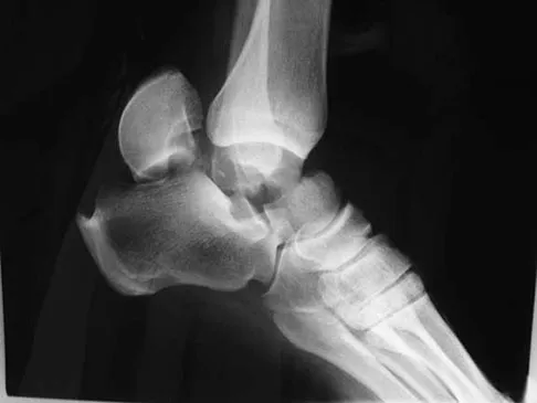
Figures 20a and 20b show the radiographs of a 14-year-old boy who sustained a twisting injury to his ankle. If attempted closed reduction is unsuccessful, what is the primary reason to proceed with surgical treatment?


Explanation

Triplane fractures generally occur in children who are near skeletal maturity. The injury is generally caused by a supination external rotation mechanism. The number of fracture fragments present (two or three) depends on what part of the physes is closed at the time of injury. Articular congruity is the major concern in the management of these injuries since the patient has almost reached skeletal maturity. The goal is to restore articular congruity to minimize the development of posttraumatic arthritis. Vaccaro A (ed): Orthopaedic Knowledge Update 8. Rosemont, IL, American Academy of Orthopaedic Surgeons, 2005, pp 757-765. Kling TF Jr, Bright RW, Hensinger RN: Distal tibial physeal fractures in children that may require open reduction. J Bone Joint Surg Am 1984;66:647-657.


Question 14

A 75-year-old woman reports foot pain and states that her foot has become progressively "flatter" in the past 3 years. Custom inserts and physical therapy have failed to provide relief. Examination reveals a flexible hindfoot and mild heel cord contracture. The patient is able to perform a single limb heel rise. Weight-bearing radiographs are shown in Figures 21a through 21d. What is the most appropriate surgical management?


Explanation

The patient has end-stage midfoot arthritis, with a secondary flatfoot deformity through the midfoot. The ability to perform a single limb heel rise indicates that the posterior tibial tendon is functioning, and the weight-bearing radiographs show normal calcaneal pitch and talar head coverage, thus confirming that the flatfoot deformity is isolated to the midfoot. Therefore, the most appropriate treatment is medial column arthrodesis and heel cord lengthening. The other listed procedures are not indicated because they are used in the management of adult flatfoot from posterior tibial tendon insufficiency. Toolan BC: Midfoot arthrodesis: Challenges and treatment alternatives. Foot Ankle Clin 2002;7:75-93.


Question 15

A 52-year-old woman who underwent cheilectomy 1 year ago for hallux rigidus now reports continued pain in the first metatarsophalangeal joint. She did not have any incision healing problems, and has not had any fevers, erythema, or drainage. Which of the following procedures will provide the best combination of pain relief and function?


Explanation

All but the Moberg osteotomy are capable of providing pain relief; however, arthrodesis offers the best long-term results and restores weight bearing and propulsion function to the first ray. Machacek F Jr, Easley ME, Gruber F, et al: Salvage of a failed Keller resection arthroplasty. J Bone Joint Surg Am 2004;86:1131-1138.


Question 16

During a posterior approach to the right Achilles tendon, the surgeon encounters a nerve running with the small saphenous vein as shown in Figure 22. This nerve innervates what part of the foot?


Explanation

The sural nerve runs with the small saphenous vein on the posterior leg just lateral to the Achilles tendon. It is formed by contributions from both the tibial and common peroneal nerves and provides sensation on the dorso-lateral aspect of the foot. Aktan Ikiz ZA, Ucerler H, Bilge O: The anatomic features of the sural nerve with an emphasis on its clinical importance. Foot Ankle Int 2005;26:560-567.


Question 17

A 23-year-old woman has had a 14-month history of ankle pain after surgical treatment of multiple injuries resulting from a motor vehicle accident. Weight bearing began 4 months after surgery. The pain occurs with weight bearing and motion, but there is very little pain at rest. She has no pertinent medical history and does not smoke. Figures 23a and 23b show current radiographs. What is the most appropriate surgical option?


Explanation

The radiographs reveal nonunion of a talar neck fracture. There is no radiographic evidence of osteonecrosis or significant degenerative arthritis. The results of talectomy are suboptimal. Arthrodesis would be indicated for degenerative arthritis. Revision ORIF is feasible and preserves motion. A vascularized graft should be considered whenever osteonecrosis is present, but the talar body appears viable in this case. Calvert E, Younger A, Penner M: Post talus neck fracture reconstruction. Foot Ankle Clin 2007;12:137-151.


Question 18

What type of physical therapy is most effective for chronic noninsertional Achilles tendinopathy?


Explanation

Eccentric gastrocsoleus strengthening (especially with heavy loads) consistently has been shown to be superior in the management of Achilles tendinopathy. Decreases in pain and increases in strength have been demonstrated despite the frequently refractory nature of this condition. Richardson EG (ed): Orthopaedic Knowledge Update: Foot and Ankle 3. Rosemont, IL, American Academy of Orthopaedic Surgeons, 2003, pp 91-102.


Question 19

A 27-year-old man was struck by a taxi cab and sustained comminuted right distal third tibia and fibula fractures; treatment consisted of placement of an intramedullary nail in the tibia the following morning. At his 6-month follow-up, he has clawing of all five toes. Examination reveals flexion deformities of the distal and proximal interphalangeal joints that are flexible with plantar flexion and rigid with dorsiflexion. Calluses are present on the dorsum and tip of the toes. Single heel rise is normal. He has a mild equinus contracture (relative to the left leg) that is not relieved with knee flexion. What is the most appropriate treatment option?


Explanation

This is an example of tethering of the flexor hallucis longus/flexor digitorum longus (FHL/FDL) to the fracture site. Additional time and/or physical therapy and bracing would not be expected to be of benefit. Release of the FHL and FDL from the fracture site or retromalleolar lengthening will address the posttraumatic claw toe deformity and Achilles tendon lengthening will address the mild equinus. Posterior tibial tendon transfer is not appropriate as the patient demonstrates a normal heel rise. Midfoot releases and hallux fusion are also not indicated. Feeny MS, Williams RL, Stephens MM: Selective lengthening of the proximal flexor tendon in the management of acquired claw toes. J Bone Joint Surg Br 2001;83:335-338.


Question 20

A 24-year-old man reports the development of a foot drop following a knee dislocation 1 year ago. The common peroneal nerve was found to be in continuity at the time of surgical reconstruction of the posterolateral corner of the knee joint. He would like to eliminate the need for an ankle-foot orthosis. What is the best option to achieve elimination of the orthosis?


Explanation

The ankle dorsiflexor muscles have been denervated for too long a period to expect reinnervation to be successful. Even if the extensor hallucis longus tendon was functional, it is unlikely to have sufficient strength to achieve dynamic ankle dorsiflexion. The tibialis posterior tendon transfer has been shown to predictably achieve these goals in a high percentage of patients. Successful ankle fusion is likely to fail with time due to the development of forefoot equinus. Pinzur MS, Kett N, Trilla M: Combined anteroposterior tibial tendon transfer in post-traumatic peroneal palsy. Foot Ankle 1988;8:27l-275.


Question 21

A 21-year-old male construction worker fell from a roof and sustained an injury to his left foot. Radiographs and CT scans are shown in Figures 24a through 24e. Compared to nonsurgical management, surgical treatment offers which of the following advantages?


Explanation

The radiographs and CT scans show a displaced intra-articular calcaneal fracture, with loss of calcaneal height and length. Recent multicenter, randomized, prospective studies suggest that surgical treatment of displaced intra-articular calcaneal fractures is associated with an almost six-fold decrease in the risk of posttraumatic subtalar arthritis (necessitating subtalar arthrodesis) compared to nonsurgical treatment. Despite ongoing controversy, surgical treatment has not been shown to be advantageous with respect to activity, time to return to work, or subtalar joint range of motion. A nonunion of a calcaneal fracture is exceedingly rare regardless of the treatment method. Buckley R, Tough S, McCormack R, et al: Operative compared with nonoperative treatment of displaced intra-articular calcaneal fractures: A prospective, randomized, controlled multicenter trial. J Bone Joint Surg Am 2002;84:1733-1744.


Question 22

A 51-year-old plumber has a failed peroneus brevis tendon repair. He reports continued pain and swelling in the distal retrofibular area. MRI shows longitudinal tears of the peroneus longus and peroneus brevis. What is the surgical treatment of choice at this time?


Explanation

A flexor digitorum longus transfer, while not as strong as the peroneals, improves the tendon balance and maintains hindfoot mobility. Subtalar fusion is a salvage procedure. Posterior tibial tendon transfer compromises inversion strength and arch height. Functional absence of the peroneals results in an imbalance that could lead to forefoot varus. Redfern D, Myerson M: The management of concomitant tears of the peroneus longus and brevis tendons. Foot Ankle Int 2004;25:695-707.


Question 23

Which of the following imaging modalities is most accurate in locating a toothpick in the plantar arch of the foot?


Explanation

Ultrasound is best at imaging abrupt changes in the density of adjacent tissue and therefore is best at imaging wood in the soft tissues of the foot. Mizel MS, Steinmetz ND, Trepman E: Detection of wooden foreign bodies in muscle tissue: Experimental comparison of computed tomography, magnetic resonance imaging, and ultrasonography. Foot Ankle Int 1994;15:437-443.


Question 24

A 35-year-old man is seen in the emergency department with a bullet wound to the foot that occurred 2 hours ago. Examination reveals a 0.5-cm entrance wound on the dorsum of the foot and a 1.5-cm exit wound on the plantar aspect. Exploration of the plantar wound in the emergency department reveals bone and metal fragments. Radiographs reveal a comminuted, unstable fracture of the base of the first metatarsal and cuneiform. Management should consist of tetanus toxoid, and


Explanation

The patient sustained a type I unstable fracture that requires debridement of superficial fragments from the sole and surgical stabilization. Low-velocity wounds less than 8 hours old are considered type I open fractures. In contrast, gunshot wounds with associated fractures more than 8 hours old are considered type II open fractures using the Gustilo and Anderson classification. Gustilo type I stable fractures due to gunshot wounds and seen within 8 hours can be treated with tetanus toxoid (if no history of immunization or booster within 5 years), surface irrigation, and casting or a hard sole shoe. Antibiotics are not required unless gross contamination is present. However, if the extent of contamination is unclear, or if a joint is penetrated, then routine antibiotic prophylaxis is recommended. Indications for surgery include: articular involvement, unstable fractures, presentation 8 or more hours after injury, tendon involvement, and superficial fragments in the palm or sole. Type I unstable fractures may be stabilized with internal or external fixation. Type II unstable fractures should be treated with external fixation and repeat debridements until clean. Holmes GB Jr: Gunshot wounds of the foot. Clin Orthop Relat Res 2003;408:86-91.

Question 25

What is the most frequent location of entrapment of the deep peroneal nerve?


Explanation

The most frequently described entrapment of the deep peroneal nerve is the anterior tarsal tunnel syndrome. This syndrome refers to entrapment of the deep peroneal nerve under the inferior extensor retinaculum. Entrapment can also occur as the nerve passes under the tendon of the extensor hallucis brevis. Compression by underlying dorsal osteophytes of the talonavicular joint and an os intermetatarseum (between the bases of the first and second metatarsals) have previously been described in runners. Kopell HP, Thompson WA: Peripheral entrapment neuropathies of the lower extremity. N Engl J Med 1960;262:56-60.

Question 26

A 50-year-old woman presents with progressive medial foot pain and loss of her arch. On examination, she has a flexible valgus hindfoot and pronounced forefoot abduction. Weight-bearing radiographs demonstrate greater than 40% talonavicular uncoverage. What is the most appropriate surgical management?


Explanation

This patient has Stage IIb posterior tibial tendon dysfunction, characterized by a flexible flatfoot with significant forefoot abduction (>40% TN uncoverage). Correcting the pronounced forefoot abduction requires a lateral column lengthening (e.g., Evans osteotomy) in addition to an FDL transfer and MDCO.

Question 27

In evaluating a supination-external rotation ankle fracture, which of the following is the most reliable radiographic parameter indicating a syndesmotic injury on a standard AP view of the ankle?


Explanation

The tibiofibular clear space, measured 1 cm above the joint line on AP and mortise views, should be less than 5 mm. It is the most reliable radiographic parameter for assessing syndesmotic widening, as it is relatively unaffected by leg rotation.

Question 28

A 45-year-old woman presents with pain and progressive medial deviation of her great toe one year following a distal chevron osteotomy and lateral soft tissue release for hallux valgus. What intraoperative technical error most commonly contributes to this complication?


Explanation

"Staking" the first metatarsal head by resecting the medial eminence past the sagittal sulcus removes the bony block to medial subluxation. Combined with a lateral release and medial capsular plication, this frequently leads to iatrogenic hallux varus.

Question 29

A 65-year-old woman presents with severe, chronic medial and lateral foot pain. On exam, she has a rigid, non-reducible hindfoot valgus deformity and fixed forefoot supination. She cannot perform a single-limb heel rise. Radiographs demonstrate advanced degenerative changes of the subtalar and talonavicular joints. What is the most appropriate surgical treatment?


Explanation

Stage III PTTD involves a rigid hindfoot valgus deformity and degenerative changes in the subtalar and/or transverse tarsal joints. A triple arthrodesis (subtalar, talonavicular, and calcaneocuboid) is the gold standard for rigid, arthritic deformity correction in this setting.

Question 30

According to the Lauge-Hansen classification, what is the initial structure injured in a supination-external rotation (SER) ankle fracture?


Explanation

The SER mechanism progresses sequentially in four stages. Stage 1 is the rupture of the anterior inferior tibiofibular ligament (AITFL), followed by a short oblique fibula fracture (Stage 2), rupture of the posterior inferior tibiofibular ligament (Stage 3), and finally, deltoid ligament failure or a medial malleolus fracture (Stage 4).

Question 31

A 50-year-old woman presents with a painful bunion. Weight-bearing radiographs reveal a hallux valgus angle of 45 degrees, an intermetatarsal angle of 18 degrees, and plantar gapping at the 1st tarsometatarsal (TMT) joint consistent with hypermobility. Which of the following procedures is most appropriate?


Explanation

The Lapidus procedure (1st TMT arthrodesis) is indicated for moderate to severe hallux valgus (IMA > 15 degrees) combined with first ray hypermobility. It provides powerful correction and stabilizes the medial column effectively.

Question 32

A 25-year-old athlete sustains a severe twisting injury to the ankle. Radiographs show widening of the medial clear space and a high fibular fracture. Which test performed intraoperatively is most reliable for evaluating syndesmotic instability after fibular fixation?


Explanation

The external rotation stress test under fluoroscopy is the most sensitive and reliable intraoperative method to detect syndesmotic instability. The Cotton test (lateral pull on the fibula) is also used but is less sensitive.

Question 33

A 45-year-old woman presents with a painful bunion. Radiographs reveal a hallux valgus angle of 42 degrees, an intermetatarsal angle of 18 degrees, and obvious hypermobility of the first tarsometatarsal joint on clinical exam. What is the most appropriate surgical treatment?


Explanation

The Lapidus procedure (first tarsometatarsal joint arthrodesis) is indicated for severe hallux valgus (IMA > 15 degrees) accompanied by first ray hypermobility. It stabilizes the medial column and prevents recurrence.

Question 34

A 50-year-old woman presents with medial ankle pain and a progressively flattening arch. She has pain and inability to perform a single-leg heel raise. The hindfoot valgus is flexible and corrects to neutral when she stands on her toes. Radiographs show uncovering of the talonavicular joint but no arthritis. What is the most appropriate surgical intervention?


Explanation

This patient has Stage II posterior tibial tendon dysfunction (flexible deformity, no arthritis). The gold standard surgical treatment is an FDL transfer to the navicular combined with a medial displacement calcaneal osteotomy.

Question 35

In a supination-external rotation (SER) ankle fracture, what is the first structure injured according to the Lauge-Hansen classification?


Explanation

In the Lauge-Hansen supination-external rotation mechanism, the anterior inferior tibiofibular ligament (AITFL) is the first structure to fail (Stage I). This is followed by a spiral fracture of the fibula (Stage II).

Question 36

A patient develops a progressive iatrogenic hallux varus deformity after a bunionectomy. Operative notes describe aggressive lateral soft tissue release and excision of the fibular sesamoid. Resection of which structure most significantly contributed to this complication?


Explanation

Excision of the fibular sesamoid often involves resection of the lateral head of the flexor hallucis brevis. This severely imbalances the dynamic stabilizers of the MTP joint, heavily predisposing the patient to a hallux varus deformity.

Question 37

A 62-year-old man presents with a painful, rigid flatfoot deformity. Examination reveals a fixed hindfoot valgus and an inability to perform a single-leg heel raise. Radiographs demonstrate advanced degenerative changes at the subtalar and talonavicular joints. What is the recommended surgical management?


Explanation

A rigid flatfoot deformity with subtalar and talonavicular arthritis defines Stage III posterior tibial tendon dysfunction. The definitive treatment for a fixed, arthritic hindfoot is a triple arthrodesis.

Question 38

A 35-year-old man sustains a severe fracture-dislocation of the ankle. Closed reduction in the emergency department is completely unsuccessful. Radiographs show a displaced fracture of the lateral malleolus, and the proximal fibular fragment appears incarcerated behind the posterior tubercle of the tibia. What is this specific injury pattern called?


Explanation

A Bosworth fracture-dislocation involves irreducible posterior dislocation of the fibula behind the posterior lateral ridge of the tibia. It requires immediate open reduction to prevent skin necrosis and definitive fixation.

Question 39

A 22-year-old woman has a painful bunion. Radiographs reveal a hallux valgus angle of 35 degrees, an intermetatarsal angle of 12 degrees, and a distal metatarsal articular angle (DMAA) of 20 degrees. The first MTP joint is congruous. Which of the following procedures is most appropriate?


Explanation

An abnormally high DMAA (>15 degrees) indicates that the distal articular surface is laterally deviated. A biplanar distal chevron osteotomy (incorporating a medial closing wedge) corrects both the intermetatarsal angle and the articular orientation.

Question 40

During surgical reconstruction for flexible adult acquired flatfoot deformity (Stage II PTTD), the surgeon explores the medial soft tissues. Which ligamentous structure is most commonly attenuated and requires imbrication or repair along with the FDL transfer?


Explanation

The plantar calcaneonavicular (spring) ligament is the primary static stabilizer of the talonavicular joint. It is frequently stretched or torn in Stage II PTTD and often requires repair or reconstruction during FDL transfer.

Question 41

A 14-year-old boy sustains a twisting injury to his ankle. Radiographs reveal a Salter-Harris III fracture of the anterolateral distal tibia. The avulsed bony fragment is attached to which of the following structures?


Explanation

This describes a juvenile Tillaux fracture, which occurs due to avulsion of the anterior inferior tibiofibular ligament (AITFL). The anterolateral physis is the last to close, making it susceptible to this specific Salter-Harris III injury.

Question 42

During a distal chevron osteotomy for hallux valgus, the surgeon must be careful to avoid avascular necrosis of the first metatarsal head. This risk is minimized by carefully preserving which of the following?


Explanation

The primary blood supply to the first metatarsal head enters via the capsular arterial network, primarily at the plantar-lateral aspect. Preserving the plantar-lateral soft tissue attachments during osteotomy and lateral release minimizes the risk of AVN.

Question 43

A 70-year-old woman with a longstanding flatfoot deformity now reports deep, aching medial ankle pain. Standing radiographs demonstrate severe hindfoot valgus, talonavicular subluxation, and talar tilt within the ankle mortise with narrowing of the lateral tibiotalar joint space. What stage of posterior tibial tendon dysfunction does this represent?


Explanation

Stage IV posterior tibial tendon dysfunction is characterized by deltoid ligament insufficiency leading to valgus tilting of the talus in the ankle mortise, often accompanied by lateral ankle impingement and arthritis.

Question 44

An ankle fracture characterized by a transverse fracture of the medial malleolus, rupture of the syndesmosis, and a short oblique or comminuted fracture of the fibula at or above the level of the syndesmosis represents which Lauge-Hansen mechanism?


Explanation

A transverse medial malleolar fracture indicates a pronation injury (tension medially). A bending force laterally causes a short oblique or comminuted fibular fracture at the syndesmosis, classic for the Pronation-Abduction mechanism.

Question 45

A patient undergoes a bunionectomy with a distal chevron osteotomy. Intraoperatively, the intermetatarsal and hallux valgus angles are completely corrected, but the great toe remains deviated laterally at the interphalangeal joint. What is the most appropriate next step?


Explanation

Residual valgus deformity of the hallux at the interphalangeal joint, despite a corrected MTP joint, is termed hallux interphalangeus. It is effectively treated with a medial closing wedge osteotomy of the proximal phalanx (Akin procedure).

Question 46

In a patient with Stage IIb posterior tibial tendon dysfunction, a flexor digitorum longus transfer and lateral column lengthening are performed. Intraoperatively, the foot is noted to have persistent forefoot supinatus with the hindfoot held in neutral. Which of the following procedures should be added?


Explanation

Persistent forefoot supinatus after hindfoot correction in flatfoot reconstruction requires medial column stabilization. This is typically achieved with an opening wedge medial cuneiform osteotomy (Cotton) or first TMT arthrodesis to plantarflex the first ray.

Question 47

A 40-year-old man sustains a trimalleolar ankle fracture. The posterior malleolus fragment involves 35% of the articular surface and remains displaced 3 mm superiorly after anatomic fibular and medial malleolar fixation. What is the most appropriate management of the posterior malleolus?


Explanation

Posterior malleolar fractures involving >25% to 33% of the articular surface or with persistent displacement >2 mm require open reduction and internal fixation to restore articular congruity and posterior stability.

Question 48

A 60-year-old woman with advanced rheumatoid arthritis presents with severe bilateral bunions, lesser toe deformities, and subluxation of the first MTP joints. Radiographs of the first MTP joint show complete loss of cartilage and erosion. What is the gold standard surgical treatment for her first ray?


Explanation

First MTP joint arthrodesis is the procedure of choice for severe, arthritic hallux valgus in rheumatoid patients. It provides definitive, long-lasting relief and creates a stable medial column to assist in lesser toe reconstruction.

Question 49

A 28-year-old professional soccer player sustains an isolated syndesmotic injury without fracture. Despite 6 weeks of conservative management, he continues to have pain and instability. Stress radiographs show a widened medial clear space. What is the most appropriate surgical management?


Explanation

Suture button fixation provides dynamic stabilization of the syndesmosis, allowing physiologic motion while maintaining reduction. This is particularly beneficial in athletes for a faster return to play and avoiding the need for hardware removal.

Question 50

A 42-year-old woman sustains a supination-external rotation ankle fracture. CT scan reveals a posterolateral tibial (posterior malleolar) fragment involving 30% of the articular surface with 3 mm of superior displacement. What is the most appropriate management for the posterior malleolus?


Explanation

A displaced posterior malleolar fracture involving >25% of the articular surface warrants fixation. A posterolateral approach allows direct visualization and stable buttress plating of the fragment, which is biomechanically superior to anterior-to-posterior lag screws.

Question 51

A 55-year-old woman presents with pain and difficulty wearing shoes 1 year after a modified McBride bunionectomy. Clinical examination reveals a rigid hallux varus deformity with severe degenerative joint disease of the first metatarsophalangeal (MTP) joint on radiographs. What is the most appropriate definitive treatment?


Explanation

In a patient with a symptomatic, rigid hallux varus deformity accompanied by severe degenerative changes of the first MTP joint, arthrodesis is the most reliable and definitive treatment to relieve pain and correct alignment.

Question 52

A 62-year-old woman presents with a flexible, acquired flatfoot deformity (Stage II PTTD). She has a positive single-leg heel rise test. Radiographs show uncovering of the talonavicular joint and a talonavicular sag. Conservative measures have failed. Which surgical combination is most appropriate?


Explanation

Stage II PTTD features a flexible flatfoot. The gold standard surgical management includes a soft-tissue reconstruction (FDL transfer) combined with a bony procedure (medial displacement calcaneal osteotomy or lateral column lengthening) to restore the arch and correct hindfoot valgus.

Question 53

A 70-year-old man presents with a painful, rigid flatfoot deformity and is unable to perform a single-leg heel rise. Examination shows fixed hindfoot valgus and forefoot abduction. Radiographs reveal advanced osteoarthritis of the subtalar and talonavicular joints, with no ankle joint arthritis. What is the most appropriate surgical treatment?


Explanation

Stage III PTTD is characterized by a rigid deformity and arthritic changes in the hindfoot joints. A triple arthrodesis (subtalar, talonavicular, and calcaneocuboid) is the most appropriate treatment to correct the rigid deformity and relieve pain.

Question 54

A 48-year-old woman has a severe hallux valgus deformity with a hallux valgus angle (HVA) of 45 degrees, an intermetatarsal angle (IMA) of 20 degrees, and hypermobility of the first tarsometatarsal (TMT) joint. What is the surgical procedure of choice?


Explanation

For severe hallux valgus (IMA >15 degrees) associated with first TMT joint hypermobility, a Lapidus procedure (first TMT arthrodesis) provides reliable correction and stabilizes the medial column to prevent recurrence.

Question 55

A 65-year-old man with poorly controlled type 2 diabetes mellitus and profound peripheral neuropathy sustains an acute, closed, displaced bimalleolar ankle fracture. What modification to the standard surgical approach is most appropriate?


Explanation

Diabetic patients with neuropathy are at a high risk for hardware failure and Charcot arthropathy following ankle fractures. Augmented fixation and an extended period of non-weight-bearing are recommended to ensure successful union and minimize complications.

Question 56

A 14-year-old girl with open physes presents with a symptomatic hallux valgus deformity (HVA 30 degrees, IMA 14 degrees). What is the most important factor to address surgically to prevent recurrence in this juvenile patient?


Explanation

Juvenile hallux valgus is frequently associated with an increased distal metatarsal articular angle (DMAA). Failure to address an abnormal DMAA with a double osteotomy often leads to high recurrence rates in adolescent patients.

Question 57

A 45-year-old woman presents with a painful bunion. Weight-bearing radiographs reveal a hallux valgus angle (HVA) of 40 degrees and an intermetatarsal angle (IMA) of 17 degrees. There is no evidence of first tarsometatarsal (TMT) joint hypermobility or degenerative changes at the first metatarsophalangeal (MTP) joint. Which of the following is the most appropriate surgical intervention?


Explanation

An IMA greater than 13-15 degrees with a large HVA requires a proximal metatarsal osteotomy or Lapidus procedure to achieve adequate correction. Since there is no TMT hypermobility, a proximal osteotomy with distal soft-tissue realignment is appropriate.

Question 58

A 55-year-old woman presents with a progressive flatfoot deformity. She is unable to perform a single-leg heel rise. Examination shows a flexible hindfoot valgus. Weight-bearing radiographs show uncovering of the talonavicular joint of 40%. Which of the following surgical procedures is most appropriate?


Explanation

This patient has Stage IIb posterior tibial tendon dysfunction (PTTD), characterized by significant forefoot abduction (>30% talonavicular uncoverage). This requires a lateral column lengthening in addition to FDL transfer and medializing calcaneal osteotomy.

Question 59

During open reduction and internal fixation of a Weber B fibula fracture, the static mortise radiograph appears normal. Which of the following intraoperative tests is most reliable for diagnosing latent syndesmotic instability?


Explanation

Intraoperative assessment using an external rotation stress test or a lateral pull test (Cotton test) under fluoroscopy is the most reliable method to detect latent syndesmotic instability.

Question 60

A 32-year-old man sustains an ankle fracture. Radiographs demonstrate a vertical fracture of the medial malleolus and a transverse fracture of the fibula at the level of the tibial plafond. According to the Lauge-Hansen classification, what is the mechanism of this injury?


Explanation

A vertical medial malleolus fracture combined with a transverse lateral malleolus fracture at or below the joint line is the hallmark of a Supination-Adduction (SAD) injury.

Question 61

A 48-year-old woman presents with a painful bunion. Clinical examination demonstrates significant hypermobility of the first tarsometatarsal (TMT) joint in the sagittal plane. Radiographs show an intermetatarsal angle (IMA) of 18 degrees and a hallux valgus angle (HVA) of 42 degrees. What is the most appropriate definitive procedure?


Explanation

The Lapidus procedure (first TMT arthrodesis) is specifically indicated for patients with moderate to severe hallux valgus accompanied by first TMT joint hypermobility.

Question 62

A 62-year-old man presents with a painful, severe flatfoot deformity. On examination, the hindfoot is in a fixed valgus position and cannot be passively inverted to neutral. Radiographs reveal degenerative changes in the subtalar and talonavicular joints. What is the most appropriate surgical treatment?


Explanation

A fixed, rigid flatfoot deformity with degenerative changes in the subtalar and talonavicular joints represents Stage III PTTD, which is best treated with a triple arthrodesis.

Question 63

A 28-year-old rugby player sustains an ankle injury. Closed reduction in the emergency department is unsuccessful. Radiographs show a posterior fracture-dislocation of the fibula, with the proximal fibular fragment trapped behind the tibia. What specific anatomical structure blocks the reduction in this Bosworth fracture-dislocation?


Explanation

A Bosworth fracture is a rare fracture-dislocation where the proximal fibular fragment becomes incarcerated behind the posterior lateral ridge (posterior tubercle) of the distal tibia, making closed reduction nearly impossible.

Question 64

A 62-year-old man with a 15-year history of poorly controlled type 2 diabetes mellitus undergoes open reduction and internal fixation for a displaced bimalleolar equivalent ankle fracture. Which of the following postoperative regimens is most appropriate to minimize complications in this specific patient?


Explanation

Diabetic patients with ankle fractures have a significantly higher risk of complications, including Charcot neuroarthropathy, hardware failure, and delayed union. Current AAOS/ABOS guidelines recommend prolonged non-weight-bearing (up to 3 months) followed by protected weight-bearing (TCC or CROW) to mitigate these risks.

Question 65

A 45-year-old woman presents with pain and medial deviation of her great toe 6 months after undergoing a distal chevron osteotomy and lateral soft tissue release for hallux valgus. Standing radiographs reveal an intermetatarsal angle (IMA) of 6 degrees and a hallux valgus angle (HVA) of -15 degrees. What intraoperative technical error is the most likely cause of this complication?


Explanation

Hallux varus is a known complication of bunion surgery. It is most commonly caused by over-resection of the medial eminence (staking the head), over-tightening of the medial capsule, over-release of the lateral structures, or excision of the fibular sesamoid.

Question 66

A 55-year-old woman presents with a painful, flexible flatfoot deformity. She is unable to perform a single-leg heel rise on the affected side. Standing radiographs demonstrate 45% uncovering of the talonavicular joint and a talonavicular uncoverage angle of 40 degrees. According to the Johnson and Strom classification (modified by Myerson), what is the most appropriate surgical management?


Explanation

This patient has Stage IIb posterior tibial tendon dysfunction, characterized by a flexible flatfoot with significant forefoot abduction (>30-40% talonavicular uncoverage). Management requires FDL transfer and medializing calcaneal osteotomy, plus a lateral column lengthening to correct the severe forefoot abduction.

Question 67

According to the Lauge-Hansen classification system, what is the third stage of injury in a Supination-External Rotation (SER) ankle fracture?


Explanation

The sequence for SER injuries is: 1) Anterior inferior tibiofibular ligament (AITFL), 2) Short oblique/spiral fibula fracture, 3) Posterior inferior tibiofibular ligament (PITFL) or posterior malleolus fracture, 4) Medial malleolus fracture or deltoid ligament tear.

Question 68

During a distal chevron osteotomy for hallux valgus, care must be taken to avoid avascular necrosis of the first metatarsal head. Which of the following vessels provides the primary blood supply to the first metatarsal head?


Explanation

The primary blood supply to the first metatarsal head arises from the first plantar metatarsal artery, which gives off capsular branches entering the head at the plantar-lateral aspect. Extensive lateral soft tissue release combined with a distal osteotomy can jeopardize this blood supply.

Question 69

A 68-year-old man presents with a long-standing flatfoot deformity. On examination, the hindfoot is in severe valgus and is completely rigid on attempted manual correction. He has significant pain over the lateral aspect of the subtalar joint. Radiographs reveal bone-on-bone arthritis of the subtalar and talonavicular joints. What is the most appropriate surgical treatment?


Explanation

This is Stage III posterior tibial tendon dysfunction, characterized by a rigid, non-correctable hindfoot deformity with subtalar/talonavicular arthritis. The gold standard surgical treatment for Stage III PTTD is a triple arthrodesis.

Question 70

A 32-year-old man presents with a closed ankle fracture-dislocation after a fall. Closed reduction in the emergency department is unsuccessful. Radiographs reveal a severe fracture-dislocation where the proximal fragment of the fibula is locked behind the posterior tubercle of the tibia. What is the eponymous name of this specific injury?


Explanation

A Bosworth fracture-dislocation is a rare injury where the proximal fragment of the fractured fibula becomes irreducibly trapped behind the posterior aspect of the tibia. This invariably requires open reduction to free the fibula.

Question 71

A 35-year-old woman presents with a painful bunion. Clinical examination demonstrates significant hypermobility of the first tarsometatarsal (TMT) joint, with dorsal elevation of the first ray on weight-bearing. Standing radiographs show a hallux valgus angle of 35 degrees and an intermetatarsal angle of 18 degrees. Which of the following is the most appropriate surgical procedure?


Explanation

The Lapidus procedure (first TMT arthrodesis) is indicated for moderate to severe hallux valgus associated with first ray hypermobility or TMT joint arthritis. It definitively addresses the apex of the deformity and stabilizes the medial column.

Question 72

In the pathogenesis of posterior tibial tendon dysfunction (PTTD), the spring ligament complex frequently attenuates. Which specific band of the spring ligament is the primary static stabilizer of the talonavicular joint and is most commonly torn?


Explanation

The superomedial calcaneonavicular ligament is the thickest and most critical component of the spring ligament complex. It acts as the primary static sling supporting the talar head, and its failure is a hallmark of progressive PTTD.

Question 73

A 24-year-old man sustains an SER-IV equivalent ankle injury with a ruptured deltoid ligament and a fibula fracture above the level of the syndesmosis. After rigid internal fixation of the fibula, how is the integrity of the syndesmosis best evaluated intraoperatively?


Explanation

The Cotton test is performed by applying a lateral traction force to the fibula with a bone hook while observing the syndesmosis under fluoroscopy. Widening of the tibiofibular clear space indicates syndesmotic instability requiring fixation.

Question 74

A patient undergoes a proximal crescentic osteotomy and distal soft tissue release for severe hallux valgus. Postoperative radiographs show the intermetatarsal angle is corrected to 7 degrees, but the great toe still deviates laterally at the interphalangeal joint with a Hallux Valgus Interphalangeus (HVI) angle of 18 degrees. What is the best next step to achieve full clinical correction?


Explanation

An Akin osteotomy (medial closing wedge of the proximal phalanx) is indicated to correct hallux valgus interphalangeus. It does not correct the intermetatarsal angle but straightens the toe when the primary MTP deformity has been addressed.

Question 75

When performing a tendon transfer for Stage II posterior tibial tendon dysfunction, the Flexor Digitorum Longus (FDL) is typically preferred over the Flexor Hallucis Longus (FHL). What is the primary functional reason for avoiding routine FHL harvest in this setting?


Explanation

While the FHL is stronger than the FDL, harvesting the FHL can lead to a significant functional deficit in great toe push-off during gait. The FDL provides sufficient strength for the transfer with highly acceptable donor site morbidity.

Question 76

A 40-year-old woman sustains a trimalleolar ankle fracture. CT scan reveals the posterior malleolar fragment involves 35% of the articular surface and is displaced 3 mm proximally, with posterior subluxation of the talus. What is the most appropriate management of the posterior malleolus?


Explanation

Posterior malleolar fractures involving >25-30% of the articular surface, exhibiting >2 mm displacement, or associated with posterior talar subluxation require ORIF. Anatomic reduction restores the joint surface and the posterior tibiofibular syndesmotic stability.

Question 77

A 16-year-old female presents with juvenile hallux valgus. Radiographs demonstrate an intermetatarsal angle of 14 degrees and a Distal Metatarsal Articular Angle (DMAA) of 25 degrees. If a standard proximal metatarsal osteotomy is performed alone without addressing the DMAA, which of the following is the most likely outcome?


Explanation

An abnormally high DMAA implies the articular surface of the metatarsal head is laterally deviated. Correcting the IMA alone without a distal procedure (like a biplanar chevron or distal closing wedge) forces the phalanx medially against a laterally directed articular surface, causing incongruency and risk of recurrence.

Question 78

A 72-year-old woman presents with severe, long-standing flatfoot. Radiographs show rigid hindfoot valgus, severe subtalar arthritis, and a valgus tilt of the talus within the ankle mortise. What is her posterior tibial tendon dysfunction stage and the most appropriate surgical treatment?


Explanation

Valgus tilt of the talus within the ankle mortise indicates deltoid ligament failure, defining Stage IV PTTD. In the presence of a rigid hindfoot and subtalar arthritis, a tibiotalocalcaneal (TTC) arthrodesis is the most reliable treatment to correct the deformity and relieve pain.

Question 79

A 28-year-old man twists his ankle. Initial non-weight-bearing radiographs show an isolated Weber B lateral malleolus fracture with a symmetric medial clear space. Clinical examination reveals exquisite tenderness over the deltoid ligament. What is the most appropriate next step to determine the need for operative fixation?


Explanation

A Weber B fracture with medial tenderness suggests a potential bimalleolar equivalent injury (deltoid rupture). A gravity stress or external rotation stress radiograph is necessary to assess medial clear space widening; if widening occurs (>4-5 mm), surgical fixation is indicated.

Question 80

A 55-year-old woman complains of progressive medial ankle pain and loss of the arch of her foot over the past year. On examination, she has a flexible flatfoot deformity and cannot perform a single-limb heel rise on the affected side. Radiographs show a talonavicular coverage angle of 15 degrees. What is the most appropriate surgical management?


Explanation

Stage II posterior tibial tendon dysfunction (PTTD) is characterized by a flexible flatfoot deformity. Joint-sparing procedures such as an FDL transfer combined with a medializing calcaneal osteotomy are the most appropriate surgical indicated treatment.

Question 81

A 62-year-old woman presents with severe flatfoot deformity. Examination reveals a rigid hindfoot in valgus and pain in the sinus tarsi. She is unable to invert her heel on double-limb heel rise. Radiographs demonstrate advanced degenerative changes in the subtalar and talonavicular joints. What is the most appropriate surgical treatment?


Explanation

Stage III PTTD involves a rigid deformity with associated hindfoot arthritis. It is best treated with a triple arthrodesis to correct the deformity and reliably alleviate arthritic pain.

Question 82

A 40-year-old woman has a painful bunion. Weight-bearing radiographs show a hallux valgus angle (HVA) of 42 degrees and an intermetatarsal angle (IMA) of 18 degrees. The first TMT joint is stable with no evidence of hypermobility or arthritis. Which of the following procedures is most appropriate?


Explanation

For a severe hallux valgus deformity (IMA >15 degrees, HVA >40 degrees) without first TMT hypermobility or midfoot arthritis, a proximal osteotomy with distal soft-tissue release provides the necessary powerful correction.

Question 83

A 45-year-old woman complains of a painful bunion. Radiographs reveal an HVA of 38 degrees and an IMA of 16 degrees. Clinical examination reveals significant sagittal plane hypermobility of the first tarsometatarsal (TMT) joint. What is the best surgical option?


Explanation

The Lapidus procedure (first TMT arthrodesis) is specifically indicated for moderate to severe hallux valgus when associated with clinical first TMT hypermobility to prevent recurrence.

Question 84

A 28-year-old man sustains a twisting injury to his ankle. Non-weight-bearing radiographs show a spiral fracture of the distal fibula at the level of the syndesmosis, with an intact medial malleolus and normal medial clear space. A subsequent external rotation stress radiograph reveals a medial clear space of 6 mm. What is the appropriate management?


Explanation

A positive external rotation stress test with widening of the medial clear space indicates a syndesmotic or deep deltoid injury (bimalleolar equivalent fracture). This requires ORIF of the lateral malleolus and evaluation with potential stabilization of the syndesmosis.

Question 85

In a Lauge-Hansen Supination-External Rotation (SER) stage IV ankle fracture, what is the correct sequential order of structural failure?


Explanation

The SER sequence classically progresses from anterolateral to posteromedial: 1) AITFL, 2) short oblique/spiral distal fibula fracture, 3) PITFL or posterior malleolus, and 4) Deltoid ligament or medial malleolus.

Question 86

A 65-year-old patient with poorly controlled type 2 diabetes mellitus and peripheral neuropathy sustains a displaced bimalleolar ankle fracture. Which of the following modifications to the surgical plan is most appropriate to minimize complications?


Explanation

Diabetic patients with neuropathy are at a significantly higher risk for Charcot arthropathy, wound complications, and hardware failure. Enhanced rigid construct fixation and at least doubling the standard non-weight-bearing period are highly recommended.

Question 87

A 35-year-old woman develops hallux varus 6 months following a bunionectomy. On examination, the deformity is flexible and she has pain with wearing shoes. Which of the following surgical steps during her index procedure most likely contributed to this complication?


Explanation

Hallux varus is an iatrogenic complication of overcorrection. Major risk factors include excessive medial eminence resection (staking the metatarsal head), over-release of the lateral soft tissues, and excision of the fibular sesamoid.

Question 88

A patient with an ankle fracture has a large posterior malleolar fragment involving 35% of the articular surface. Posterior subluxation of the talus is evident on the lateral radiograph. What is the primary biomechanical advantage of open reduction and internal fixation of this posterior malleolar fragment?


Explanation

Anatomical fixation of a significant posterior malleolus fragment (>25% or with subluxation) directly restores the PITFL insertion. This offers superior biomechanical stability to the syndesmosis compared to isolated syndesmotic screws.

Question 89

Which of the following structures is the primary static stabilizer of the talonavicular joint and is most commonly attenuated or torn in conjunction with posterior tibial tendon dysfunction?


Explanation

The superomedial calcaneonavicular (spring) ligament is the primary static restraint to talar head plantarflexion. It is frequently attenuated or torn as PTTD progresses.

Question 90

A 22-year-old woman presents with bilateral foot pain due to juvenile hallux valgus. Radiographs show an HVA of 30 degrees, an IMA of 12 degrees, and a distal metatarsal articular angle (DMAA) of 25 degrees (normal <10). What surgical procedure is critical to achieve a congruent first MTP joint in this patient?


Explanation

Juvenile hallux valgus is frequently associated with an increased DMAA. Failure to correct the DMAA using a biplanar or double osteotomy will result in an incongruent joint and a very high rate of recurrence.

Question 91

A 50-year-old female undergoes a distal chevron osteotomy for a mild hallux valgus deformity. During the procedure, the lateral soft tissues are aggressively released intra-articularly to correct sesamoid subluxation. What is the most significant risk associated with this specific combination of maneuvers?


Explanation

The primary blood supply to the first metatarsal head is the first plantar metatarsal artery. Combining a distal osteotomy with an extensive intra-articular lateral soft-tissue release disrupts the capsular blood supply, significantly increasing the risk of avascular necrosis.

Question 92

A 45-year-old man presents with an ankle fracture. Radiographs show a transverse fracture of the medial malleolus and a short oblique fracture of the fibula starting at the level of the mortise and extending proximally. Based on the Lauge-Hansen classification, what is the mechanism of injury?


Explanation

A transverse medial malleolus fracture (or deltoid rupture) followed by a short oblique/transverse fibula fracture at or slightly above the joint line represents a Pronation-Abduction mechanism.

Question 93

Which clinical sign on physical examination is most specific for diagnosing a severe, rigid Stage III posterior tibial tendon dysfunction compared to a flexible Stage II?


Explanation

Stage III PTTD is clinically differentiated from Stage II by the presence of a fixed, rigid hindfoot valgus deformity that cannot be passively reduced to a neutral alignment.

Question 94

A 30-year-old female presents with a hallux valgus deformity and a symptomatic hallux valgus interphalangeus (HVI) angle of 20 degrees. Following a first metatarsal osteotomy, the IMA and HVA are corrected, but the big toe still abuts the second toe due to the HVI. What is the most appropriate next step?


Explanation

The Akin osteotomy (a medial closing wedge osteotomy of the proximal phalanx) is indicated specifically to correct hallux valgus interphalangeus after the primary IMA and joint congruency have been addressed.

None

Clinic OS
Medically Verified Content by
Prof. Clinic OS
Consultant Orthopedic & Spine Surgeon
Chapter Index