Arthroscopic Subacromial Decompression & Acromioplasty: A Masterclass in Shoulder Impingement Management
Key Takeaway
Join us in the OR for an immersive masterclass on arthroscopic subacromial decompression and acromioplasty. We'll meticulously review patient selection, precise portal placement, diagnostic arthroscopy, and the nuanced technique for subacromial bursectomy and acromial bone resection. Learn to navigate critical anatomy, manage potential pitfalls, and optimize patient outcomes for refractory shoulder impingement.
Comprehensive Introduction and Patho-Epidemiology
Arthroscopic subacromial decompression (ASAD) and anterior acromioplasty represent a foundational pillar in the surgical armamentarium of the orthopedic shoulder surgeon. When appropriately indicated, this procedure offers profound and durable relief for patients suffering from chronic shoulder pain and dysfunction secondary to extrinsic mechanical impingement. The evolution of this technique from an open, morbid procedure to a minimally invasive arthroscopic intervention has revolutionized our approach to shoulder pathology. However, the success of ASAD is entirely dependent upon a rigorous understanding of the underlying patho-epidemiology, meticulous patient selection, and flawless surgical execution. Before introducing instruments into the subacromial space, the modern surgeon must possess a rock-solid grasp of the complex interplay between shoulder biomechanics and structural anatomy.
The conceptual framework for subacromial impingement was meticulously defined by Charles Neer in 1972. Neer described impingement syndrome as a chronic, progressive mechanical process involving the compression of the rotator cuff tendons beneath the rigid coracoacromial arch. Fundamentally, extrinsic impingement occurs when the supraspinatus tendon is repetitively compressed against the undersurface of the anterior acromion, the coracoacromial (CA) ligament, or the acromioclavicular (AC) joint during arm elevation and internal rotation. This repetitive microtrauma initiates a predictable cascade of localized inflammation, bursal hypertrophy, and tendinosis, ultimately culminating in partial or full-thickness rotator cuff tears if left untreated. Neer’s seminal work shifted the paradigm away from intrinsic tendon degeneration as the sole cause of cuff pathology, highlighting the critical role of mechanical outlet narrowing.
Neer’s classification system remains an indispensable clinical tool for staging the progression of impingement and guiding therapeutic decision-making. Stage I impingement is typically observed in athletic populations under the age of 25 and is characterized by acute, reversible edema and hemorrhage within the subacromial bursa and rotator cuff tendons. Stage II impingement generally manifests in patients aged 25 to 40, driven by repeated episodes of mechanical friction that lead to irreversible fibrotic thickening of the bursa and chronic tendinopathy of the supraspinatus. Stage III is the most advanced pathological state, occurring almost exclusively in patients over the age of 40, characterized by structural failure of the tendon (partial or complete rotator cuff tears), associated biceps lesions, and reactive bony alterations such as osteophyte formation at the anterior acromion and greater tuberosity. While Stage I and early Stage II lesions often respond favorably to structured nonoperative modalities, refractory Stage II and virtually all symptomatic Stage III lesions typically necessitate surgical decompression and/or repair.
A critical predisposing factor in the pathogenesis of impingement is the intrinsic vascularity of the rotator cuff. The supraspinatus tendon possesses a distinct watershed area of hypovascularity—often referred to as Codman's critical zone—located approximately 1 to 1.5 centimeters medial to its insertion on the greater tuberosity. Microangiographic studies by Rathbun and Macnab demonstrated that this zone is highly susceptible to ischemia, particularly when the arm is adducted and the tendon is placed under tension over the humeral head. This inherent hypovascularity renders the tendon uniquely vulnerable to degeneration and impairs its intrinsic healing capacity when subjected to the repetitive microtrauma of chronic outlet impingement. Understanding this vascular vulnerability is paramount for the orthopedic surgeon, as it underscores why mechanical decompression alone may be insufficient if the tendon tissue is already severely degenerated.
Detailed Surgical Anatomy and Biomechanics
A meticulous, three-dimensional understanding of the regional anatomy is the absolute prerequisite for safe and effective arthroscopic decompression. The surgical anatomy of the subacromial space is complex and unforgiving; a lack of spatial awareness can lead to devastating iatrogenic complications. The primary anatomical construct we are addressing is the coracoacromial arch, which forms the rigid superior boundary of the supraspinatus outlet. This arch is comprised of the anterior undersurface of the acromion, the coracoid process, and the taut coracoacromial ligament spanning between them. This unyielding roof dictates the volume of the subacromial space, beneath which the rotator cuff tendons, the subacromial bursa, and the proximal humerus must seamlessly glide. In a healthy shoulder, the acromiohumeral interval averages 9 to 14 millimeters. Any pathological reduction in this critical dimension—whether secondary to acromial spurring, AC joint osteophytes, or ligamentous hypertrophy—precipitates mechanical impingement during forward flexion and internal rotation.
Acromial morphology plays a decisive role in the etiology of extrinsic impingement. Bigliani and colleagues developed a universally adopted classification system based on the sagittal morphology of the acromion, which is vital for preoperative templating. Type I represents a flat acromion; Type II is a smoothly curved acromion; and Type III is a hooked acromion with an abrupt downward angulation of the anterior third. A Type IV acromion, characterized by a convex undersurface, was later added but is exceedingly rare. Clinical and cadaveric studies have demonstrated a profound correlation between a Type III hooked acromion and the incidence of full-thickness rotator cuff tears (up to 73% in some series). The primary objective of an arthroscopic acromioplasty is to surgically alter this morphology, converting a pathological Type II or III acromion into a benign Type I configuration, thereby expanding the supraspinatus outlet and eliminating the mechanical block.
Immediately subjacent to the coracoacromial arch lies the subacromial-subdeltoid bursal complex. Under normal physiological conditions, this bursa is a paper-thin, highly lubricated synovial sac that facilitates frictionless gliding of the proximal humerus beneath the deltoid and acromion. However, in the setting of chronic impingement, the bursa undergoes a dramatic pathological transformation. It becomes markedly hypertrophic, highly vascularized, and densely fibrotic. This thickened bursal tissue not only acts as a primary pain generator due to dense nociceptive innervation but also physically occupies valuable volume within the already compromised subacromial space, exacerbating the mechanical impingement. Consequently, a radical and meticulous subacromial bursectomy is not merely a step to improve arthroscopic visualization; it is a critical therapeutic component of the decompression itself.
Neurovascular vigilance during ASAD cannot be overstated. The axillary nerve, which innervates the deltoid and teres minor, courses through the quadrangular space and wraps around the surgical neck of the humerus. While generally safe during standard subacromial work, it is at significant risk during aggressive inferior or lateral dissection, particularly if working blindly past the lateral edge of the acromion. The suprascapular nerve, passing through the suprascapular notch, is typically well-protected during isolated ASAD but must be respected during extensive posterior capsular releases or when managing massive, retracted cuff tears. Furthermore, the acromial branch of the thoracoacromial artery provides the primary blood supply to the anterior acromion. Aggressive bony resection during acromioplasty routinely transects these intraosseous vessels, necessitating meticulous hemostasis with radiofrequency ablation to maintain a clear visual field and prevent postoperative hematoma formation.
Exhaustive Indications and Contraindications
The decision to proceed with arthroscopic subacromial decompression must be predicated on a rigorous, evidence-based assessment of indications and contraindications. ASAD is not a panacea for all shoulder pain; diagnostic precision is the cornerstone of surgical success. The primary indication for isolated ASAD is Neer Stage II or early Stage III impingement syndrome that has proven entirely refractory to a comprehensive, minimum 3-to-6-month trial of nonoperative management. This conservative regimen must include targeted physical therapy focusing on rotator cuff strengthening and periscapular stabilization, judicious use of nonsteroidal anti-inflammatory drugs (NSAIDs), and subacromial corticosteroid injections. Surgical intervention is warranted only when these modalities fail to provide durable relief and the patient continues to experience unacceptable pain and functional limitation, particularly during overhead activities or at night.
ASAD is also frequently indicated as an essential adjunctive procedure during the surgical management of other shoulder pathologies. When performing an arthroscopic repair of a partial or full-thickness rotator cuff tear, an acromioplasty is routinely performed if there is clear evidence of extrinsic mechanical wear, such as a Type II or III acromion, or "kissing lesions" (fraying on the bursal surface of the cuff corresponding to a roughened anterior acromion). Furthermore, in cases of symptomatic calcific tendinitis where the calcium deposit causes a mechanical block, or in the setting of symptomatic AC joint osteoarthritis with inferior osteophytes encroaching on the supraspinatus outlet, an ASAD combined with a distal clavicle excision (coplaning) is highly effective.
Conversely, recognizing absolute and relative contraindications is vital to prevent catastrophic surgical failures. The most critical contraindication to an isolated ASAD is the presence of a massive, irreparable rotator cuff tear. In these scenarios, the coracoacromial ligament serves as the primary secondary restraint to superior translation of the humeral head. Resecting the CA ligament and anterior acromion in a patient with a deficient rotator cuff will inevitably lead to anterosuperior escape of the humeral head, resulting in pseudo-paralysis and a functionally devastating outcome. Other contraindications include active glenohumeral or subacromial infection, severe glenohumeral osteoarthritis (where pain is intra-articular, not subacromial), and profound cervical radiculopathy mimicking shoulder impingement.
| Category | Specific Conditions | Clinical Rationale |
|---|---|---|
| Primary Indications | Refractory Neer Stage II Impingement | Failure of 3-6 months of comprehensive conservative therapy (PT, NSAIDs, injections). |
| Refractory Neer Stage III Impingement | Structural tendinopathy or partial tears failing conservative management. | |
| Symptomatic Acromial Spurring | Type II or III acromion with corresponding clinical impingement signs. | |
| Adjunctive Indications | Rotator Cuff Repair | Performed concurrently to protect the repaired tendon from ongoing extrinsic mechanical friction. |
| Symptomatic AC Joint Arthrosis | Combined with distal clavicle excision when inferior osteophytes narrow the supraspinatus outlet. | |
| Absolute Contraindications | Massive Irreparable Cuff Tears | Resection of the CA ligament removes the secondary restraint, leading to catastrophic anterosuperior humeral escape. |
| Active Infection | Septic arthritis or active subacromial bursitis requires irrigation and debridement, not elective decompression. | |
| Adhesive Capsulitis (Frozen Shoulder) | Primary pathology is capsular contracture. ASAD will not restore motion and may exacerbate stiffness. | |
| Relative Contraindications | Cervical Radiculopathy | Pain is referred from the C-spine; ASAD will not address the neurological root cause. |
| Primary Glenohumeral Instability | "Internal impingement" in overhead athletes requires labral repair/capsular plication, not subacromial decompression. |
Pre-Operative Planning, Templating, and Patient Positioning
Surgical excellence begins long before the patient enters the operating theater. Preoperative planning for an ASAD requires a synthesis of a meticulous clinical examination and advanced radiographic interpretation. The clinical evaluation must definitively isolate the subacromial space as the primary pain generator. Patients typically present with a painful arc of motion between 60° and 120° of forward elevation, marked tenderness over the greater tuberosity, and positive provocative maneuvers including the Neer and Hawkins-Kennedy signs. The most definitive clinical tool is the Neer impingement test: the injection of 10cc of 1% lidocaine directly into the subacromial space. Complete abolition of pain and normalization of provocative testing following the injection virtually confirms the diagnosis of extrinsic impingement and is a highly reliable predictor of a successful surgical outcome.
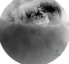
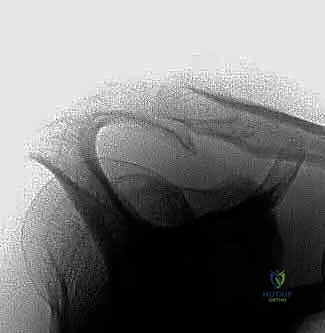
Radiographic evaluation is mandatory and must include a standardized trauma series: a true anteroposterior (Grashey) view, an axillary lateral view, and a supraspinatus outlet (Y-scapular) view. The supraspinatus outlet view is the most critical image for surgical templating. Taken with the beam angled 10° to 15° caudally, this view projects the acromion in a true sagittal plane, allowing the surgeon to precisely classify the acromial morphology (Bigliani Type I, II, or III) and quantify the exact amount of anterior and inferior bone resection required to achieve a flat (Type I) configuration. The axillary view is essential for identifying an os acromiale—an unfused acromial apophysis—which, if present, alters the surgical approach, as an aggressive acromioplasty can destabilize the anterior fragment.
While plain radiographs are sufficient for diagnosing osseous impingement, Magnetic Resonance Imaging (MRI) is the gold standard for evaluating the soft tissue envelope. An MRI is highly recommended to assess the integrity of the rotator cuff, quantify the degree of tendinosis, evaluate for concurrent labral or biceps pathology, and assess muscle trophism (fatty infiltration). Even in cases of presumed isolated impingement, an MRI can reveal occult partial-thickness articular-sided tears or interstitial tearing that may dictate a change in the operative plan. Ultrasound is a viable, cost-effective alternative in experienced hands, offering dynamic assessment of impingement, though it lacks the comprehensive intra-articular diagnostic capability of an MRI.
Patient positioning is a matter of surgeon preference, with both the beach-chair and lateral decubitus positions offering distinct advantages. The beach-chair position (approximately 45° to 60° of upright elevation) provides an anatomic orientation that is highly intuitive, facilitates easy conversion to an open approach if necessary, and allows for dynamic intraoperative assessment of arm range of motion. It is particularly advantageous for evaluating the clearance of the greater tuberosity beneath the acromion post-decompression. Conversely, the lateral decubitus position, utilizing 10 to 15 pounds of longitudinal and lateral traction, maximizes joint distraction, providing superior visualization of the glenohumeral joint and the subacromial space. Regardless of the position chosen, meticulous padding of all bony prominences and careful attention to head and neck alignment are critical to prevent devastating positioning-related neurapraxias.
Step-by-Step Surgical Approach and Fixation Technique
The execution of an arthroscopic subacromial decompression requires a systematic, reproducible approach to ensure complete pathology resolution while minimizing iatrogenic trauma. The procedure begins with the establishment of standard arthroscopic portals. A standard posterior viewing portal is established 2 cm inferior and 1 cm medial to the posterolateral corner of the acromion. An anterior working portal is established via an outside-in technique through the rotator interval, lateral to the coracoid process. A thorough diagnostic arthroscopy of the glenohumeral joint is mandatory to rule out concurrent intra-articular pathology (e.g., SLAP tears, biceps tendinopathy, chondral defects) before redirecting the arthroscope into the subacromial space.
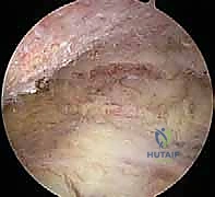
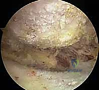
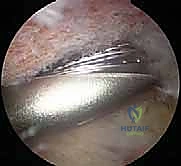
Once the arthroscope is redirected into the subacromial space, the initial view is often obscured by dense, hypertrophic bursal tissue. A lateral working portal is established 2 to 3 cm lateral to the acromial edge, in line with the posterior aspect of the clavicle. The first critical surgical step is a radical subacromial bursectomy. Utilizing a 4.0mm or 5.0mm full-radius shaver and a radiofrequency ablation wand, the surgeon must systematically clear the bursa to expose the entire undersurface of the acromion, the coracoacromial ligament, and the bursal surface of the rotator cuff. Meticulous hemostasis is vital here; the acromial branch of the thoracoacromial artery often bleeds vigorously when the anterior bursa is resected. The bursectomy is complete only when the "bare area" of the acromion and the distinct fibers of the CA ligament are clearly visualized.
Following the bursectomy, the CA ligament is identified and released. Using the radiofrequency wand, the ligament is systematically detached from its insertion on the anterior-inferior aspect of the acromion. It is critical to peel the ligament off the bone rather than transecting it mid-substance, as this exposes the anterior acromial spur (the "keel") that requires resection. Care must be taken to preserve the superior deltoid fascia; violating this fascia can lead to extravasation of fluid into the deltoid muscle and potential postoperative dehiscence. Once the anterior acromion is skeletonized, the bony architecture is thoroughly assessed to confirm the preoperative templating.
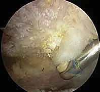
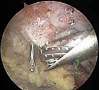
The acromioplasty is performed using a 5.5mm or 4.0mm barrel burr. The goal is to resect the anterior-inferior prominence, converting a Type II or III acromion into a flat Type I configuration. The "cutting block" technique is highly effective: the burr is introduced through the lateral portal, and resection begins at the anterolateral corner, sweeping medially towards the AC joint. The posterior aspect of the acromion serves as a depth gauge; the burr is moved from posterior to anterior, resecting bone until the anterior undersurface is coplanar with the posterior undersurface. The resection should extend approximately 5 to 8 millimeters posteriorly from the anterior edge.
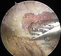
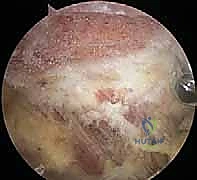
Following the bony resection, the adequacy of the decompression must be dynamically verified. The arthroscope is swept across the newly created flat acromial undersurface to ensure no residual ridges or anterior "hooks" remain. The arm is then taken through a full range of motion—specifically forward elevation and internal rotation—while visualizing the subacromial space. The greater tuberosity and the rotator cuff should glide smoothly beneath the acromion without any mechanical contact. If the distal clavicle is found to be contributing to impingement (via inferior osteophytes), a coplaning procedure or a formal arthroscopic distal clavicle excision is performed concurrently.
Complications, Incidence Rates, and Salvage Management
While arthroscopic subacromial decompression is generally considered a safe and highly successful procedure, complications can and do occur. The most frequent cause of a "failed" ASAD is not a technical complication, but rather an error in preoperative diagnosis. Operating on a patient whose primary pathology is a frozen shoulder, cervical radiculopathy, or occult glenohumeral instability will invariably lead to persistent, and often exacerbated, postoperative pain. Rigorous adherence to strict diagnostic criteria is the most effective preventative measure against surgical failure.
From a technical standpoint, inadequate decompression is the most common iatrogenic error. Failure to resect the anterior-most extent of the acromial hook or leaving residual medial osteophytes near the AC joint will result in persistent mechanical impingement. Patients will present postoperatively with the exact same clinical symptoms they had preoperatively. Diagnosis of inadequate resection is confirmed via a postoperative supraspinatus outlet radiograph. Salvage management in these cases requires a revision arthroscopic decompression to complete the bony resection. Conversely, over-resection of the acromion is a less common but far more devastating complication. Aggressive burring that violates the superior cortical bone of the acromion can lead to an acromial stress fracture or catastrophic detachment of the anterior deltoid origin.
Another profound complication is the iatrogenic creation of anterosuperior escape. As previously discussed, releasing the coracoacromial ligament in a patient with an unrecognized massive, irreparable rotator cuff tear removes the essential secondary stabilizer of the glenohumeral joint. The humeral head will migrate superiorly, articulating directly with the acromion and resulting in severe pain and pseudo-paralysis. Salvage options for this catastrophic error are highly complex and often require major reconstructive procedures, such as superior capsular reconstruction (SCR) or, more reliably in older patients, a reverse total shoulder arthroplasty (rTSA).
| Complication | Estimated Incidence | Etiology / Risk Factors | Salvage Management |
|---|---|---|---|
| Inadequate Decompression | 5% - 10% | Failure to resect the anterior hook; retained medial AC osteophytes. | Revision arthroscopic acromioplasty; aggressive physical therapy. |
| Diagnostic Error | 3% - 5% | Operating on unrecognized adhesive capsulitis or cervical radiculopathy. | Treat the underlying primary pathology (e.g., C-spine epidural, aggressive stretching for frozen shoulder). |
| Acromial Fracture | < 1% | Over-resection violating the superior cortex; aggressive burring in osteoporotic bone. | Conservative (sling immobilization) for non-displaced; ORIF for displaced fractures with deltoid compromise. |
| Anterosuperior Escape | < 1% | Releasing the CA ligament in the presence of a massive, irreparable cuff tear. | Superior Capsular Reconstruction (SCR) or Reverse Total Shoulder Arthroplasty (rTSA). |
| Deltoid Detachment | < 0.5% | Violation of the superior deltoid fascia during aggressive anterior resection. | Open surgical repair of the deltoid origin to the acromion. |
| Postoperative Stiffness | 2% - 5% | Inadequate postoperative rehabilitation; underlying mild adhesive capsulitis. | Aggressive physical therapy; intra-articular corticosteroid injection; rarely, arthroscopic capsular release. |
Phased Post-Operative Rehabilitation Protocols
The surgical intervention is merely the first step in the patient's journey to recovery; a meticulously structured, phased postoperative rehabilitation protocol is equally critical to achieving an optimal outcome. Because an isolated ASAD does not involve the structural repair of tendon or labral tissue, the rehabilitation timeline can be relatively accelerated compared to a rotator cuff repair. However, aggressive early mobilization must be balanced against the need to control postoperative inflammation and allow for soft tissue healing at the portal sites and the resected acromial bed.
Phase I: Protection and Early Passive Motion (Weeks 0-2)
The primary goals of Phase I are to control pain, minimize swelling, and prevent the development of postoperative adhesive capsulitis. Patients are placed in a standard shoulder sling for comfort, but strict immobilization is discouraged. Starting on postoperative day one, patients are instructed to remove the sling 3 to 4 times daily to perform gentle pendulum exercises and passive range of motion (PROM) exercises within a pain-free arc. Cryotherapy is utilized aggressively to manage hemarthrosis and surgical inflammation. Active use of the elbow, wrist, and hand is highly encouraged to prevent distal stiffness and promote venous return.
Phase II: Active-Assisted to Active Motion (Weeks 2-6)
As the acute surgical pain subsides, the protocol transitions to restoring full, functional range of motion. The sling is typically discontinued by the end of week two. Patients begin active-assisted range of motion (AAROM) exercises utilizing pulleys, T-bars,
