Revision THA: Unlocking Stability with Porous Tantalum Cups
Key Takeaway
This topic focuses on Revision THA: Unlocking Stability with Porous Tantalum Cups, Porous tantalum cups and augments are vital for revision total hip arthroplasty, especially when managing significant acetabular bony defects due to implant wear and aseptic loosening. These implants provide robust structural support and promote biological fixation, effectively restoring acetabular integrity and stability in complex reconstructive cases, such as those with extensive osteolysis.
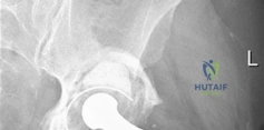
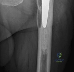
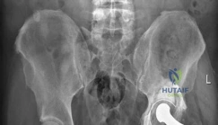
A 72-year-old patient presents with a painful, loose total hip arthroplasty performed 15 years ago. Imaging reveals significant acetabular bone loss. You are considering a revision. Describe the biomechanical advantages of using porous tantalum components in this revision scenario compared to traditional metallic implants.
Candidate: Porous tantalum is better because it is more porous, which helps bone grow into it. It has a lower modulus of elasticity, which reduces stress shielding, and it is very rough, which gives a good "scratch fit" for stability.
Providing a vague description of "bone ingrowth" without quoting the specific material properties. Failing to quantify the modulus of elasticity compared to standard alloys or neglecting the critical distinction between primary mechanical stability (the scratch fit) and secondary biological fixation.
Structure the answer around three key biomechanical pillars: 1. Volumetric Porosity: 75-80% porosity mimics cancellous bone, providing an expansive surface area for osteoconduction. 2. Low Modulus of Elasticity: At ~3 GPa, it closely matches subchondral bone, reducing stress shielding compared to Titanium (~110 GPa) or CoCr (>200 GPa). 3. High Coefficient of Friction: ~0.98, which facilitates a superior "scratch fit." This primary mechanical stability is the absolute prerequisite for long-term secondary biologic integration.
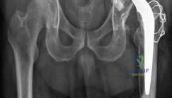
During the procedure, you identify a Paprosky Type IIIB acetabular defect. How does this influence your management, and what is the role of augments in this setting?
Candidate: I would use a large porous tantalum shell. If there is a big defect, I might use an augment. I would try to get as much screw fixation as possible into the remaining host bone.
Failing to mention the restoration of the center of rotation (COR). Ignoring the need to assess the pelvic columns and not addressing the specific surgical technique required (e.g., cementing the augment or using specialized screws).
Acknowledge that Type IIIB defects require reconstruction of the acetabular rim to restore the anatomic center of rotation (15-20mm superior to the teardrop). - Augment Usage: Used for segmental deficiencies; they are secured to host bone with screws and the augment-shell interface is bonded with PMMA cement. - Technical nuance: Ensure >50% host bone contact for the cup. If the defect is too massive, mention the threshold for moving toward a cup-cage or triflange construct to address potential pelvic discontinuity.
What are the potential risks if you achieve less than optimal primary stability in a revision acetabular reconstruction? How do you assess your intraoperative screw placement to avoid neurovascular injury?
Candidate: If it's not stable, it will loosen. For screws, I try to stay in the upper part of the cup to be safe.
Giving imprecise anatomical advice for screw placement. Failing to cite the Wasielewski safe zones. Ignoring the biological consequence (fibrous tissue interposition instead of bone ingrowth).
If stability is compromised (<150 micrometers of motion is the goal), you risk fibrous tissue ingrowth leading to aseptic loosening. Wasielewski Safe Zones: Screw placement must be restricted to the posterosuperior and posteroinferior quadrants. - Avoid: Anterosuperior (external iliac vessels) and anteroinferior (obturator nerve/vessels) quadrants. Always verify screw purchase in the ilium or ischium radiographically or via direct palpation.
