Revision THA: Acetabular Impaction Grafting for Severe Bone Loss
Key Takeaway
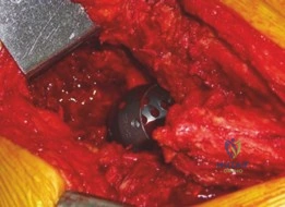
Learn more about Revision THA: Acetabular Impaction Grafting for Severe Bone Loss and how to manage it. Acetabular impaction grafting is a surgical technique used in revision total hip arthroplasty to reconstruct significant acetabular bone loss. It involves tailoring and impacting structural bone allografts, often combined with morselized graft, to restore bone stock and stabilize defects. This creates a robust foundation for the secure implantation and cementation of a new acetabular cup.
You are reviewing this patient in the clinic, a 68-year-old female who is 12 years post-primary THA. She presents with a 6-month history of progressive groin pain and a sensation of "instability." Look at these radiographs.

Describe the radiological findings and how would you classify this defect to guide your surgical planning?
Candidate: The radiograph shows significant osteolysis around the acetabular cup with superior migration of the implant and thinning of the medial wall. It looks like a Paprosky Type III defect. I would plan for a revision THA, likely using some form of bone grafting or a reconstruction cage.
Failure to be systematic. Candidates often guess the Paprosky grade without justifying it via anatomical landmarks (rim integrity, ischial line, teardrop). They also ignore the "stability" aspect of the clinical history, failing to mention the need for CT imaging to rule out pelvic discontinuity.
Systematically describe: 1) Implant position (superior migration/protrusio), 2) Bone loss (acetabular rim, medial wall/Kohler's line), 3) Associated pathology (implied polyethylene wear/osteolysis). Define the defect using the Paprosky classification: IIIA involves superior migration > 3cm with rim deficiency; IIIB involves migration > 3cm with significant ischial lysis. Mention the need for CT with metal artifact reduction to assess for pelvic discontinuity and remaining host bone for fixation.
Let's assume this is a Paprosky IIB defect with an intact acetabular rim. You decide to perform Acetabular Impaction Grafting (AIG). Walk me through your intraoperative technique for the graft preparation and impaction.
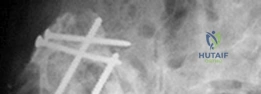
Candidate: I would use morselized allograft, ideally 3-10mm. I would remove all membrane and necrotic bone to expose bleeding bone. Then I impact the graft in layers, starting from the periphery moving centrally using hemispherical tampers until it is firm and hard to the touch.
Neglecting the "containment" principle. Candidates often forget to mention how they ensure the graft stays in place, or they use too large particles which impede the "biological bed" effect. They also forget the crucial step of pulsatile lavage to clean the bone bed.
Detail the steps: 1) Thorough debridement to bleeding bone. 2) Containment check (if not fully contained, use a mesh/cage). 3) Use of 3-10mm cancellous morselized allograft. 4) Layer-by-layer impaction starting peripherally, moving to central, using progressively larger tampers. 5) Assessment of "mechanical end-point" (the graft should be stable/firm). 6) Finally, cementation of the liner using contemporary techniques (lavage, cement restrictor, retrograde filling).
Post-operatively, the patient asks about her recovery. What is the rationale behind your weight-bearing protocol for this patient, and what are the specific clinical risks of violating it?
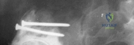
Candidate: I would recommend partial or toe-touch weight-bearing for 6-12 weeks. The graft needs time for revascularization and creeping substitution. If she puts full weight on it too early, the graft could collapse or shift, leading to cup loosening.
Focusing only on the "mechanical" stability and ignoring the "biological" incorporation. A poor answer misses the distinction between "primary stability" (provided by the impaction/cage) and "secondary stability" (achieved through graft remodeling).
Structure the answer: 1) The biological rationale is to prevent shear forces across the graft-host interface during the critical period of revascularization and creeping substitution. 2) Clinical risks: Early loading causes micromotion, leading to fibrous non-union instead of osseous integration, graft resorption, and catastrophic failure (subsidence/loosening). 3) Mention the use of radiographic monitoring to confirm graft consolidation before advancing weight-bearing status.
