Acetabular Revision Cupcage: A Challenging Case Solved
Key Takeaway
This topic focuses on Acetabular Revision Cupcage: A Challenging Case Solved, An acetabular revision cupcage is a surgical construct employed in complex hip revision for severe acetabular bone loss, like Paprosky IIIB aseptic cup loosening. It typically involves a porous tantalum cup protected by a Burch-Schneider ring, with a cemented UHMWPE liner. This method restores hip stability, alleviates pain, and improves function in challenging revision cases.
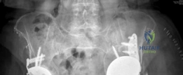
You are presented with a 68-year-old patient who is 15 years post-primary THA and 5 years post-revision THA. He complains of severe left hip pain and a "clunking" sensation. Examination reveals a 3.5cm leg length discrepancy and significant abductor weakness. Review the following radiograph and describe your immediate assessment and top differential diagnoses.
Candidate: The radiograph shows a failed acetabular component with significant superior and medial migration. There is evidence of massive periacetabular osteolysis and a failed allograft. My main differential is aseptic loosening with severe bone loss (Paprosky IIIB), but I must rule out periprosthetic joint infection (PJI) and potential pelvic discontinuity.
Failing to mention the clinical correlation (e.g., the "clunking" suggestive of discontinuity). Candidates often jump to a surgical plan (like "I would do a revision") before establishing a diagnosis or ruling out infection. They may also ignore the crucial status of the femoral component.
A structured response is essential: 1. Radiographic findings: Aseptic loosening, superior/medial migration, massive osteolysis (DeLee/Charnley zones), and failed structural allograft. 2. Diagnosis: Highly suspicious of a Paprosky IIIB defect with clinical features of pelvic discontinuity. 3. Workup: I would order ESR/CRP to rule out PJI, Judet views to assess columns, and a MARS-CT to delineate the discontinuity and volumetric bone loss before proceeding to a definitive reconstruction plan.
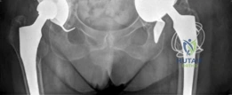
The CT scan confirms a pelvic discontinuity. Explain the biomechanical rationale for choosing a "Cup-Cage" reconstruction over other options such as structural allograft or a custom triflange component.
Candidate: The cup-cage construct is superior because it provides both biological and mechanical fixation. The porous cup allows for ingrowth, while the cage acts as a bridge to neutralize shear forces across the discontinuity. Unlike structural allografts, which have high failure rates due to resorption, this provides immediate rigid stability.
Focusing only on the implant and ignoring the biology. Failing to mention why custom triflanges are often avoided (cost, manufacturing time, lack of intraoperative adjustability) displays a lack of real-world surgical awareness.
Structure the answer by mechanism: 1. Biological: Porous trabecular metal promotes osseointegration into remaining host bone (ilium/ischium). 2. Mechanical: The ilioischial cage bridges the discontinuity, neutralizing shear to facilitate union. 3. Clinical Advantage: Cementing a dual-mobility liner into the cage allows for independent control of version and inclination, correcting biomechanics regardless of the underlying host anatomy, unlike fixed-geometry custom triflanges.
During the procedure, you perform the exposure and confirm the massive defect. What are the critical surgical steps to ensure a successful outcome for this specific reconstruction? Refer to the surgical site intraoperatively.
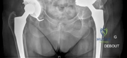
Candidate: The keys are protecting the sciatic nerve, thorough debridement of all necrotic/osteolytic tissue, and securing the ischial flange of the cage. I would use multiple screws to lock the cup and cage together to create a single stable construct.
Overlooking the sciatic nerve safety (it is often tethered in superior migration). Forgetting to mention the importance of the ischial fit—the cage fails if the inferior flange is not seated securely in the ischium.
Prioritize these: 1. Neurolysis: Early identification and protection of the sciatic nerve (often tethered). 2. Debridement: Aggressive removal of membrane and failed allograft to reach bleeding bone. 3. Ischial Seating: Precise preparation of the ischium to seat the cage flange—the linchpin of stability. 4. Construct Integration: Locking the cup and cage together with screws into both columns, followed by cementation of the liner for version/inclination control.
