Part of the Master Guide

Orthopedic Prometric Exam Preparation MCQs - Part 1

Orthopedic Prometric Exam Preparation MCQs - Part 13

25 Apr 2026 46 min read 17 Views
Orthopedic Prometric Exam Preparation MCQs - Part 13

Orthopedic Prometric Exam Preparation MCQs - Part 13

Comprehensive 100-Question Exam


00:00

Start Quiz

Question 1

Arthroscopic resection/debridement posterior to the midline of the radio- capitellar joint can result in damage to __ ligament, resulting in ______ instability.


Explanation

Resection posterior to the midpoint of the radiocapitellar joint can result in damage to the lateral collateral ligament and subsequent development of posterolateral rotator instability.

Question 2

The muscle that flexes the interphalangeal joint of the thumb is innervated by which roots of the brachial plexus:


Explanation

The interphalangeal joint of the thumb is flexed secondary to actions of the flexor pollicis longus (FPL). The FPL is innervated by the anterior interosseous nerve, which is the longest branch of the median nerve. The median nerve is formed by the lateral (roots C 5, C 6, and C 7) and medial (roots C 8 and T1) cords of the brachial plexus.

Question 3

The anterior interosseous nerve (AIN) originates from the median nerve at what distance from the medial epicondyle:


Explanation

The AIN, the largest branch of the median nerve, originates 5 cm to 8 cm distal to the medial epicondyle from the posteroradial aspect of the median nerve just distal to the proximal border of the superficial head of the pronator teres.

Question 4

man presents to the emergency department with a 6-week history of difficulty writing and pain after playing tennis. She also reports a recent inability to abduct and adduct her fingers. What is the mechanism of her symptoms:


Explanation

Approximately 17% of the population has a Martin-Gruber interconnection, and 50% of these patients may show additional denervation of normally ulnar nerve-innervated intrinsic muscles. The Martin-Gruber anomaly is a motor neural connection between the anterior interosseous nerve and ulnar nerves that is located adjacent to the ulnar artery in the proximal forearm.

Question 5

What is the innervation of the indicated muscle in the image (Slide):


Explanation

Orthopedic Prometric Exam Question 
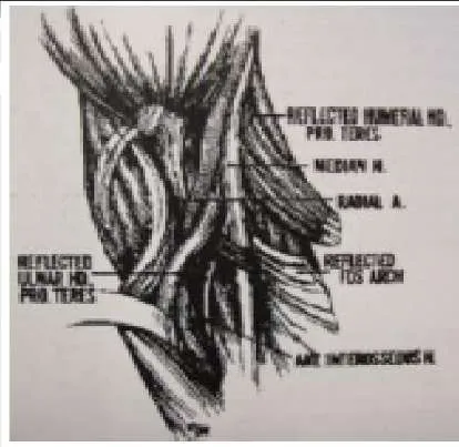
The arrow in the photograph (Slide) is pointing to the pronator teres â one of the most common sites for compression of the anterior interosseous nerve. The pronator teres is innervated by the median nerve.

Question 6

Sites of potential compression of the median nerve include all of the following except:


Explanation

Around the elbow, the median nerve may be compressed by the pronator teres (causing either anterior interosseous nerve syndrome or pronator syndrome) or the ligament of Struthers originating from a supracondylar process (causing pronator syndrome). In the wrist, the median nerve may be compressed by the transverse carpal ligament or a mass within the carpal canal. The ulnar nerve, not the median nerve, can be compressed by the pisohamate ligament.

Question 7

What position of the wrist most commonly produces scaphoid fractures:


Explanation

Frykman performed biomechanical studies to evaluate the wrist position in falls that produce scaphoid fractures. The results showed that wrist extension greater than 90° and radial deviation consistently resulted in fracture of the scaphoid.

Question 8

Which of the following blood vessels supplies the majority of the scaphoid:


Explanation

Gelberman and Menon used injection studies to demonstrate that the majority of scaphoid blood flow stems from branches of the radial artery entering the scaphoid at the distal pole. Of these, the branch entering the dorsal ridge supplies 70% to 80% of the intraosseous vascularity of the scaphoid bone. The proximal pole is completely dependent on the intraosseous blood supply and is vulnerable to avascular necrosis when fracture disrupts this vascular source.

Question 9

During a posterior (dorsal) approach to percutaneous screw fixation for a scaphoid fracture, many structures are close to the guidewire insertion location and are at risk for injury. Which of the following structures is the closest to the guidewire insertion location according to a recent cadaveric study:


Explanation

Adamany and colleagues performed a cadaveric study to evaluate the dorsal structures at risk with truly percutaneous headless screw placement for scaphoid fractures. They noted that the posterior interosseous nerve and the extensor digitorum communis to the index finger were an average of 2.2 mm from the guidewire and therefore at greatest risk from this approach.

Question 10

Which of the following is a concerning risk factor for a dorsal open approach to the scaphoid:


Explanation

The dorsal approach is advantageous in obtaining central screw placement. The scapho-trapezial-trapezoid joint is at risk during a volar approach, not a dorsal approach, and infection has not been shown to occur more frequently in one approach over the other. The LT ligament is not seen in either approach. The risk of the open dorsal approach is compromise of the main blood supply to the scaphoid, entering through the dorsal ridge.

Question 11

A volar approach to the scaphoid is ideal in which of the following fractures:


Explanation

The volar approach to the scaphoid is optimal in distal pole fractures because it allows direct visualization of the fracture line and exact reduction and fixation. A volar approach is not recommended for fractures or avascular necrosis of the proximal pole, where dorsal screw placement is best. Scapholunate ligament tears are generally repaired from a dorsal approach because the ligament is stoutest dorsally.

Question 12

Which finger is most commonly involved in a flexor digitorum profundus (FDP) avulsion injury:


Explanation

An FDP avulsion is caused by forceful extension of the distal interphalangeal (DIP) joint, occurring at the same time as a maximum contraction of the FDP tendon. Jersey finger is often seen in athletes, especially football or rugby players, who commonly get their fingers caught in an opposing playerâ s jersey, thus the name. In most cases, this injury affects the ring finger.

Question 13

Which type of flexor digitorum profundus (FDP) avulsion is considered the most severe:


Explanation

Type I is the most threatening scenario because the FDP tendon retracts into the palm, and vincular and diffusional blood supply is lost. The sheath may be noncompliant after a few days and may not allow passage of the FDP tendon through the sheath in an attempt to repair the stump to its insertion. Additionally, proximal muscle contracture prevents tendon stump advancement.

Question 14

When performing pollicization to correct a hypoplastic thumb, the surgeon should rotate the index finger:


Explanation

After the index finger is rotated 150º, the index finger will be in the ideal location as it mimics the position of where the thumb would have naturally been. This position allows for the greatest amount of grip and pinch strength possible.

Question 15

For which types of thumb hypoplasia is pollicization the best option:


Explanation

Reconstruction is possible and is therefore the best option for thumb hypoplasia in patients with type I, type II, and type IIIA. These three types of hypoplasia can be corrected because the thumb still has most of the bones and muscles intact. Corrective surgery is necessary to correct weak muscles or a tight web space between the thumb and index finger. When a type IIIB exists, reconstruction is not possible and pollicization must be performed.C orrect Answer: Type I, type II, and type IIIA

Question 16

What is the most critical step in pollicization to create a normal-looking thumb:


Explanation

It is necessary to create a hand with a natural first web space, shorter tendons that allow for natural movement, a shorter metacarpal that ensures the finger will not grow to an unnatural length, and a hyperextended joint to create the most naturallooking hand possible.

Question 17

All of the following may be present in a child with type IIIA hypoplasia except:


Explanation

Type IIIA hypoplasia includes web space narrowing, thenar atrophy, MP joint laxity, and extrinsic tendon abnormalities. Type IIIA is distinguished from a type IIIB by the fact that a stable C MC joint exists. Because a stable CMC joint exists, a reconstruction is the treatment of choice. When the C MC joint is unstable, as in type IIIB hypoplasia, a pollicization is necessary to restore thumb stability.

Question 18

The main 3-4 viewing portal for wrist arthroscopy lies in between which two tendons:


Explanation

The 3-4 portal is the main viewing portal and is located between the third and fourth compartment. This portal is bordered by the extensor digitorum communis (EDC ) to the index finger, and the extensor pollicis longus (EPL) can be palpated in the â soft spotâ 1 cm distal to Listers tubercle. This portal is usually the first portal to be made during wrist arthroscopy.

Question 19

C omplications after wrist arthroscopy occur in what percentage of patients:


Explanation

The complication rate after routine wrist arthroscopy is between 2% and 5%.

Question 20

C omplications after wrist arthroscopy occur in what percentage of patients:


Explanation

The complication rate after routine wrist arthroscopy is between 2% and 5%.

Question 21

The fracture fragment in Bennettâ s fracture is located in which of the following areas of the hand:


Explanation

As an axial load is placed on the thumb tip, it drives the thumb metacarpal (MC ) base in a dorsal-radial direction. As the thumb MC base moves dorsoradially, a fracture is created in the volar, ulnar quadrant of the thumb MC base. Gedda and Moberg describe this as a ligament fracture avulsion. The volar, ulnar quadrant piece usually remains stationary, perhaps migrating a small amount distal the thumb metacarpal base moves dorsoradially, creating a fracture in the volar, ulnar quadrant of the trapezium.C orrect Answer: Ulnopalmar thumb metacarpal base

Question 22

Which of the following two main soft tissue forces are disrupted by Bennets fracture subluxation:


Explanation

The volar, ulnar quadrant piece usually remains stationary due to the volar beak ligament. The thumb metacarpal base tends to sublux dorsoradially due to unopposed pull of the abductor pollicis longus and adductor pollicis. The intact volar beak ligament is usually the counterforce the to these two muscles in the static situation. The extensor pollis longus, flexor pollicis brevis, and abductor pollis longus do not have significant involvement in the Bennettâ s fracture subluxation. Although the dorsal radial ligament is important for carpometacarpal stability, it is not the ligament attached to the fractures fragment.

Question 23

The greatest amount of step-off that is well-tolerated in a Bennettâ s fracture is:


Explanation

Studies by Livesley, Kjaer-Petersen, and others have shown that patients with fractures with more than a 1-mm step-off after reduction were more likely to develop arthritis at the thumb carpometacarpal joint. Although some studies have not shown functional outcome correlating with the presence of arthritis, Oosterbos and De Boer found that all their patients with fair and poor overall results had nonanatomic reductions. Although a cadaveric study by C ullen has shown that a 2-mm step-off may be acceptable, this contrasts with the clinical evidence currently available.

Question 24

When fracture step-off is greater than the accepted limits, which of the following complications is the most common:


Explanation

Studies by Livesley, Kjaer-Petersen, and others have shown that patients with fractures with more than a 1-mm step-off after reduction were more likely to develop arthritis at the thumb carpometacarpal joint. Pain, decreased range of motion, and decreased pinch strength also correlated with these poor outcomes.

Question 25

C linically, what is the upper limit of acceptable fracture angulation for a fifth metacarpal neck fracture:


Explanation

Although this is controversial, conservatively treated patients with angulations less than 70° fared well in two prospective studies. Fourteen percent of patients will have a cosmetic deformity, but operatively treated patients exhibited extensor lag and increased rehabilitation times.

Question 26

In cadaveric models, when does the biomechanics of fifth finger flexion consistently change in relationship to metacarpal neck fracture angulation:


Explanation

Thirty degrees of angulation is the maximum deformity for acceptable fifth finger grip strength. Ali et al showed that fracture angulation of 30° results in a significant decrease in the distance between the origin and the insertion of the flexor digiti minimi (FDM). This shortening creates more slack in the FDM muscle and more excursion is wasted as muscle shortening prior to the initiation of metacarpophalangeal (MP) flexion.C orrect Answer: 30°

Question 27

Up to how much angulation can be tolerated in the small finger metacarpal shaft fracture:


Explanation

The small finger carpometacarpal joint is mobile, which allows the small finger metacarpal to tolerate deformity better than the fixed index and middle finger rays. Thus, 41° to 50° of angulation can be accommodated by the mobile carpometacarpal joint.

Question 28

If a metacarpal shaft fracture shortens 4 mm, what will the theoretical amount of extensor lag be at the metacarpophalangeal joint:


Explanation

For each 2 mm of shortening, a 7° extensor lag exists. Thus, with 4 mm of shortening, there will be a 14° extensor lag at the metacarpophalangeal joint.

Question 29

In a short oblique metacarpal shaft fracture without comminution or bone loss, what is usual amount of maximal shortening that will occur:


Explanation

In a cadaveric study, shortening beyond 5 mm was prevented by the tethering effect of the transverse metacarpal ligaments and adjacent metacarpals.

Question 30

Which of the following statements is true regarding metacarpophalangeal joint anatomy:


Explanation

The collateral ligaments are lax in extension and tight in flexion. The joint volume is highest in extension. The metacarpal head is cam-shaped. The collateral ligaments originate dorsal to the axis of flexion. Due to the tightening of the collateral ligaments over the cam-shaped metacarpal head in flexion, joint stability is maximized.

Question 31

Which of the following fracture patterns and mechanisms is incorrectly paired:


Explanation

Biomechanically and clinically, fracture patterns are often associated with certain types of force. Transverse fractures occur with a direct blow, comminuted fractures occur with axial compression and bending, spiral fractures occur in torsion, and oblique fractures occur with torsion and axial load.

Question 32

Giant cell tumor of tendon sheath commonly occurs in which of the following age groups:


Explanation

Giant cell tumor of tendon sheath is most commonly found in patients in the fourth through sixth decades; therefore, age 30-40 years is the most appropriate answer choice.

Question 33

Which of the following clinical features is common in giant cell tumor of tendon sheath:


Explanation

Giant cell tumor of tendon sheath is painless. Giant cell tumor of tendon sheath does not transilluminate, as ganglion cyst does. No overlying skin color changes occur. Giant cell tumor of tendon sheath only increases in size and does not fluctuate like a ganglion cyst; it does not present with a rapid increase in size.

Question 34

After plain radiographs of giant cell tumor of tendon sheath are obtained, the following imaging study should be obtained:


Explanation

Magnetic resonance imaging provides anatomic detail of the soft tissue mass, helps generate a differential diagnosis, and determines if the mass is unifocal or multifocal and where it originates. Giant cell tumor of tendon sheath is a soft-tissue tumor. Computed tomography is best for bone-based tumors. Ultrasound helps localize lesions but does not provide anatomic detail to help determine the type of mass. Although angiograms are useful for vascular tumors such as renal cell carcinoma or arteriovenous malformations, they are not necessary in the evaluation of a soft tissue mass in the hand with features suggestive of giant cell tumor of tendon sheath. A bone scan is useful when malignant bone tumors are suspected rather than benign soft tissue masses.

Question 35

Which of the following cell types is not typically found in giant cell tumors of tendon sheath:


Explanation

Multinucleated giant cells, histiocytes, monocytes, and fibroblasts are commonly found in pathologic giant cell tumor of tendon sheath specimens. Polymorphonuclear lymphocytes are typically associated with bacterial infections.

Question 36

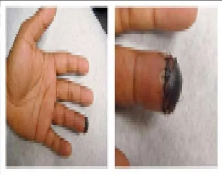
A 25-year-old, right-hand-dominant male truck driver presents to the emergency department (Slide 1, Slide 2). The tip of his left ring finger was amputated in a bicycle accident 2 weeks prior. The amputated piece was â sewn back onâ in the emergency department immediately after the accident, but â turned blackâ over the next week. There is no evidence of infection. He states that the appearance of his finger is embarrassing, and he would like it taken care of as soon as possible. Which of the following procedures is the most appropriate:


Explanation

The Atasoy-Kleinert V-Y advancement flap is the best option for transversely oriented fingertip amputations/defects and also for defects with more dorsal than volar tissue loss. The apex of the V is positioned at, or just distal, to the distal interphalangeal join crease on the volar side of the digit. After incising the V marking, the flap is advanced distally to cover the defect, and the incisions are closed in a Y pattern. Local, or chemical, debridement and allowing the resulting defect to heal by secondary intention are a viable option, but the patient stated that he would prefer an aggressive treatment protocol because the appearance of his fingertip is so embarrassing. The Kutler (lateral) V-Y advancement flap is typically used to cover tip defects that demonstrate more volar than dorsal tissue loss. The procedure involves creating V-Y advancement flaps laterally on either side of the affected digit and advancing them toward each other in the midline thereby covering the defect. The Moberg flap is typically used for reconstruction of thumb amputations. This procedure involves the creation of volar tissue flap that includes the neurovascular bundles on either side of the digit. Its use is cautioned in very distal amputations because excess stretch on the vascular pedicles may lead to necrosis at the tip of the flap. Its use is also cautioned in the fingers because of the difference in orientation of the blood supply compared to the thumb. A full-thickness, rather than a split-thickness, skin graft is a viable option to manage this patient. Skin grafts for hand reconstruction should be harvested with the â like replaces likeâ principle in mind, especially when reconstructing the volar skin. Volar hand skin is much thicker and of unique quality when compared with the rest of the body, and therefore, the most appropriate place to harvest a skin graft is the volar surface of the hand.

Question 37

A 52-year-old, right-hand-dominant watchmaker arrives at the emergency department 30 minutes after the volar soft tissue of his right thumb and index finger was avulsed while using a bandsaw. Physical examination shows 2 cm 3 2 cm wounds involving the distal phalanx of each affected digit. No exposed tendon or bone is present, and no involvement of the joints is noted. The patient requests a treatment option that will retain the most sensation so he can effectively continue in his occupation. Which of the following options is the most appropriate management of this patientâ s wounds:


Explanation

Local flaps such as cross finger flaps are good options but require at least two surgeries (inset then division) and can often result in stiffness secondary to the requisite period of immobilization. In addition, local flaps have lesser return of sensibility than the other techniques listed. Return of tactile sensibility is excellent after healing by secondary intention, but dressing changes for wounds that measure 2 cm 3 2 cm would take months to completely heal. Skin grafting is the next available option with acceptable sensory return. It can be performed during local anesthesia, requires only one operation, and allows for early motion thereby avoiding stiffness. Studies have shown that full-thickness skin grafts recover sensation better than split-thickness skin grafts. A radial forearm flap will be excessively bulky, has unacceptable donor site morbidity in this situation, and results in inadequate sensory recovery.

Question 38

Which of the following is not considered a part of the triangular fibrocartilage complex:


Explanation

The triangular fibrocartilage complex is made up of the dorsal and palmar radioulnar ligaments, the meniscal homologue, the articular disk, the ulnolunate, and the ulnotriquetral ligaments. The radiolunate ligament is not part of the complex.

Question 39

Which of the following arterial branches does not supply the peripheral 25% of the triangular fibrocartilage complex:


Explanation

The triangular fibrocartilage complex is supplied by both branches of the anterior interosseous artery and the ulnar artery; it is not supplied by the dorsal branch of the radial artery.

Question 40

Which of the following statements is true:


Explanation

Cadaveric studies have been performed to determine the amount of load across the wrist with various relationships between the radius and ulna lengths. In wrists with neutral ulnar variance (in which the radius and ulna are equal in length), 20% of the load is transmitted across the ulna and 80% is transmitted across the radius. In wrists with negative ulnar variance (in which the ulnar is shorter than the radius), more load is transmitted across the radius and less is transmitted across the ulna. The opposite is true with positive ulnar variance (the ulna is longer than the radius).C orrect Answer: In a wrist with neutral ulnar variance, 20% of the axial load is transmitted across the ulna.

Question 41

Which of the following parameters is not a determinant of the Palmer classification of triangular fibrocartilaginous complex injuries:


Explanation

The Palmer classification divides triangular fibrocartilage complex lesions into traumatic and degenerative. Traumatic subclassifications are based on the location of the ligament tear. In the degenerative tear, subclassifications are based on the degree of injury to the triangular fibrocartilage complex and associated chondral and ligamentous injury. Thus, the size of the lesion is not a parameter in the Palmer classification.

Question 42

What are the components of a Galeazzi fracture-dislocation:


Explanation

Only one in vitro study examined the soft tissue constraints of the Galeazzi fracture-dislocation pattern. Moore and colleagues performed a radial osteotomy at the pronator teres insertion of nine cadaveric forearms and then sectioned the TFC C and the interosseous membrane in alternating orders. They found that all three structures (TFC C , interosseous membrane, and radialshaft) must be injured to create a radial shortening of more than 10 mm in relationship to the distal ulna.C orrect Answer: Triangular fibrocartilage complex (TFC C ) tear, interosseous membrane tear, and radial shaft fracture

Question 43

Who are the most common athletes to get medial epicondylitis of the elbow:


Explanation

Although medial epicondylitis is called golferâ s elbow, tennis players are more likely to have this condition. Medial epicondylitis can occur in any sport such as baseball pitching, javelin throwing, swimming, and gymnastics in which athletes place a significant valgus flexion force on their elbow.

Question 44

Indications for replantation include:


Explanation

A sharp amputation, particularly of the thumb, is the best indication for replantation. C rushed digits, prolonged ischemia time, poor condition of the severed part, and single digit loss proximal to the FDS insertion on the middle phalanx are relative contraindications to replantation.

Question 45

An amputation through the wrist is an indication for attempted replantation.


Explanation

Question 46

Care of an amputated part prior to replantation includes:


Explanation

The appropriate care of an amputated part includes wrapping it in saline- dampened gauze and placing it on ice in a watertight bag. These actions preserve the tissues and slow cellular death until replantation is attempted. The part should not be immersed, painted with povidone- iodine, or kept next to the body.

Question 47

Replants are monitored by:


Explanation

Monitoring of replanted parts postoperatively is accomplished by clinical checks of color or turgor to indicate blood flow. Additional objective monitoring is performed by using Doppler probes to check flow or by measuring temperature differences between the replanted part as compared to other digits.

Question 48

C omplications post-replantation include:


Explanation

Complications after replantation include cold intolerance, which may improve over time, and stiffness of the replanted digit, which is generally due to immobility, tendon adhesions, and joint contracture.

Question 49

Placing some tension on a flexor tendon repair increases the ultimate tensile strength of the repair.


Explanation

Question 50

Flexor tendon nutrition is derived from:


Explanation

Flexor tendon nutrition in the uninjured state is derived via the vincula, which contain blood vessels for nutrition. Injured tendons obtain nutrition via diffusion of synovial fluid. The pulley system does not contribute to flexor tendon nutrition.

Question 51

A 35-year-old carpenter presents with an inability to make an "OK" sign with his thumb and index finger. A diagnosis of Anterior Interosseous Nerve (AIN) syndrome is suspected. Which of the following muscles will have NORMAL function in this patient?


Explanation

The AIN innervates the FPL, the FDP to the index and middle fingers, and the pronator quadratus. The flexor carpi radialis is innervated by the proper median nerve before the AIN branches off.

Question 52

During an in situ ulnar nerve decompression at the elbow, the surgeon releases the tissue spanning the two heads of the flexor carpi ulnaris (FCU). Which anatomical structure forms the primary roof of the cubital tunnel in this region?


Explanation

Osborne's ligament (or Osborne's fascia) forms the roof of the cubital tunnel, bridging the olecranon and the medial epicondyle over the two heads of the FCU. The Arcade of Struthers is a potential compression site located approximately 8 cm proximal to the medial epicondyle.

Question 53

The radial nerve is at risk during a lateral approach to the distal humerus. At approximately what distance proximal to the lateral epicondyle does the radial nerve pierce the lateral intermuscular septum to transition from the posterior to the anterior compartment?


Explanation

The radial nerve courses from posterior to anterior by piercing the lateral intermuscular septum approximately 10 cm proximal to the lateral epicondyle. This anatomical landmark is critical when extending surgical exposures of the distal humerus.

Question 54

A 24-year-old athlete undergoes posterior cruciate ligament (PCL) reconstruction. The surgeon must understand the biomechanics of the PCL's two primary bundles. Which bundle is most taut in knee flexion?


Explanation

The PCL is composed of two main bundles: the anterolateral (AL) and posteromedial (PM) bundles. The larger AL bundle is taut in flexion and lax in extension, whereas the PM bundle is taut in extension.

Question 55

A patient with refractory lateral epicondylitis undergoes open surgical debridement. The primary pathological tissue targeted during this procedure is the origin of which of the following structures?


Explanation

Lateral epicondylitis primarily involves angiofibroblastic hyperplasia of the origin of the Extensor Carpi Radialis Brevis (ECRB). Surgical management focuses on excising this degenerative tissue while sparing the overlying ECRL.

Question 56

A 45-year-old typist complains of aching pain in the proximal forearm and numbness in the radial three and a half digits. Which physical examination finding most reliably distinguishes pronator syndrome from carpal tunnel syndrome?


Explanation

The palmar cutaneous branch of the median nerve branches off proximal to the carpal tunnel, supplying sensation to the thenar eminence. Consequently, sensation over the thenar eminence is spared in carpal tunnel syndrome but affected in pronator syndrome.

Question 57

The 'terrible triad' of the elbow is notoriously difficult to manage due to profound instability. This injury pattern typically involves a posterior elbow dislocation, a radial head fracture, and a fracture of which of the following structures?


Explanation

The terrible triad of the elbow consists of an elbow dislocation, a radial head fracture, and a coronoid fracture. Restoration of the coronoid and radial head is critical for re-establishing anterior osseous buttressing and elbow stability.

Question 58

A 42-year-old male undergoes a single-incision anterior approach for the repair of an acute distal biceps tendon rupture. Postoperatively, he complains of numbness along the lateral aspect of his forearm. Which nerve was most likely injured?


Explanation

The lateral antebrachial cutaneous nerve (LABCN) is the terminal sensory branch of the musculocutaneous nerve. It exits the deep fascia lateral to the biceps tendon and is the most commonly injured nerve in an anterior single-incision distal biceps repair.

Question 59

During shoulder arthroscopy for instability, the surgeon identifies a Buford complex. This normal anatomical variant is defined by a thickened, cord-like middle glenohumeral ligament and the absence of which structure?


Explanation

A Buford complex is present in about 1.5% of shoulders and consists of a cord-like middle glenohumeral ligament (MGHL) and an absent anterosuperior labrum. It must not be mistakenly repaired to the glenoid, as doing so will severely restrict external rotation.

Question 60

A 6-year-old child sustains a displaced extension-type supracondylar humerus fracture.

Based on typical displacement patterns, what is the most common neurological deficit associated with this specific injury?


Explanation

The anterior interosseous nerve (AIN) is the most commonly injured nerve in extension-type supracondylar humerus fractures, particularly those with posterolateral displacement. Flexion-type fractures place the ulnar nerve at higher risk.

Question 61

A 50-year-old male sustains a displaced femoral neck fracture. Which artery is the primary contributor to the blood supply of the adult femoral head, placing it at high risk for avascular necrosis in this injury?


Explanation

The medial femoral circumflex artery (MFCA), specifically its lateral epiphyseal terminal branches, provides the predominant blood supply to the adult femoral head. The artery of the ligamentum teres supplies a negligible amount of blood in adults.

Question 62

In an anterior cruciate ligament (ACL) reconstruction, positioning the femoral tunnel is critical for restoring rotational stability. For a right knee, placing the tunnel at which clock face position optimally mimics the native anatomical footprint?


Explanation

For a right knee, placing the femoral tunnel at the 10 o'clock position (or 2 o'clock for a left knee) optimally addresses the native ACL footprint. Vertical placement at 12 o'clock restores AP stability but fails to control rotational translation.

Question 63

A 13-year-old obese boy presents with an insidious onset of knee pain and a limp. A pelvic radiograph is obtained.

Which of the following radiographic lines is most appropriate to evaluate for Slipped Capital Femoral Epiphysis (SCFE)?


Explanation

Klein's line is drawn along the superior margin of the femoral neck on an AP and frog-leg lateral radiograph. In a normal hip, it intersects the lateral portion of the femoral epiphysis; in SCFE, the epiphysis falls below this line.

Question 64

A 4-month-old infant is being treated with a Pavlik harness for Developmental Dysplasia of the Hip (DDH). The mother notes that the child has stopped kicking the affected leg. On exam, the child lacks active knee extension. Which nerve is most likely compressed due to hyperflexion?


Explanation

Femoral nerve palsy is a known complication of Pavlik harness treatment when the hip is placed in excessive flexion. The harness should be temporarily discontinued, and function almost always recovers spontaneously.

Question 65

During a posterior approach to the shoulder, the axillary nerve is visualized emerging through the quadrangular space. Which muscle forms the inferior border of this anatomical space?


Explanation

The quadrangular space is bordered superiorly by the teres minor, inferiorly by the teres major, medially by the long head of the triceps, and laterally by the surgical neck of the humerus. It transmits the axillary nerve and the posterior circumflex humeral artery.

Question 66

Flexor tendon injuries in 'Zone 2' of the hand are historically referred to as 'no man's land' due to the high risk of adhesion formation. What are the anatomical boundaries of Zone 2?


Explanation

Zone 2 of the flexor tendons begins at the proximal edge of the A1 pulley (distal palmar crease) and ends at the insertion of the Flexor Digitorum Superficialis (FDS) on the middle phalanx. In this zone, the FDP and FDS tendons lie closely together within the tight fibro-osseous sheath.

Question 67

A 22-year-old male sustains a scaphoid waist fracture. Which of the following best describes the predominant arterial supply to the scaphoid, explaining its propensity for proximal pole avascular necrosis?


Explanation

The scaphoid is predominantly supplied by the dorsal carpal branch of the radial artery, which enters at the distal pole and provides retrograde blood flow to the proximal pole. Fractures at the waist or proximal pole disrupt this retrograde supply, risking avascular necrosis.

Question 68

A 28-year-old manual laborer presents with dorsal wrist pain. Radiographs reveal sclerosis and fragmentation of the lunate (Kienbock's disease). Which of the following radiographic anatomical variants is most highly associated with this condition?


Explanation

Negative ulnar variance is strongly associated with Kienbock's disease. The shortened ulna relative to the radius leads to increased radiolunate contact stresses, precipitating lunate avascular necrosis.

Question 69

A patient undergoes a radiograph 8 weeks after an undisplaced talar neck fracture. A subchondral radiolucent band is seen in the talar dome (Hawkins sign). What does this finding signify?


Explanation

The Hawkins sign represents subchondral osteopenia in the talar dome, typically appearing 6 to 8 weeks post-injury. It indicates intact vascular supply to the talar body, virtually excluding the development of avascular necrosis.

Question 70

During hip arthroscopy, establishing the anteroinferior portal carries the highest risk of injury to which of the following neurological structures?


Explanation

The anteroinferior portal in hip arthroscopy places the lateral femoral cutaneous nerve (LFCN) at greatest risk. The nerve courses near the anterior superior iliac spine (ASIS) and can be easily injured if portal placement is poorly controlled.

Question 71

A 25-year-old male sustains a high-energy trauma resulting in the hip injury shown.

During the surgical approach for fixation, understanding the vascular anatomy is critical to prevent avascular necrosis. The deep branch of the medial femoral circumflex artery (MFCA) consistently runs between which two structures?


Explanation

The deep branch of the MFCA is the predominant blood supply to the femoral head. It consistently passes anterior to the quadratus femoris and posterior to the obturator externus, making it vulnerable during posterior approaches if the quadratus femoris is released too aggressively.

Question 72

An 8-year-old child presents with an extension-type supracondylar humerus fracture as shown.

If the distal fragment demonstrates significant posteromedial displacement, which nerve is at the greatest risk of injury due to tethering across the fracture site?


Explanation

Posteromedial displacement of the distal fragment in an extension-type supracondylar fracture puts the radial nerve at greatest risk as the proximal fragment displaces anterolaterally. Conversely, posterolateral displacement predominantly endangers the median nerve and anterior interosseous nerve (AIN).

Question 73

Highly cross-linked ultra-high-molecular-weight polyethylene (UHMWPE) is widely used in total hip arthroplasty to reduce volumetric wear. Which step in its manufacturing process significantly reduces the risk of in vivo oxidation but comes at the cost of decreasing the ultimate tensile strength?


Explanation

Remelting highly cross-linked polyethylene eliminates free radicals, thereby preventing long-term oxidation and structural degradation. However, this thermal process decreases crystallinity, which reduces the material's ultimate tensile and yield strength.

Question 74

A 19-year-old male presents with severe, aching pain in his mid-tibia that classically worsens at night. He reports complete relief of symptoms within 30 minutes of taking aspirin. Radiographs reveal a cortical lucency surrounded by sclerosis. The central nidus of this lesion predominantly secretes high levels of which biochemical mediator?


Explanation

The clinical presentation is classic for an osteoid osteoma. The central nidus produces a high concentration of Prostaglandin E2 (PGE2), which causes severe pain that is exquisitely sensitive to COX inhibitors like aspirin and NSAIDs.

Question 75

A 24-year-old athlete undergoes a posterolateral corner (PLC) reconstruction of the knee utilizing a fibular-based technique. The surgeon isolates a nerve that winds posterior to the biceps femoris tendon. A motor deficit resulting from injury to this specific nerve would manifest primarily as weakness in which muscle?


Explanation

The common peroneal nerve lies posterior to the biceps femoris tendon and wraps around the fibular neck. Injury to it affects the deep peroneal nerve, leading to weakness in the tibialis anterior and resulting in foot drop.

Question 76

A 35-year-old male falls from a ladder and sustains an L2 burst fracture. Imaging shows 40% canal compromise. He is neurologically intact, and MRI confirms an intact posterior ligamentous complex. According to the Thoracolumbar Injury Classification and Severity Score (TLICS), what is his total score and the generally recommended management?


Explanation

Under the TLICS system, a burst fracture scores 2 points, intact neurological status is 0 points, and an intact posterior ligamentous complex is 0 points, resulting in a total score of 2. A score of 3 or less is an indication for non-operative management.

Question 77

A hypotensive trauma patient is diagnosed with an Anteroposterior Compression Type III (APC-III) pelvic ring disruption. A pelvic binder is applied, and the patient undergoes immediate pre-peritoneal pelvic packing. This intervention primarily aims to tamponade bleeding from which of the following anatomic structures?


Explanation

In severe pelvic ring disruptions, the vast majority (up to 80-90%) of bleeding is venous in origin, primarily from the presacral venous plexus and prevesical veins. Pre-peritoneal packing effectively tamponades this low-pressure venous bleeding.

Question 78

A 45-year-old male presents with recurrent posterolateral rotatory instability (PLRI) of the elbow. During surgical reconstruction of the lateral ulnar collateral ligament (LUCL), the isometric point on the lateral epicondyle must be identified for graft placement. Where is this point anatomically located?


Explanation

The isometric point for LUCL reconstruction is located on the lateral epicondyle at the center of the capitellum's axis of rotation. Improper graft placement here leads to laxity or stiffness during different arcs of elbow motion.

Question 79

A patient presents with an inability to make an "OK" sign with their thumb and index finger following a forearm injury. Which of the following muscles is primarily innervated by the affected nerve?


Explanation

The anterior interosseous nerve (AIN) innervates the flexor pollicis longus, the radial half of the flexor digitorum profundus, and the pronator quadratus. It is a pure motor nerve and does not provide cutaneous sensory innervation.

Question 80

A 32-year-old female fell on an outstretched hand and sustained the injury shown in the radiograph.

Assuming this is a "terrible triad" injury of the elbow, which of the following is the standard evidence-based surgical sequence for management?


Explanation

The terrible triad of the elbow consists of an elbow dislocation, radial head fracture, and coronoid fracture. The standard surgical approach works deep to superficial: coronoid fixation, radial head repair/replacement, followed by LCL complex repair.

Question 81

During an electrodiagnostic evaluation for suspected cubital tunnel syndrome, a Martin-Gruber anastomosis is identified. This anatomical variant involves a neural connection between which two structures?


Explanation

The Martin-Gruber anastomosis is a motor crossover from the median nerve (often via the AIN) to the ulnar nerve in the forearm. It can confound nerve conduction studies by producing spuriously normal ulnar motor amplitudes.

Question 82

An obese 13-year-old boy presents with left knee pain and an obligate external rotation of the hip during flexion. The radiograph is shown below.

When treating this condition with in situ percutaneous pinning, what is the most significant iatrogenic risk factor for the development of chondrolysis?


Explanation

In Slipped Capital Femoral Epiphysis (SCFE), unrecognized intra-articular screw penetration is a major risk factor for chondrolysis. Surgeons must use the "approach-withdraw" fluoroscopic technique to confirm the screw tip is entirely within the femoral head.

Question 83

A 28-year-old pitcher complains of right upper extremity heaviness, fatigue, and numbness in the ulnar digits after throwing. Examination reveals a positive Adson's test and reproduction of symptoms with hyperabduction. If neurogenic thoracic outlet syndrome is confirmed, the neurovascular bundle is most commonly compressed between which structures?


Explanation

Neurogenic thoracic outlet syndrome most frequently involves compression at the interscalene triangle. This anatomical space is bordered by the anterior scalene muscle, the middle scalene muscle, and the first rib.

Question 84

A 22-year-old soccer player sustains a twisting knee injury. Radiographs reveal a small avulsion fracture of the lateral tibial plateau (Segond fracture). This pathognomonic finding is an avulsion of the capsule and the anterolateral ligament (ALL). It is most highly associated with a tear of which other structure?


Explanation

A Segond fracture is an avulsion of the anterolateral capsule and the anterolateral ligament (ALL) from the lateral tibia. It is considered highly pathognomonic for an underlying anterior cruciate ligament (ACL) rupture.

Question 85

A 6-month-old infant with developmental dysplasia of the hip (DDH) failed Pavlik harness treatment and is scheduled for a closed reduction and spica casting. During the procedure, the surgeon assesses the "safe zone" of Ramsey. How is this zone defined?


Explanation

Ramsey's safe zone is the arc of motion between maximum abduction (limited by the adductors) and the angle at which the hip re-dislocates as it is adducted. Immobilization within this zone prevents both avascular necrosis (from excessive abduction) and re-dislocation.

Question 86

A 45-year-old male sustains a C5-C6 bilateral interfacetal dislocation from a motor vehicle accident. He is awake, alert, cooperative, and has a complete C5 spinal cord injury (ASIA A). What is the most appropriate initial step in management before surgical stabilization?


Explanation

In an awake, alert, and cooperative patient with a cervical facet dislocation and a neurological deficit, urgent closed reduction using cranial traction is indicated. MRI is required before reduction only if the patient is uncooperative, obtunded, or fails closed reduction.

Question 87

During secondary fracture healing, the cartilaginous soft callus is eventually replaced by hard woven bone. Which of the following transcription factors is most essential for the initial differentiation of multipotent mesenchymal stem cells into the osteoblastic lineage?


Explanation

Runx2 (Cbfa1) is the master transcription factor responsible for committing mesenchymal stem cells to the osteoblast lineage. In contrast, Sox9 is the primary transcription factor driving chondrogenic differentiation during soft callus formation.

None

Dr. Mohammed Hutaif
Medically Verified Content by
Prof. Dr. Mohammed Hutaif
Consultant Orthopedic & Spine Surgeon
Chapter Index