Part of the Master Guide

Orthopedic Prometric Exam Preparation MCQs - Part 1

Orthopedic Prometric Exam Preparation MCQs - Part 15

27 Apr 2026 61 min read 15 Views
Orthopedic Prometric Exam Preparation MCQs - Part 15

Orthopedic Prometric Exam Preparation MCQs - Part 15

Comprehensive 100-Question Exam


00:00

Start Quiz

Question 1

Which of the following statements concerning Ehlers-Danlos syndrome (EDS) is true:


Explanation

Ehlers-Danlos syndrome (EDS) types I, II, III, and VII are commonly seen by orthopedic surgeons. Type VII is characterized by congenital hip and knee dislocations. EDS type III is the mildest form of the disease; the main symptom of this type is hyperlaxity. EDS may be inherited via any of the Mendelian patterns. Children with EDS should be encouraged not to use their hyperlaxity as a â trickâ because of potential long-term joint damage that may occur. Knowing the subtype of the disease is often helpful in management and perioperative planning because than the surgeon may anticipate operative risks and potential complications.

Question 2

Which subtype of Ehlers-Danlos syndrome (EDS) is caused by lysyl hydroxylase deficiency:


Explanation

Many of the mutations responsible for the differing subtypes of Ehlers- Danlos syndrome (EDS) have not been identified (EDS types I, II, and III). Lysyl hydroxylase deficiency has been identified as the cause of type IV. A mutation in type I collagen has been identified as the cause of EDS VII.C orrect Answer: EDS type IV

Question 3

The most common type of chronic inflammatory arthritis in childhood is:


Explanation

Forty percent to 60% of children afflicted with juvenile rheumatoid arthritis (JRA) have the pauciarticular subtype. Polyarticular JRA is the second most common type occurring in 30%-40%. Systemic onset JRA is the least common form and occurs in approximately 20% of children. Seronegative spondyloarthropathy is more rare in patients <20 years old. Polyarticular JRA comprises about 30%-40% of JRA. Systemic onset JRA is the least common form of JRA. Seronegative spondyolarthropathy is uncommon in childhood. Reactive arthropathy is not a chronic inflammation.

Question 4

A 14-year-old left-handed boy suffers an avulsion of the medial epicondyle of the distal humerus when landing from a fall. The epicondyle is displaced 7 mm. His physical demands include swimming and lifting boxes. The recommended treatment for this injury is:


Explanation

Unless the fragment is entrapped or significant valgus loading is anticipated, nonoperative treatment is indicated. Range of motion should be started within 1 week. Open reduction is only indicated, if the epicondyle was entrapped in the joint, or if significant valgus loading was anticipated. The results of nonoperative treatment are just as good as any invasive treatment. Excision of the fragment is only indicated, if operative treatment is indicated, and the epicondyle is fragmented.

Question 5

Which of the following statements is true about the medial humeral epicondyle:


Explanation

The medial epicondyle may have several ossification centers. The medial epicondyle is the third center to ossify, beginning at age 4 to 6 years old. The medial epicondyle usually fuses near skeletal maturity, at approximately age 15. The medial epicondyle is located posteromedially. The medial epicondyle is the origin of the flexor-pronator muscles.

Question 6

Which of the following statements is true regarding ankylosing spondylitis:


Explanation

Ankylosing spondylitis is an inflammatory enthesopathy that rarely affects children. Diagnosis is often difficult because the features may be similar to pauciarticular juvenile rheumatoid arthritis. It typically affects males >8 years of age. It is associated with haplotype HLA-B27. The long-term prognosis in these children is generally good.

Question 7

A 4-year-old child injures his elbow and presents with swelling and limitation of voluntary movement. The radiographs show no obvious fracture, but it does show a Baumann angle of 71° and an elevation of the posterior fat pad. You tell the parents that this most likely represents:


Explanation

Occult supracondylar fracture was the most common diagnosis assigned after careful study of a clinical series of elevated pediatric posterior fat pads. The value for Baumann angle is normally 81° - 64°. Nothing in this description suggests a congenital anomaly. Medial epicondyle fractures are extremely rare before 9 years of age. Although a Salter I physeal separation is a possibility, it is a rare injury. With an elevation of the posterior fat pad, there is increasing recognition that a fracture exists.

Question 8

A 6-year-old boy sustains a supracondylar fracture of the humerus. The 2 fragments are not completely displaced, but there is some overlap of the medial column and a gap on the lateral column of the distal humerus. Baumannâ s angle measures 85°-89º.The alignment on the lateral film shows no significant translation, but approximately 15° of increased extension. The recommended treatment is:


Explanation

Closed reduction should allow regain of alignment and percutaneous pin fixation will allow it to be maintained. The elbow is in 10°-15° of varus and this will be an objectionable appearance in the future. Supination will increase the varus. Extension will exacerbate the deformity seen on the lateral and will cause further loss of contact of the fracture fragments. Medial and lateral plate fixation is needed in adolescents and adults with intercondylar fractures to allow early range of movement but is excessive treatment for this fracture in young children.

Question 9

A 6-year-old girl presents with a fracture of the radial neck that is angulated 25° compared to the other side. No other abnormalities are seen. The recommended treatment is:


Explanation

Fractures with angulation <30° have an excellent chance of remodeling, and can be left as they are with early range of motion. Reduction using an intramedullary K-wire introduced from a retrograde approach (Metaizeau technique) is indicated mainly for fractures angulated >30°. Reduction using a percutaneously placed K-wire with intraosseous fixation, while effective, is not needed unless the fracture is angulated >30°. Open reduction and internal fixation are indicated only if the fracture is angulated >50° and does not reduce by one of the manipulative techniques.

Question 10

A 7-year-old boy falls and suffers a Salter type IV fracture of the proximal radius. The size of the displaced fragment is 40% of the radial head, and it is translated distally by 2 mm. The optimum treatment is:


Explanation

Because the displacement is likely to be >2 mm, open reduction may lessen the risk of problems with growth and mobility. This is suggested by clinical series. The displacement is likely to be greater than the radiograph shows, and growth disturbance is likely. Range of motion at two weeks is too early. Percutaneous fixation in situ would still carry a risk of growth disturbance, because the displacement is likely to be more than the plain radiographs show due to the largely cartilaginous nature of the radial head. Excision of a fragment this large is likely to produce incongruity of the radio-capitellar joint.

Question 11

A 12-year-old boy sustains a Salter type II fracture of the proximal humerus during a fall. The fracture has an apex angulation of 40° anteriorly and laterally. The neurovascular examination is normal. The recommended treatment is:


Explanation

Recommended treatment involves no formal reduction attempt, rather placement of the arm in a sling. This simple treatment is adequate for all patients with at least 2 years of growth remaining. This is due to the tendency to self-align, the remodeling potential, and the ability to tolerate some deformity in the region with no functional consequence. The abduction splint is cumbersome. It is not necessary because simpler means are effective due to the young age and remodeling potential. Closed reduction and pin fixation are not needed because adequate remodeling is expected. Any residual deformity is well tolerated in this region. The pins can sometimes cause significant soft tissue irritation in the bulky area of the shoulder. Plate fixation is not feasible because of the open physis. It is also not necessary because simpler means are available. Skeletal traction is not needed because the humerus will align itself better with time in a dependent position.

Question 12

The most common cause of a pediatric pathologic fracture of the proximal humerus is:


Explanation

The most common cause of a pathologic fracture of the proximal humerus is unicameral bone cyst. This fluid-filled cyst expands and weakens the humerus, often causing a fracture as its first evidence of existence. Although the proximal humerus is one of the more common sites of their occurrence, osteochondromas do not significantly weaken the humerus. Osteogenic sarcoma is a relatively rare bone tumor, and it does not weaken the bone until it is at an advanced stage. Codman tumor (chondroblastoma) is classically described in the proximal humerus, it is rare and typically epiphyseal in location, and does not appreciably weaken the bone. Although fibrous cortical defects are common tumors, they rarely present in this way.

Question 13

A 6-year-old boy has a painful elbow, with swelling over the region of the olecranon. Radiographs reveal a thin sliver of bone that is displaced 4 mm from the proximal border of the olecranon. Treatment should consist of:


Explanation

Open reduction and tension band fixation is the best method to hold the proximal ulnar apophysis. The patient has a â sleeveâ fracture that should be reduced because it is attached to the olecranon apophysis. The osseous fragment is attached to the entire olecranon apophysis, which develops a secondary ossification center at age 9. With early range of motion, further displacement and/or nonunion may develop.

Question 14

Which of the following statements is true about the radiographic development of the proximal ulna:


Explanation

There is a secondary ossification center developing in children approximately 9 years of age. There is no ossification center in the proximal ulna until the child reaches 9 years old.

Question 15

A 14-year-old boy sustains an intercondylar fracture of the distal humerus. There is a single fracture line into the joint between the capitellum and the trochlea. The medial column of the distal humerus is comminuted, but the lateral column is not. All fragments are highly displaced. Neurovascular status is normal. The recommended treatment is:


Explanation

A posterior approach (Bryan-Morrey or olecranon osteotomy) will facilitate anatomic reduction and rigid fixation sufficient for early range of motion. Prolonged traction and cast will result in an incomplete reduction and excessive stiffness. A cast alone will result in an incomplete reduction and excessive stiffness. Rigid fixation with plates, rather than pins, is required to maintain reduction of these fractures and allow early range of motion. An anterior approach will not allow adequate exposure of the distal humerus for articular fixation.

Question 16

In treating which of the following elbow fractures is it most important to begin early range of motion:


Explanation

Intercondylar fractures have a significant risk of loss of motion because of the magnitude of injury, intra-articular extension, and older age of patient. Salter I physeal fractures typically occur in young children. They usually pose no difficulty with regaining motion after 4 to 6 weeks of immobilization. Supracondylar fractures usually are followed by regaining motion after healing despite immobilization of up to 6 weeks or more. Patients with this fracture usually regain their motion after healing. Because this is a nonarticular fracture, loss of motion is not a high risk.

Question 17

A previously healthy 3-year-old girl presents with 3 weeks of painful torticollis and facial asymmetry. A birth history reveals a normal vaginal delivery with no perinatal complications. The girl has no history of esophagitis or gastrointestinal problems. Her mother reports that approximately 1 month ago, the young girl had an upper respiratory tract infection that has since resolved. The most likely diagnosis is:


Explanation

Grisel syndrome is an abnormal rotation of the atlantoaxial joint that produces painful torticollis and often follows an upper respiratory tract infection. Muscular torticollis is unlikely in this patient because this condition is usually attributed to a difficult delivery, breech presentation, or some type of in utero positioning problem. Furthermore, muscular torticollis usually presents itself within the first 4-6 weeks and has no association with respiratory infections. Symptoms associated with os odontoideum consist of neck pain, paresthesia, transient paresis, or myelopathy. Patients with os odontoideum do not have torticollis. Sandifer syndrome is a painful torticollis and the abnormal trunk rotation is associated with gastroesophageal reflux disease. Pseudosubluxation does not produce painful torticollis.

Question 18

A computerized tomography (C T) scan of the neck reveals an atlantoaxial rotatory displacement with 6 mm of anterior translation. The most likely associated anatomic defect is:


Explanation

In order to have anterior displacement of C 1 on C 2 >5 mm, there must be disruption of both the transverse ligament of C 1 and the alar ligaments. Odontoid fracture does not disrupt the articulation between the dens and the atlas, therefore, there would be no abnormal diastasis between the atlas and the dens. The anterior and posterior longitudinal ligaments attach to the anterior and posterior aspects of the vertebral bodies respectively. Insufficiency does not affect the atlantoaxial articulation. Disruption of the ligamentum flavum alone is not thought to result in translation of C 1 on C 2. An ossiculum terminale is a persistent growth center at the tip of the odontoid, but is not indicative of any pathological condition.

Question 19

A 14-year-old ice hockey player had a jersey pulled over his head in a brawl during a game. He finished the game without incident and denies any other traumatic event. The boy presents the following day with a stiff neck tilted to the right side and an inability to bring his head to a neutral position. On more careful physical examination, the boys head is tilted to the right 20°, rotated to the left 20°, and slightly flexed. Attempts at passive rotation to a neutral position produce pain. The exam is otherwise unremarkable. C omputerized tomography scans show atlantoaxial rotatory displacement with no anterior displacement of C 1 on C 2. Treatment should include:


Explanation

A soft collar, exercises, and nonsteroidal anti-inflammatory drugs (NSAIDs) should be tried for 1 week if the diagnosis of atlantoaxial rotatory displacement is made within a week of its onset. The patientâ s progress must be followed closely. If NSAIDs and a collar do not work after 1 week, the patient should be hospitalized and head halter traction should be administered along with muscle relaxants. If head halter traction successfully reduces the deformity, the patient should be placed in a Philadelphia collar with Minerva casting for 6 weeks. If the patient has no neurologic findings and no anterior displacement, the condition is likely to resolve with conservative measures alone. If surgery becomes necessary, the occiput should not be included in surgical treatment of atlantoaxial rotatory displacement.

Question 20

C ongenital pseudarthrosis of the clavicle occurs most commonly on which side:


Explanation

Ninety percent of cases are noted on the right side. Ten percent of cases are bilateral and have been associated with bilateral cervical ribs. Only a few cases of left-sided pseudarthrosis have been described and have been associated with dextrocardia. Only <5% of cases of congenital pseudarthrosis are on the left. Congenital pseudarthrosis is not related to fibrous dysplasia. Congenital pseudarthrosis is not related to proximal focal femoral dysplasia.

Question 21

The most common structure to be injured in conjunction with an elbow dislocation is:


Explanation

With an injury rate of approximately 6%, the ulnar nerve is the most common injured structure in an elbow dislocation. Median nerve injuries are rare with elbow dislocation. Such injuries may be due to nerve entrapment. If chronic, this may produce the Matev sign of a groove in the distal humerus. The radial nerve and biceps tendon are not commonly injured with an elbow dislocation. The brachial artery is rarely injured with an elbow dislocation.

Question 22

A 4-year-old girl is brought in for examination by her mother because of a bump on the lateral side of her elbow. The girl is unable to extend her elbow. She falls as much as any child, but no particular injury to the elbow is recalled. Radiographs show a dislocated, enlarged radial head that is convex proximally. There is a proximal radioulnar synostosis. Recommended treatment includes:


Explanation

This child has a congenital dislocation of the radial head, and no treatment is indicated unless the forearm is fixed in a position of extreme malrotation. Radial head excision should only be performed if there is pain and the child is skeletally mature. Silastic radial head arthroplasty has a significant risk of particulate synovitis.

Question 23

A 6-year-old child falls and suffers a fracture of the elbow. The tenderness is mostly lateral. A fracture line may be seen separating a fragment of the humeral metaphysis and physis 3 mm from the lateral portion of the elbow, but the distal extension of the fracture cannot be visualized because of lack of ossification at this age. Treatment should consist of:


Explanation

Closed reduction and pin fixation should be attempted for all lateral condyle fractures that are displaced more than 2 mm. If closed reduction is not successful, open reduction should be performed. This is a lateral condyle fracture; nonunion may result with no immobilization and early range of motion. With more than 2 mm of displacement, reduction and fixation should be carried out. Plate fixation is not feasible because of the presence of the physis. Rigid fixation beyond the use of pins is not required.

Question 24

The following is the most common complication of lateral condyle fractures:


Explanation

Nonunion occurs more commonly after lateral condyle fracture than following most childrenâ s fractures because the fracture line is predominantly intra-articular and not always adequately reduced and immobilized. Radial nerve injury, compartment syndrome, heterotopic ossification, and posterior interosseous nerve injury are uncommon with lateral condyle fractures.

Question 25

The most common nerve injury in a Monteggia fracture and the type of Monteggia fracture with which it is most commonly found is:


Explanation

The posterior interosseous branch of the radial nerve is injured in 10%-20% of Monteggia fractures. Most cases occur at the time of fracture, but some occur later. Spontaneous resolution is the usual outcome. The ulnar nerve has only been reported injured in one case. The posterior interosseous nerve is the most commonly injured, at 10-20%, but type I and type III are the most common types affected. The median nerve is the second most commonly injured, after the radial nerve.

Question 26

A 7-year-old girl is seen because of a persistent anterior dislocation of the radial head that occurred 2 months ago with an ulna fracture. The ulna has healed but has 25° of angulation. Her family would like to have this fixed to remove the prominence in the hope of preventing future joint degeneration. The recommended treatment is:


Explanation

The combination of open reduction, annular ligament reconstruction, and ulnar osteotomy should correct all of the components of the deformity. Closed reduction is not successful beyond 1-2 weeks after injury. Because of the 25° ulnar bow, recurrence is likely, and ulnar osteotomy is indicated. An ulnar osteotomy should be included to prevent recurrence.

Question 27

A 6-year-old patient has an acute proximal ulnar fracture with an apex posteriorly, as well as a radial head dislocation. Treatment at this stage should consist of:


Explanation

Extension will reduce the dislocation and the fracture. This type II Monteggia proximal fracture is best immobilized in extension. An intramedullary rod is indicated only if closed treatment fails. Open reduction of the radial head and plate fixation of the ulna are indicated only if closed treatment fails. Annular ligament reconstruction is indicated only if the dislocation is unreduced for more than 1 to 2 weeks.

Question 28

A 5-year-old patient sustains a fracture of the ulna with apex anteriorly, as well as an anterior dislocation of the radial head. The recommended treatment is:


Explanation

Flexion to at least 90° helps redirect the radial head, as does partial supination. Extension of the elbow will increase the tendency to dislocate. Pronation of the forearm will increase the tendency to dislocate. An intramedullary rod is needed only if closed treatment fails. Open reduction is required only if closed treatment fails.

Question 29

A 2-year-old boy fell 4 feet from a countertop and landed on his outstretched hand. There is circumferential swelling and tenderness. Radiographs show no fracture, but the posterior fat pad elbow is elevated and the radius and ulna are translated slightly laterally on the anteroposterior view, and posteriorly on the lateral view. The most likely diagnosis is:


Explanation

The separation occurs through cartilage, so it is not visible as a fracture on plain films. However, the translation indicates a problem and elbow dislocation does not occur with any frequency at this age. An undisplaced supracondylar fracture would not produce translation of the radius and ulna. A lateral condyle fracture would not produce translation of the ulna. Traumatic dislocation is extremely uncommon in this age group. C ongenital elbow subluxation would not account for the swelling and tenderness. This is an extremely rare condition.

Question 30

A physeal fracture-separation of the distal humerus is seen in an 18-month- old boy. When the parents ask about the prognosis after this injury, you tell them that the most common complication is:


Explanation

Cubitus varus occurred in seven out of twelve physeal fracture separations of the distal humerus in one series. This most likely occurred due to malalignment or avascular necrosis of the trochlea. Median nerve injury, ulnar nerve injury, and brachial artery injury are extremely uncommon after this injury because the surfaces of the metaphysis and the physis are relatively smooth in comparison to supracondylar fractures. Also, these injuries are relatively low-energy. Growth plate damage causing undergrowth is rare after this fracture.

Question 31

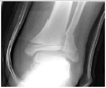
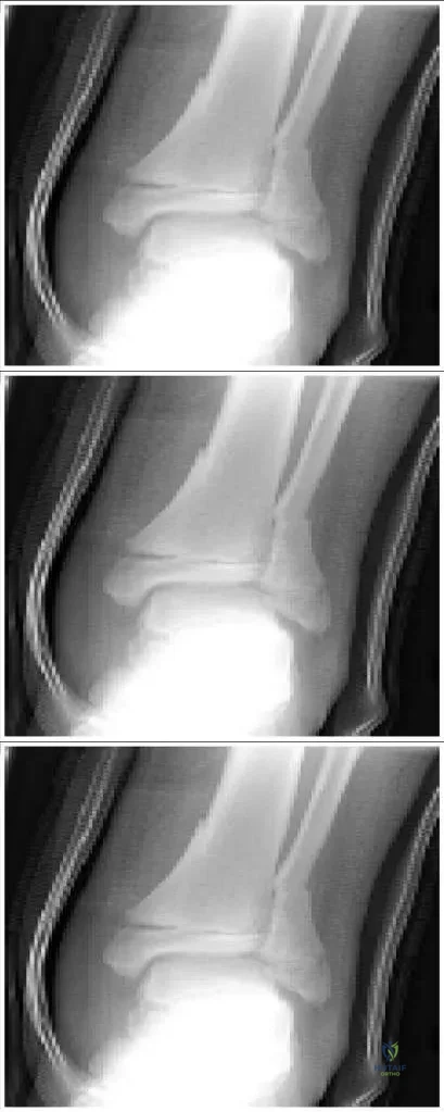
Slide 1 A 12-year-old boy presents to the emergency department after being struck by a car (slide 1). His only complaint at the time of presentation is right ankle pain. After obtaining an excellent reduction and casting the leg, the risks of a future growth disturbance through the involved physis must be discussed with the family. What are the chances of a significant growth disturbance of his leg:


Explanation

Orthopedic Prometric Exam Question This is a Salter-Harris type II fracture of the distal tibia. The distal tibia is at moderate risk for growth arrest after physeal injury. The average incidence of growth disturbance is 15% for all physeal injuries in this area. The marked displacement and mechanism of injury in this patient increase the risk of permanent physeal damage. The patientâ s age and remaining growth also increase the likelihood of a growth arrest causing a significant deformity or leg length discrepancy.C orrect Answer: 10% to 15%

Question 32

Three years after a Salter-Harris type I physeal fracture of the right distal femur, a 12-year-old boy presents with complaints of knee pain and a limp. On examination, the boy has a valgus alignment of his right knee and a 2-cm leg length discrepancy with the right leg shorter than the left. Plain radiographs and a scanogram showed 30% growth plate closure with a femoral-tibial angle of 12° of valgus and 2.5 cm of shortening of the right femur. What is the best treatment:


Explanation

The bar resection has a reasonable chance of success. The angulation can be corrected by the osteotomy and the length would be partially corrected. Correction of the length (2 cm) is less important than prevention of future angulation and shortening. At 12 years old, if varus osteotomy of the right femur and bilateral distal femur epiphyseodesis were performed, the patient would be sacrificing 6 cm of leg length which most patients would consider unacceptable. Right medial hemiepiphyseal stapling and completion of distal femoral epiphyseal closure would neither produce significant length equalization nor correct the angulation.

Question 33

Two years after a Salter-Harris type II fracture of the right distal femur, a 12-year-old girl presents to the clinic with knee pain. On examination, she is found to have a valgus alignment of the right knee and a 3-cm leg length discrepancy with the right leg shorter than the left leg. The scanogram confirms 3 cm of shortening in the right femur. The next step in the management of this patient is:


Explanation

A magnetic resonance image of the right distal femoral physis provides the best detail of the physis if the correct region and sequences are ordered. A computed tomography scan is not the preferred method to allow visualization of the physis. A bone scan or photon emission computed tomography scan will not highlight a bar of the physis. Waiting 9 months would allow for worsening of the deformity.

Question 34

The mutations underlying Stickler syndrome have been identified in which of the following molecules:


Explanation

Type II collagen is abnormal in the classic form of Stickler syndrome, but in a few patients, type XI collagen has been found to be the causative mutation. Type 1 collagen is predominant in bone and is abnormal in osteogenesis imperfecta, for example. Stickler syndrome is characterized by a defect in type II collagen. Fibrillin is abnormal in Marfan syndrome, but in Stickler syndrome, the causative abnormalities have been found in type II collagen. Sulfate transport protein has been found abnormal in diastrophic dysplasia Fibroblast growth factor proteins are abnormal in achondroplasia and hypochondroplasia, but the basic defect in Stickler syndrome is type II collagen.

Question 35

A 15-month-old child has bowing of the legs. Examination reveals a 3 cm distance between the femoral condyles and a thighfoot angle of 20° internal. Radiographs reveal 10° varus of the mechanical axis, no evidence of skeletal dysplasia, and a metaphyseal-diaphyseal angle of 9° on each side. Recommended treatment is:


Explanation

Although a diagnosis of Blount disease cannot be made at this time, the disease cannot be ruled out. The patient should be rechecked in 6 months by an experienced examiner. However, if the parents are reliable, they can be told that they do not need to return if the childâ s legs develop a normal shape in 6 months. The diagnosis of pathologic varus cannot be made; therefore, no treatment is indicated. The findings described are not outside of normal limits; surgery is not necessary. No pathologic diagnosis has yet been made. In addition, stapling is not an indicated form of treatment for such a young child. Although a pathologic condition has not been diagnosed, one cannot be ruled out either at this stage.

Question 36

An 11-year-old girl is observed for legs that have been bowed for the past 5 years. She has a mechanical axis that is in 26° of varus, a medial tibial plateau slope of 8°, a tibial joint angle of 15° varus, and a femoral joint angle of 10° varus. Her physis appears open. Recommended treatment is:


Explanation

Both a tibial and a femoral valgus osteotomy are necessary to correct significant deformities in these areas. A tibial valgus osteotomy is necessary but not sufficient. The femoral joint angle is off by 13° from the normal angle. A femoral valgus osteotomy is necessary but not sufficient. The tibial joint angle is off by 15° of varus. A tibial plateau elevation is not necessary for this mild degree of plateau depression. Brace treatment is not recommended for patients older than 3 years of age.

Question 37

The radiographic feature that is most characteristic of infantile Blount disease is:


Explanation

Bowing is focally located at the proximal medial tibia in infantile Blount disease. The physis may be widened medially, but it is normal laterally. Bone mineralization is not impaired in Blount disease. Bowing is focally located at the proximal tibia, not the distal femur. Blount disease is associated with internal, not external, tibial torsion.

Question 38

Marfan syndrome is now recognized as a defect in the following molecule:


Explanation

Marfan syndrome is due to an abnormality in fibrillin-1, a component of elastic connective tissue. Type I collagen is most commonly abnormal in osteogenesis imperfecta. Type I collagen is normal in Marfan syndrome. Type II collagen is a homotrimer (encoded by a single gene) and is mainly located in cartilaginous tissues. Type II collagen is abnormal in spondyloepiphyseal dysplasia congenita and Stickler syndrome. The sulfate transport protein is important for cartilage formation. The sulfate transport protein is impaired in diastrophic dysplasia. Type IX collagen is also found in cartilage. Type IX collagen is abnormal in some forms of multiple epiphyseal dysplasia.

Question 39

A 15-year-old basketball player has mild scoliosis, pes planus, pectus carinatum, and long slender fingers. In order to help determine if he has Marfan syndrome and should be allowed to continue playing basketball, it is most useful to order a:


Explanation

A cardiac echo will give information about enlargement of the ascending aorta, as well as the valves. This is the most important information for activity recommendations and patient prognosis. A magnetic resonance image (MRI) of the lumbosacral spine will either rule in or out dural ectasia, but a MRI has no clinical importance for activity recommendations. A slit lamp examination is not as important as a cardiac echo in making sport recommendations. A chest radiograph may be helpful, if the patient is suspected of having a pneumothorax. A dermal fibroblast assay is useful in assessing collagen, such as in osteogenesis imperfecta, but it is not helpful in diagnosing Marfan syndrome.

Question 40

The following skeletal feature helps to establish a diagnostic level of major skeletal involvement in Marfan syndrome:


Explanation

Pectus carinatum has a high diagnostic value for Marfan syndrome. Hyperextension of the knees is not common in patients with Marfan syndrome. This skeletal feature is seen in many other conditions; therefore, it has low diagnostic specificity. Atlantoaxial subluxation is rare in Marfan syndrome, but is common in some of the skeletal dysplasias. Although patients with Marfan syndrome are generally tall, height is too nonspecific to be a helpful diagnostic criterion. Kyphosis is not a skeletal diagnostic criterion.

Question 41

A 12-year-old girl has a scoliosis of 36° from T2-T7 and 15° from T7- L1. She is premenarchal. The following treatment is recommended:


Explanation

The Milwaukee brace offers the best chance of controlling this curve. Charleston bending braces are not effective for curves larger than 35°. Boston overlap braces are not effective for curves with an apex above T8. Lateral electrical spinal stimulation has been proven ineffective for scoliosis. This curve is not large enough to pose a recommend fusion.

Question 42

All of these findings are features of patients with Scheuermann kyphosis, except:


Explanation

Obesity is not any more common in patients with Scheuermann kyphosis than in the general population. Vertebral wedging is a common feature of Scheuermann kyphosis. Endplate irregularity is a common feature of Scheuermann kyphosis. Schmorl nodes are a manifestation of the disordered behavior of the vertebral endplates under load. Back pain is common in Scheuermann kyphosis.

Question 43

An 18-year-old man is seen in the office because of back pain in the thoracic region. He has a kyphosis of 65°, a slight wedging in the midthoracic spine, and a Risser sign of 4. Recommended treatment includes:


Explanation

Exercises must be the first treatment for this patient because he is too mature for brace treatment. Brace treatment is not indicated for someone of this skeletal maturity. Surgery is not indicated unless the patient fails conservative treatment.

Question 44

Prior to treatment, this pathologic finding characterizes clubfoot:


Explanation

The talar head and neck are deviated medially. This deformity cannot be corrected surgically but may improve with growth. The dome of the talus is not originally flattened, but it may become this way after repeated manipulation. The navicular is positioned more medially than normal and may touch the medial malleolus. The foot has either a normal arch or a cavus deformity (the opposite of a rocker bottom). The rocker bottom foot may develop as a result of over-vigorous manipulation of a clubfoot against a tight heelcord. The tendoachilles inserts are more medially positioned than normal on the calcaneus.

Question 45

In the surgical correction of a clubfoot, the following clinical or radiographic finding indicates that a child should have a plantar release:


Explanation

Plantarflexion of the first ray, especially if the first ray is not colinear with the talus on the lateral radiograph, should serve as an indication for a plantar release. Anterior extrusion of the talus is common because of the tightness of the Achilles tendon and the posterior capsule. Inferior subluxation of the calcaneocuboid joint is not a feature of clubfoot. Medial subluxation of the calcaneocuboid joint, in combination with the clinical appearance of a curved lateral border of the foot, should serve as an indication for a lateral release, not a plantar release. Inability to dorsiflex the ankle is an indication for a posterior release.

Question 46

Hip subluxation is most likely to occur in patients with this type of cerebral palsy:


Explanation

Patients with total involvement have the highest degree of axial muscle imbalance; therefore, they have the highest risk of hip subluxation (greater than 50%). Hip dysplasia is uncommon in athetoid cerebral palsy (CP). Diplegic C P has the second highest incidence of neuromuscular hip dysplasia, but total involvement has the highest rate. Hip subluxation is not common in hemiplegic CP. Monoplegia is a rare form of CP with a low rate of subluxation.

Question 47

A 13-year-old boy sustains a Salter II fracture of the proximal humeral epiphysis. On radiograph, there is a 40° varus angulation and a 30° apex anterior angulation. Recommended treatment includes:


Explanation

Spontaneous partial reduction will occur when the patient becomes upright, and there is good remodeling potential due to growth. Closed reduction and abduction cast is not necessary because of the patientâ s age, remodeling potential, and range of motion available in the joint. A formal attempt at closed reduction is not necessary because there is no way of holding it without internal fixation. This process will occur naturally to a large degree when the patient becomes upright. Open reduction and percutaneous pin fixation poses a risk of pin tract infection because of the large amount of muscle traversed. This procedure is not necessary because of the good results with conservative treatment.

Question 48

A 12-year-old girl sustains a closed type III Monteggia fracture. One week after closed reduction, the radial head resubluxates and the ulna bows. The next step of treatment is:


Explanation

Once the ulna is predictably reduced, the radial head has a good chance of staying reduced. Repeat closed reduction and a long arm cast has a significant chance of redisplacement. If further time passes, radial head reduction will be difficult. Annular ligament reconstruction is only indicated if there is late subluxation (after several weeks) that does not reduce with closed means. Once the ulna is realigned and fixed, closed reduction of the radius must be tried next. Pinning of the radial head should be a last resort because of the risk of breakage and stiffness.

Question 49

The following parameter is the most useful in predicting the need for surgical correction of developmental coxa vara:


Explanation

The Hilgenreiner-epiphyseal angle is a measure of the shear stress on the femoral neck. A value greater than 60° is an indication for surgery. The range of abduction does not directly reflect the mechanical stresses on the femoral neck that may need correction. The epiphyseal extrusion index is a parameter used in Perthes disease. The acetabular index is used in developmental hip dysplasia. It is usually essentially within normal limits, even in severe coxa vara.

Question 50

A 7-month-old girl is newly seen for a dislocation of the left hip. The newborn exam was unremarkable; there was no history of trauma or evidence of spasticity. Recommended treatment includes:


Explanation

An exam under anesthesia and arthrogram should be performed. C losed reduction must be performed if the hip reduces with a medial dye pool less than 4 mm and a safe zone greater than 20°. The Pavlik harness is not strong enough to hold an infant older than 4 to 6 months. Open reduction is reserved for hips that do not reduce by closed means. A Salter osteotomy is only indicated if the hip is still unstable after an open reduction.

Question 51

A 13-year-old obese male presents with right groin pain and an obligatory external rotation of the hip when it is passively flexed. If this condition is treated with forceful closed reduction, what is the most significant and devastating complication?


Explanation

The presentation is classic for Slipped Capital Femoral Epiphysis (SCFE). Forceful closed reduction of a SCFE significantly increases the risk of avascular necrosis of the femoral head, which is the most devastating complication.

Question 52

In the early stages of osteoarthritis, which of the following best describes the characteristic biochemical changes that occur in the articular cartilage?


Explanation

The hallmark biochemical change in early osteoarthritis is the breakdown of the collagen matrix, which leads to increased permeability and an influx of water, alongside a decrease in proteoglycan concentration.

Question 53

Which of the following ligaments is the strongest in the body and provides the primary stability to the posterior pelvic ring against vertical shear forces?


Explanation

The interosseous sacroiliac ligament is the strongest ligament in the body. It plays the primary role in stabilizing the posterior pelvic ring, particularly against vertical shear stresses.

Question 54

During anterior cruciate ligament (ACL) reconstruction using a bone-patellar tendon-bone (BTB) autograft, what is structurally the weakest link of the reconstruction during the first 6 weeks of graft healing?


Explanation

Following a BTB autograft reconstruction, the graft undergoes avascular necrosis and subsequent revascularization (ligamentization). During the first 6 to 8 weeks, the intra-articular graft substance is at its weakest point.

Question 55

A 15-year-old boy presents with a destructive diaphyseal bone lesion exhibiting an 'onion-skin' periosteal reaction. Genetic testing reveals a t(11;22) chromosomal translocation. Which of the following is the specific fusion protein associated with this tumor?


Explanation

The clinical and radiographic presentation is highly characteristic of Ewing sarcoma. The t(11;22) translocation results in the EWS-FLI1 fusion protein, which is found in over 85% of Ewing sarcoma cases.

Question 56

A 65-year-old woman is undergoing a total hip arthroplasty (THA). Which of the following bearing surface combinations is associated with the lowest linear wear rate but carries a risk of catastrophic component fracture?


Explanation

Ceramic-on-ceramic bearing surfaces provide the lowest linear wear rates among THA options. However, their primary disadvantage is the risk of catastrophic brittle fracture of the ceramic components.

Question 57

A 55-year-old male presents with bilateral hand clumsiness and a broad-based gait. MRI demonstrates severe cervical canal stenosis. Which of the following physical exam findings is an upper motor neuron sign highly indicative of cervical myelopathy?


Explanation

The inverted radial (or supinator) reflex is a classic upper motor neuron sign of cervical myelopathy. It occurs when striking the brachioradialis tendon produces paradoxical finger flexion due to loss of upper motor neuron inhibition.

Question 58

A patient presents with a swollen, painful index finger. Which of the following is NOT one of Kanavel's four cardinal signs of acute suppurative flexor tenosynovitis?


Explanation

Kanavel's signs include fusiform swelling, tenderness along the sheath, pain on passive extension, and a flexed resting posture. Erythema extending proximal to the wrist is not a cardinal sign and may suggest a more widespread cellulitis or proximal spread.

Question 59

In the Eichenholtz classification of Charcot neuroarthropathy, which of the following radiographic findings is most characteristic of Stage 2 (Coalescence)?


Explanation

Eichenholtz Stage 2 (Coalescence) is marked by the absorption of fine intra-articular debris, early fusion of larger bone fragments, and subchondral sclerosis. Stage 1 is fragmentation, and Stage 3 is remodeling.

Question 60

A 9-year-old boy presents with mild right arm pain after a minor fall. Radiographs are obtained.

Assuming the radiograph demonstrates a central, completely lytic metaphyseal lesion with a 'fallen leaf' sign, what is the most appropriate initial management?


Explanation

A central, lytic metaphyseal lesion with a 'fallen leaf' sign is pathognomonic for a Unicameral Bone Cyst (UBC). The initial treatment of choice for symptomatic or fracture-prone UBCs is aspiration and corticosteroid injection.

Question 61

A 25-year-old male sustains a severe closed tibial shaft fracture. Which of the following pressure measurements represents the most widely accepted threshold for performing a four-compartment fasciotomy to treat acute compartment syndrome?


Explanation

A delta pressure (diastolic blood pressure minus intracompartmental pressure) of less than 30 mmHg is the standard threshold indicating inadequate tissue perfusion and the need for emergent fasciotomy.

Question 62

A 3-month-old infant is treated with a Pavlik harness for developmental dysplasia of the hip (DDH). What is the most common nerve palsy associated with excessive hip flexion in this device?


Explanation

Femoral nerve palsy is a known complication of treating DDH with a Pavlik harness when the hips are placed in excessive hyperflexion, which compresses the nerve against the inguinal ligament.

Question 63

During a posterior-stabilized total knee arthroplasty (TKA), the surgeon uses spacer blocks and notes that the knee is tight in flexion but well-balanced in extension. Which of the following is the most appropriate next step to balance the knee?


Explanation

If the knee is tight in flexion but balanced in extension, downsizing the femoral component will increase the flexion gap by reducing the posterior condylar offset, without altering the extension gap.

Question 64

In a child with an extension-type supracondylar humerus fracture presenting with significant posteromedial displacement of the distal fragment, which nerve is most commonly at risk of injury?


Explanation

In a posteromedially displaced supracondylar humerus fracture, the proximal fragment displaces anterolaterally, putting the radial nerve at the highest risk of injury. Posterolateral displacement primarily endangers the median nerve and AIN.

Question 65

A 28-year-old male sustains a displaced fracture of the proximal pole of the scaphoid. The predominant blood supply to the scaphoid, which determines its risk for avascular necrosis, enters primarily through which of the following areas?


Explanation

The primary blood supply to the scaphoid enters through the dorsal ridge at the waist and flows in a retrograde fashion to the proximal pole. This retrograde flow is why proximal pole fractures have a high rate of avascular necrosis.

Question 66

During the process of endochondral ossification in secondary fracture healing, which type of collagen is most predominantly synthesized by chondrocytes in the soft callus phase?


Explanation

The soft callus is primarily composed of cartilage, where chondrocytes predominantly synthesize Type II collagen. As healing progresses to hard callus, osteoblasts replace this with Type I collagen.

Question 67

A 22-year-old athlete sustains a midfoot injury. Weight-bearing radiographs demonstrate a 3 mm widening between the base of the first and second metatarsals. The injured Lisfranc ligament normally connects which two osseous structures?


Explanation

The Lisfranc ligament is an interosseous ligament that connects the lateral aspect of the medial cuneiform to the medial base of the second metatarsal, providing critical stability to the midfoot.

Question 68

Which of the following historical features most reliably distinguishes neurogenic claudication (due to lumbar spinal stenosis) from vascular claudication?


Explanation

Neurogenic claudication is classically relieved by lumbar flexion (e.g., the 'shopping cart sign'), which increases the cross-sectional area of the spinal canal. Vascular claudication is typically relieved simply by resting or standing still.

Question 69

A 35-year-old male sustains a Hawkins Type III talar neck fracture. What is the approximate reported rate of avascular necrosis (AVN) of the talar body associated with this specific injury pattern?


Explanation

A Hawkins Type III fracture involves a talar neck fracture with dislocation of both the subtalar and tibiotalar joints. Because this disrupts the major blood supplies to the talus, the rate of AVN is exceedingly high, approaching 80-100%.

Question 70

Which of the following benign bone tumors is characteristically located within the epiphysis of long bones in skeletally immature patients?


Explanation

Chondroblastoma is a rare, benign cartilaginous tumor that classically presents in the epiphysis or apophysis of long bones in children and adolescents before physeal closure.

Question 71

A 15-year-old boy presents with knee pain. Radiographs show the lesion seen in the image.

Assuming a malignant primary bone tumor, what is the most likely diagnosis given a 'sunburst' periosteal reaction in the metaphysis?


Explanation

Osteosarcoma often presents in the metaphysis of long bones in adolescents with a 'sunburst' periosteal reaction and Codman's triangle. Ewing sarcoma typically affects the diaphysis and has an 'onion skin' appearance.

Question 72

Which of the following genetic defects is most commonly associated with Marfan syndrome?


Explanation

Marfan syndrome is an autosomal dominant connective tissue disorder caused by mutations in the FBN1 gene on chromosome 15, which encodes fibrillin-1. This leads to skeletal, cardiovascular, and ocular abnormalities.

Question 73

Achondroplasia is the most common form of short-limb dwarfism. It is caused by a gain-of-function mutation in which of the following genes?


Explanation

Achondroplasia is an autosomal dominant disorder caused by a gain-of-function mutation in the FGFR3 gene. This mutation inhibits chondrocyte proliferation in the proliferative zone of the physis.

Question 74

Osteogenesis imperfecta (OI) type I is characterized by which of the following defects?


Explanation

Type I OI is a mild form characterized by a decreased production of structurally normal type I collagen (quantitative defect). In contrast, types II, III, and IV involve qualitative defects in collagen structure.

Question 75

Which of the following is the most important prognostic factor in a patient with Legg-Calvé-Perthes disease?


Explanation

Age at clinical onset is the most critical prognostic factor in Legg-Calvé-Perthes disease. Children who develop the condition before 6 to 8 years of age have a better prognosis due to greater potential for acetabular remodeling.

Question 76

What is the approximate rate of bilateral involvement in patients presenting with Slipped Capital Femoral Epiphysis (SCFE)?


Explanation

Bilateral involvement in SCFE occurs in 20-40% of cases, with up to half presenting simultaneously. Patients with endocrinopathies or who present at an atypically young age have an even higher risk of bilaterality.

Question 77

In the pathophysiology of acute compartment syndrome, what is the initial event at the microvascular level?


Explanation

The initial microvascular event in compartment syndrome is venous outflow obstruction due to increased tissue pressure. This causes secondary increases in capillary pressure, transudation of fluid, and ultimately compromised arterial inflow.

Question 78

Which of the following is NOT one of Kanavel's cardinal signs for flexor tenosynovitis?


Explanation

Kanavel's four cardinal signs are: flexed posture of the digit, fusiform swelling, tenderness along the flexor sheath, and severe pain with passive (not active) extension. Pain with active motion is less specific.

Question 79

A 65-year-old man presents with severe back pain and anemia. A skeletal survey shows multiple lytic 'punched-out' lesions in the skull and spine. Which of the following laboratory findings is most specific for this condition?


Explanation

Multiple myeloma classically presents with lytic bone lesions, hypercalcemia, anemia, and renal involvement. A monoclonal M-protein spike on serum or urine protein electrophoresis is highly characteristic.

Question 80

Which of the following total hip arthroplasty bearing surfaces is associated with the lowest linear wear rate?


Explanation

Ceramic-on-ceramic bearings have the lowest linear and volumetric wear rates of all available articulations. However, they carry risks of squeaking and catastrophic ceramic fracture.

Question 81

During the ligamentization process of a hamstring autograft used in ACL reconstruction, when is the graft mechanically at its weakest?


Explanation

The graft undergoes avascular necrosis and subsequent revascularization and cellular repopulation. It is mechanically at its weakest during the revascularization phase, typically around 6 to 8 weeks postoperatively.

Question 82

Where is the anatomically correct position to apply a pelvic binder to effectively reduce a suspected 'open book' pelvic ring injury?


Explanation

A pelvic binder must be centered over the greater trochanters to directly close the pelvic ring through the pubic symphysis. Placement over the iliac crests is incorrect and can paradoxically open the pelvis further.

Question 83

Which of the following clinical findings most reliably indicates the end of spinal shock?


Explanation

Spinal shock is a temporary state of flaccid paralysis and areflexia following spinal cord injury. The return of the bulbocavernosus reflex marks the end of spinal shock, allowing accurate determination of the neurologic deficit.

Question 84

Which type of collagen is predominantly synthesized during the soft callus phase of secondary fracture healing?


Explanation

During the soft callus (chondrogenesis) phase of secondary fracture healing, chondrocytes predominantly produce Type II collagen. As the hard callus forms, osteoblasts replace this with Type I collagen.

Question 85

Which ligament is specifically disrupted in a classic Lisfranc injury?


Explanation

The Lisfranc ligament connects the medial cuneiform to the base of the second metatarsal. Its disruption leads to instability of the tarsometatarsal joint complex, as there is no direct intermetatarsal ligament between the first and second metatarsals.

Question 86

Which of the following structures is the most volar structure within the carpal tunnel?


Explanation

The median nerve is the most volar (superficial) structure within the carpal tunnel, lying immediately deep to the transverse carpal ligament. The FDS tendons to the middle and ring fingers are immediately dorsal to the nerve.

Question 87

In the Graf classification for developmental dysplasia of the hip (DDH) using ultrasound, what does the alpha angle measure?


Explanation

The alpha angle in Graf's ultrasound evaluation measures the concavity of the bony acetabular roof. A normal alpha angle is typically greater than 60 degrees. The beta angle assesses the cartilaginous roof.

Question 88

The major blood supply to the body of the talus is provided by the artery of the tarsal canal, which is a branch of which artery?


Explanation

The artery of the tarsal canal is a branch of the posterior tibial artery and provides the dominant blood supply to the body of the talus. Disruption of this supply in talar neck fractures carries a high risk of avascular necrosis.

Question 89

A 30-year-old woman presents with knee pain. Radiographs reveal an eccentric, lytic epiphyseal lesion in the proximal tibia without a sclerotic margin. Histology shows multinucleated giant cells. Which of the following is the standard medical treatment targeting the underlying pathophysiology?


Explanation

Giant cell tumors of bone express high levels of RANKL, which drives the formation of osteoclast-like giant cells. Denosumab, a monoclonal antibody against RANKL, is specifically used for unresectable or recurrent cases.

Question 90

Following a total knee arthroplasty, a patient exhibits a persistent lateral patellar tilt and subluxation. Which of the following technical errors is most likely responsible?


Explanation

Internal rotation of the femoral or tibial components increases the Q-angle and leads to lateral patellar maltracking. To optimize patellar tracking, the femoral component is typically placed in slight external rotation (e.g., 3 degrees).

Question 91

Which of the following gene mutations is most commonly associated with Osteogenesis Imperfecta type I?


Explanation

Osteogenesis imperfecta type I is an autosomal dominant condition most commonly caused by mutations in the COL1A1 or COL1A2 genes. These mutations lead to a quantitative defect in type I collagen, resulting in bone fragility, blue sclerae, and hearing loss.

Question 92

A 14-year-old boy presents with scoliosis, pectus excavatum, and a highly arched palate. He is diagnosed with a condition caused by a defect in fibrillin-1. Which of the following cardiovascular anomalies is the leading cause of mortality in this patient population?


Explanation

Marfan syndrome is an autosomal dominant connective tissue disorder caused by a mutation in the FBN1 gene affecting fibrillin-1. Aortic root dilation and subsequent dissection is the leading cause of morbidity and mortality in these patients.

Question 93

A 3-year-old boy presents with disproportionate short stature, frontal bossing, and rhizomelic shortening. A radiograph is shown.

What is the underlying genetic mechanism for this condition?


Explanation

Achondroplasia is caused by a gain-of-function mutation in the FGFR3 gene, which inhibits chondrocyte proliferation in the proliferative zone of the physis. This results in rhizomelic dwarfism, characteristic facial features, and spinal stenosis.

Question 94

A 4-year-old girl is diagnosed with pauciarticular (oligoarticular) juvenile idiopathic arthritis (JIA). Which of the following routine screening examinations is highly recommended due to the high risk of asymptomatic, sight-threatening complications?


Explanation

Patients with oligoarticular JIA, especially those who are Antinuclear Antibody (ANA) positive, are at a very high risk for developing chronic, asymptomatic anterior uveitis. Routine slit-lamp screening by an ophthalmologist is mandatory to prevent blindness.

Question 95

Morquio syndrome (Mucopolysaccharidosis type IV) is an autosomal recessive disorder characterized by severe skeletal dysplasias. Which of the following cervical spine abnormalities is a hallmark of this condition and requires careful evaluation before any surgical procedure?


Explanation

Morquio syndrome typically presents with odontoid hypoplasia leading to severe atlantoaxial instability. Prophylactic cervical spinal fusion is often required early in childhood to prevent fatal spinal cord compression.

Question 96

A 6-year-old boy with delayed motor milestones routinely uses his hands to push on his legs to stand up from the floor. Muscle biopsy shows absent dystrophin. What is the typical inheritance pattern of this disease?


Explanation

Duchenne Muscular Dystrophy (DMD) is an X-linked recessive disorder caused by nonsense or frameshift mutations in the dystrophin gene. The described Gowers' sign is a classic clinical manifestation of the resultant proximal muscle weakness.

Question 97

Multiple Epiphyseal Dysplasia (MED) is characterized by delayed and irregular ossification of the epiphyses. Which of the following genes is NOT typically associated with MED?


Explanation

RUNX2 mutations are associated with Cleidocranial Dysplasia, not MED. MED is typically caused by mutations in the genes encoding COMP, matrilin-3 (MATN3), or type IX collagen (COL9A1, COL9A2, COL9A3).

Question 98

Which of the following variants of Ehlers-Danlos syndrome is caused by a mutation in the COL3A1 gene (resulting in a type III collagen defect) and carries a high risk of fatal arterial rupture?


Explanation

Vascular Ehlers-Danlos syndrome is caused by mutations in the COL3A1 gene. It is considered the most severe and life-threatening form due to the propensity for spontaneous rupture of medium and large arteries, the bowel, and the uterus.

Question 99

A 10-year-old child presents with delayed closure of the cranial sutures, dental anomalies, and absent clavicles allowing extreme shoulder protraction. This condition is caused by a mutation in which of the following transcription factors?


Explanation

Cleidocranial dysplasia is an autosomal dominant condition caused by a mutation in the RUNX2 (CBFA1) gene, which is an essential transcription factor for osteoblast differentiation. Patients characteristically present with absent or hypoplastic clavicles and delayed fontanelle closure.

Question 100

A 7-year-old boy with a history of multiple café-au-lait spots and axillary freckling presents with a rapidly progressive spinal deformity. Anteroposterior and lateral radiographs demonstrate a short-segment, sharply angulated thoracic curve. Which of the following is the most likely diagnosis?


Explanation

Dystrophic scoliosis in Neurofibromatosis type 1 typically presents as a short-segment, sharply angulated curve with severe vertebral wedging, spindling of transverse processes, and rib penciling. It is prone to rapid progression and often requires early surgical stabilization with combined anterior and posterior fusion.

None

Dr. Mohammed Hutaif
Medically Verified Content by
Prof. Dr. Mohammed Hutaif
Consultant Orthopedic & Spine Surgeon
Chapter Index