Part of the Master Guide

Orthopedic Prometric Exam Preparation MCQs - Part 1

Orthopedic Prometric Exam Preparation MCQs - Part 12

27 Apr 2026 47 min read 18 Views
Orthopedic Prometric Exam Preparation MCQs - Part 12

Orthopedic Prometric Exam Preparation MCQs - Part 12

Comprehensive 100-Question Exam


00:00

Start Quiz

Question 1

Lateral epicondylitis is associated with a tear in the fibers of which muscle:


Explanation

Current consensus is that tennis elbow is associated with a strain or microtear of the EC RB origin, which lies beneath the EC RL.

Question 2

The gold standard for diagnosis of lateral epicondylitis is considered:


Explanation

The clinical diagnosis of lateral epicondylitis is supported by specific provocative tests. The gold standard for diagnosis is the history and physical examination. Tenderness on examination is localized to the lateral epicondyle, which can radiate into the forearm; the area of maximum tenderness is approximately 2 mm to 5 mm distal and anterior to the midpoint of the lateral epicondyle. There is usually a history of overuse or of a repetitive activity. The pain is aggravated, with the elbow extended, by resisted wrist and finger extension or with passive finger and wrist flexion.

Question 3

The amount of time that nonoperative management should be followed for lateral epicondylitis is closest to:


Explanation

Ninety to 95% of all patients with tennis elbow respond to nonoperative treatment, and it remains the mainstay for treatment of lateral epicondylitis. Operative treatment may be indicated for debilitating pain in patients without other pathologic causes of pain for whom nonoperative treatment has failed after a reasonable length of time. This time period is usually a minimum of 6 to 12 months.

Question 4

Extracorporeal shock wave therapy ____ in the treatment of lateral epicondylitis in high-quality trials.


Explanation

Current studies have found no benefit of extracorporeal shock wave therapy in the treatment of lateral epicondylitis.

Question 5

All of the following medications are indicated in the early treatment of frostbite injury except:


Explanation

Peripheral beta-blockade has minimal effect on peripheral vasculature and is not typically used in patients with frostbite injury. Ibuprofen, tetanus booster, antibiotic prophylaxis, and appropriate pain medication are routinely used in the treatment of frostbite injuries.

Question 6

Orthopedic sequelae of frostbite injury include all of the following except:


Explanation

People who have had previous frostbite injuries are at increased risk of thermal injury, whether cold or heat related. Joint contractures, localized osteoporosis, punched-out subchondral bony lesions, and cold intolerance are often present after frostbite injury.

Question 7

Initial treatment of an acute frostbite injury should include:


Explanation

Rapid rewarming in a 40° C to 42° C circulating water bath is the most effective early treatment of frostbite injury. Slow or fast rewarming in other temperatures and/or rewarming in air is not indicated.

Question 8

All of the following except _ increase the risk of frostbite injury.


Explanation

High altitudes, prolonged exposure, and anything that would cause peripheral vasoconstriction increase the risk of frostbite injury. Humidity does not play a significant role in the development of frostbite injury.

Question 9

Treatment for frostbite injury includes:


Explanation

Treatment of frostbite includes rapid rewarming (even when reperfusion pain occurs), early active motion, elevation, and avoidance of dry heat that can dessicate tissues. Massaging the frostbitten area is not recommended because it may induce additional trauma via shearing forces.

Question 10

Arthritis of the wrist is estimated to effect what percentage of the U.S. population:


Explanation

Arthritis of the wrist is estimated to affect 5.3% of the U.S. population, based on radiographic assessments of 4,000 wrists.1 After having rheumatoid arthritis (RA) for 10 years, 90% of patients experience arthritis in their wrist joints.2

Question 11

The accessory ulnar collateral ligament inserts on the:


Explanation

The accessory ulnar collateral ligament inserts into the volar plate, whereas the proper collateral inserts into the base of the proximal phalanx.

Question 12

Which of the following nerves is not a primary articular nerve of the wrist:


Explanation

Fukumoto and colleagues have used Wykeâ s definition to explain primary and accessory innervation of the wrist. Primary articular nerves consist of small nerves that pass to each joint as independent branches of adjacent peripheral nerves. There are three primary articular nerves: the PIN, the lateral antebrachial cutaneous nerve, and the articular branches from the ulnar nerve. Accessory nerves originate from small, twig branches of intramuscular or cutaneous nerves that innervate the skin around the wrist joint. The accessory articular nerves have been identified as the anterior interosseous nerve (AIN), the palmar cutaneous branch of the median nerve, the deep and dorsal branches of the ulnar nerve, and the superficial branch of the radial nerve to the first intercarpal space.

Question 13

Which of the following nerves provides principal innervation to the central dorsal portion of the wrist:


Explanation

The PIN is found on the deep radial wall of the fourth dorsal compartment, 1.2 cm ulnar to Listerâ s tubercle. As the PIN approaches the radiocarpal joint, it is covered in fascia and gives one branch to the radioscaphoid joint and three to four terminal branches to the intercarpal joints. The PIN is the principal innervation to the central dorsal portion of the wrist. The AIN innervates the radial volar lip of the distal radius. The dorsal branch of the ulnar nerve contributes to innervation of the triangular fibrocartilage complex. The lateral antebrachial cutaneous nerve innervates the thumb carpometacarpal joint and the scaphotrapezotrapezoid joint.

Question 14

What is the area of innervation of the anterior interosseous nerve (AIN):


Explanation

The AIN is a branch of the median nerve. Its muscular innervations include the flexor pollicis longus, the radial half of the flexor digitorum profundus, and the pronator quadratus. The AIN terminates as a sensory branch to the volar radial surface of the distal radius. The TFC C is innervated by components of the ulnar nerve. The dorsal radiocarpal joint is innervated by the posterior interosseous nerve. The thumb carpometacarpal is innervated by the sensory branch of the radial nerve and the lateral antebrachial cutaneous nerve.

Question 15

When performing complete wrist denervation as described by Wilhem, what pain pathology did not have predictable results:


Explanation

In 1983, Ekerot and colleagues reported his results in 48 patients. They used the technique described by Wilhelm but only denervated the radial side of the wrist for patients with scaphoid or lunate pathology. However, the entire wrist was denervated in patients with global degenerative wrist disease or wrist pain with an unknown etiology. Pain relief occurred in only 56% of the patients. They noted the best results occurred in patients with scaphoid nonunion, osteonecrosis of the lunate, and primary radiocarpal arthritis.

Question 16

What two nerves are resected through a single dorsal incision for wrist denervation:


Explanation

Kupfer and colleagues presented a podium presentation of a single-incision approach to the resection of the PIN and AIN for denervation of the radial side of the wrist. Weinstein and Berger published their results in 2002 with a similar technique. They described a 2-cm long dorsal incision that was 3 to 5 cm proximal to the ulnar head. They then resected a 2-cm segment of the PIN and AIN. In their group of 20 patients, 85% were satisfied with their procedure after an average follow-up of 2.5 years. If failure were to occur, it occurred within the first year.

Question 17

What muscle is at risk for denervation when a single dorsal incision is used to denervate the radial side of the wrist:


Explanation

The single dorsal incision approach to wrist denervation involves resection of the posterior interosseous nerve (PIN) and the anterior interosseous nerve (AIN). Distally, the PIN is purely sensory and does not give off motor branches in the vicinity of the wrist joint. The terminal portion of the AIN has both motor and sensory components. A majority of this is motor, and it innervates the pronator quadratus right up to the radiocarpal articulation. Resection of the AIN close to the radiocarpal joint has a high probability of denervating the pronator quadratus. The extensor indicis is usually the last motor branch of the PIN, but this terminal portion of this branch is more than 5 cm proximal from the distal radioulnar joint. The flexor pollicis longus is innervated by the AIN, but motor branches to this muscle are more proximal than branches to the pronator quadratus. The flexor digitorum profundus muscle is innervated by the AIN and ulnar nerve. However, the motor branches are more proximal than the incision for dorsal innervation. C orrect Answer: Pronator quadratus

Question 18

A 62-year-old man presents with weakness in finger extension in his right hand. He has had the weakness for 1 month but denies any significant traumatic event. The patient maintains an active lifestyle, including golf and tennis. He denies pain or numbness in his hand and is otherwise neurologically intact. Which of the following is the most likely diagnosis:


Explanation

Posterior interosseous nerve palsy is described as painless finger drop. This syndrome is commonly associated with trauma to the lateral elbow.

Question 19

Which of the following are characteristic signs of PIN palsy:


Explanation

Painless finger drop is characteristic of posterior interosseous nerve palsy. This syndrome may also involve elbow tenderness in the absence of other clinical findings. Pain in the dorsum of the hand is not associated with this condition because the posterior interosseous nerve contains no sensory component.

Question 20

What is the most common site of posterior interosseous nerve entrapment:


Explanation

The most common site of posterior interosseous nerve entrapment is at the arcade of Frohse, which is a fibrotendinous ring found within the fibers of the supinator muscle as the posterior interosseous nerve originates from the radial nerve.

Question 21

Which of the following muscles is innervated by the posterior interosseous nerve:


Explanation

The posterior interosseous nerve innervates a number of muscles involved primarily in finger extension, including the extensor carpi ulnaris, extensor digitorum, extensor digiti minimi, extensor pollicis brevis and longus, abductor pollicis longus, and extensor indices.

Question 22

Posterior interosseous nerve palsy affects finger extension at the metacarpophalangeal and interphalangeal joints.


Explanation

Question 23

A 53-year-old woman presents with bilateral hand numbness and tingling. Her right hand is more affected than her left. The numbness wakes her up at night and is relieved when she shakes her hand. In addition, the patient has had increasing difficulty with fine motor tasks, such as shirt buttoning, over the past 2 to 3 months. Upon close inspection, muscle atrophy is present at the base of her thumbs. Which of the following is the most likely diagnosis:


Explanation

This patient displays the classic signs and symptoms of bilateral carpal tunnel syndrome, which involves median nerve entrapment at the base of the palm. This entrapment leads to numbness and dysesthesias that are worse at night and upon exertion. Pain is typically relieved by shaking the hand. Furthermore, the median nerve innervates several muscles of the hands, and entrapment may lead to muscle atrophy.

Question 24

All of the following muscles are innervated by the median nerve except:


Explanation

Lumbricals 1 and 2 are innervated by the median nerve, in addition to the opponens pollicis brevis, abductor pollicis brevis, and flexor pollicis brevis.

Question 25

All of the following are true regarding the transverse carpal ligament except:


Explanation

All of the above statements are true regarding the transverse carpal ligament.

Question 26

All of the following structures pass through the carpal tunnel except:


Explanation

The palmar cutaneous branch of the median nerve originates proximally to the carpal tunnel and travels superficial to the tunnel.

Question 27

Dupuytrenâ s contracture characteristically involves which part of the hand:


Explanation

Dupuytrens contracture most frequently involves the ring and small fingers. Although Dupuytrenâ s cords at the thumb have been described, they are rare.

Question 28

The use of clostridial collagenase for Dupuytrenâ s contracture is performed by:


Explanation

Clostridial collagenase works by breaking the collagen connections. The Dupuytrenâ s cord is ruptured manually; surgery is not necessary.

Question 29

Dupuytrens cord tissue is characterized by what change from normal:


Explanation

Compared to normal palmar fascia, the fibrous bands in Dupuytrenâ s disease have an increased ratio of type III to type I collagen, and an overall increase in the amount of type III collagen.

Question 30

A 29-year-old man with a remote history of wrist trauma and chronic pain presents with a palpable clunk on radio-ulnar deviation of the wrist. The most sensitive technique for identifying a scapholunate injury is:


Explanation

Magnetic resonance imaging is commonly used among patients with concern for ligamentous injuries of the wrist, particularly in the presence of an abnormal physical exam when plain radiographs are normal. However, the sensitivity of MRI has been shown to be less than 40% in comparison with arthroscopy. Arthroscopy has become the gold standard for the diagnosis of ligamentous injuries to the wrist. A classification scheme has been proposed based on both radiocarpal and midcarpal arthroscopic findings.

Question 31

The radiographic abnormality seen on the lateral radiograph characteristic of scapholunate instability is:


Explanation

On a lateral view of the wrist, when the lunate slips into a statically dorsiflexed position greater than 10°, the condition is defined as dorsal intercalated segmental instability (DISI). DISI deformity is also present when the scapholunate angle is greater than 60 degrees (45+/- 15 degrees is normal). The VISI deformity is seen on the lateral radiograph is characteristic of lunotriquetral dissociation. The other signs are seen on the anteroposterior projection.

Question 32

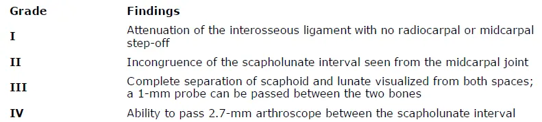
A 40-year-old woman with radial sided wrist pain for the last 2 years presents to the clinic. Plain radiographs are normal. Because of continued discomfort despite conservative therapies and occasional â clickingâ of the wrist, she is taken to the operating room for diagnostic arthroscopy. At the time, fraying of the membranous portion of the scapholunate (SL) ligament is seen, with mild incongruity from the midcarpal joint. The surgeon is unable to pass a 1-mm probe through the defect. This is most consistent with:


Explanation

Arthroscopy has become the gold standard for the diagnosis of ligamentous injuries to the wrist. A classification scheme has been proposed by Geissler and colleagues, based on both radiocarpal and midcarpal arthroscopic findings (Table). Table. Arthroscopic C lassification of Interosseous Ligament Injury

Question 33

A 33-year-old woman with a history of a traumatic fall onto her wrist and tenderness over the scapholunate (SL) interval presents to the clinic. Radiographs are normal, and magnetic resonance imaging reveals a partial tear of the SL ligament. The remaining wrist ligaments are normal. If conservative therapy is attempted, then it should consist of:


Explanation

Orthopedic Prometric Exam Question Conservative management includes a period of splinting and activity modification, followed by proprioception training of the flexor carpi radialis to act as a dynamic scaphoid stabilizer.

Question 34

Congenital thumb duplication:


Explanation

Experts recommend treating congenital thumb duplication before the age of 6 months, when the potential for growth and remodeling is greatest. The condition usually presents unilaterally, lacking association with other systemic abnormalities, and usually presents with a hypoplastic radial duplicate and dominant ulnar duplicate.

Question 35

Complete bifurcation of two distal phalanges articulating with a wide epiphysis of a single proximal phalanx is classified as:


Explanation

Wassel II (also categorized as IP in the universal classification system) occurs when the duplication begins at the interphalangeal joint of the thumb, resulting in complete bifurcation of two distal phalanges that articulate proximally with a single proximal phalanx.

Question 36

One of the more common complications of congenital thumb duplication reconstruction is:


Explanation

A Z-deformity, with ulnar deviation at the MC P joint and radial deviation at the IP joint, is one of the most common complications after reconstruction. Weakness, paresthesias, and wound complications are uncommon possible complications.

Question 37

Ultrasound therapy delivers superficial heat to the tissue and has a penetration depth of 5 mm.


Explanation

Question 38

Thermal ultrasound is used for all of the following purposes EXC EPT:


Explanation

Thermal uses of ultrasound include increasing pain threshold, decreasing scar, and improving extensibility of the ligaments and joint capsule. Ultrasound has not been shown to have an effect in Dupuytrenâ s contracture.

Question 39

Phonopheresis is:


Explanation

Phonopheresis is delivery of medicine through the skin using ultrasound. Although there is some question as to whether the medications are more effectively absorbed or delivered with ultrasound use, this is a described modality.

Question 40

Iontophoresis delivers medications such as analgesics or steroids through the skin using an electrical charge.


Explanation

Question 41

Iontophoresis has been effectively used in all of the following EXC EPT:


Explanation

Iontophoresis is effective in soft tissue conditions such as rotator cuff bursitis and lateral epicondylitis.

Question 42

Types of nerve tissues surrounding the axons include all of the following EXC EPT:


Explanation

The structures surrounding the axons and Schwann cells include the endoneurium, perineurium, and epineurium. The mesoneurium is an adventitial layer in addition.

Question 43

The Seddon grades of nerve injury include all of the following EXC EPT:


Explanation

Neuropraxic injuries are stretch injuries to the nerve. Axonotmetic injury involves a more severe injury with loss of continuity of axons, and connective tissue elements remain intact. Neurotmesis is a complete nerve discontinuity. Schwann cell disruption occurs in neurotmesis but is not among the grades of Seddon nerve injury.

Question 44

Younger age is associated with worse outcomes with nerve repair.


Explanation

Question 45

Optimum conditions for nerve healing after direct repair include:


Explanation

Tension-free repair is the optimal technique to improve the potential for nerve recovery. Gapping, failure to splint to prevent tension on the nerve with motion, and failure to excise scarred or devitalized nerve tissue are impairments to successful nerve repairs. Suture repairs through the deep nerve segments can damage the axons. Sutures should be placed through the epineurium or, in a grouped fascicular repair, through the perineurium around the fascicles.

Question 46

Gunshot or missile wounds can frequently cause neuropraxic injuries to peripheral nerves.


Explanation

Question 47

The anatomic location of the pathologic lesion of lateral epicondylitis is the:


Explanation

While the EC RL and EDL can sometimes be involved, the primary location of most cases of tennis elbow show characteristic changes at the origin of the EC RB.

Question 48

Which of the following injectable substances have shown benefit in the treatment of lateral epicondylitis:


Explanation

All of these substances have been shown to have efficacy in the treatment of tennis elbow. However, placebo saline injections have also been proven to add some benefit compared to no treatment.

Question 49

The nerve most at risk during arthroscopic debridement of lateral epicondylitis is the:


Explanation

While the ulnar nerve is most at risk during elbow arthroscopy in general, debridement of the lateral capsule posterior to the midpoint of the radiocapitellar joint places the posterior interosseous branch of the radial nerve at risk.

Question 50

Common concomitant intra-articular pathology that can be found and addressed at arthroscopy for lateral epicondylitis include all of the following, except:


Explanation

While all of the other answers are intra-articular lesions that have been reported in elbow arthroscopies, medial epicondylitis is an extra-articular condition and must be addressed in an open fashion given the proximity of the ulnar nerve.

Question 51

A 45-year-old male presents with a terrible triad injury of the elbow after a fall on an outstretched hand.

According to standard surgical protocols, what is the most appropriate sequence of repair to restore elbow stability?


Explanation

The standard inside-out surgical sequence for a terrible triad injury is to fix the coronoid first to restore the anterior buttress. This is followed by radial head fixation or replacement, and finally repair of the lateral collateral ligament (LCL) complex.

Question 52

A 6-year-old child sustains an extension-type supracondylar fracture of the humerus. On examination, the child is unable to form an "A-OK" sign with the thumb and index finger. Which nerve is most likely injured?


Explanation

The anterior interosseous nerve (AIN) is the most commonly injured nerve in extension-type pediatric supracondylar humerus fractures. Injury to the AIN results in weakness of the flexor pollicis longus and flexor digitorum profundus to the index finger, preventing the "A-OK" sign.

Question 53

During an open reduction and internal fixation of a complex, intra-articular distal humerus fracture (OTA 13C3), an olecranon osteotomy is planned. At which specific anatomical location should the osteotomy be directed to minimize articular damage?


Explanation

An olecranon osteotomy for distal humerus exposure is classically a chevron-type osteotomy directed through the "bare area" of the greater sigmoid notch. This area has devoid articular cartilage, minimizing postoperative articular incongruity and arthritis.

Question 54

Medial epicondylitis is primarily associated with tendinosis and microtearing of the origin of which of the following muscle groups?


Explanation

Medial epicondylitis (Golfer's elbow) involves the common flexor origin. The most commonly affected muscles are the pronator teres and the flexor carpi radialis (FCR).

Question 55

A patient is undergoing in situ decompression for cubital tunnel syndrome. Which of the following structures forms the roof of the cubital tunnel and must be released?


Explanation

The roof of the cubital tunnel is formed by Osborne's ligament (the cubital tunnel retinaculum) and the aponeurosis of the two heads of the flexor carpi ulnaris. Struthers' ligament is a potential site of median nerve compression proximal to the elbow.

Question 56

According to the Bado classification, a Type III Monteggia fracture-dislocation is characterized by a proximal ulna fracture with which associated radial head displacement?


Explanation

In the Bado classification of Monteggia injuries, Type I is anterior, Type II is posterior, Type III is lateral or anterolateral, and Type IV involves fractures of both the radius and ulna with an anterior radial head dislocation.

Question 57

A 28-year-old male sustains a Galeazzi fracture. Following rigid plate fixation of the radial shaft, the distal radioulnar joint (DRUJ) is noted to be highly unstable in supination but stable in pronation. What is the most appropriate next step in management?


Explanation

If the DRUJ remains unstable after rigid anatomical fixation of the radius in a Galeazzi fracture, it should be pinned in the position of maximum stability. If it is unstable in supination (and stable in pronation), pinning the DRUJ in the stable position or supination depending on reducibility is required to allow ligamentous healing.

Question 58

The primary blood supply to the scaphoid, which accounts for the high rate of avascular necrosis in proximal pole fractures, enters the bone through which surface?


Explanation

The primary blood supply to the scaphoid is derived from the dorsal carpal branch of the radial artery, which enters at the dorsal ridge and supplies the proximal 80% of the bone via retrograde flow.

Question 59

A 35-year-old manual laborer presents with Lichtman Stage IIIB Kienbock's disease (lunate collapse, fixed scaphoid rotation, no extensive arthritic changes). Radiographs reveal an ulnar minus variance of 3 mm. Which of the following is the most appropriate surgical treatment?


Explanation

In early to mid-stage Kienbock's disease (up to Stage IIIB) in a patient with ulnar negative variance, a joint-leveling procedure such as a radial shortening osteotomy is indicated to offload the lunate. Proximal row carpectomy or arthrodesis is reserved for later stages with secondary degenerative changes.

Question 60

In the classification of flexor tendon injuries of the hand, Zone II (often referred to historically as "no man's land") extends anatomically from:


Explanation

Flexor tendon Zone II begins at the proximal edge of the A1 pulley and extends to the insertion of the flexor digitorum superficialis (FDS) tendon on the middle phalanx. It is characterized by the FDS and FDP tendons running together within a tight fibro-osseous sheath.

Question 61

Stenosing tenosynovitis (trigger finger) most commonly results from thickening and nodule formation associated with which of the following pulleys?


Explanation

Trigger finger is caused by a size mismatch between the flexor tendon and the first annular (A1) pulley, leading to catching and locking. Surgical release involves transecting the A1 pulley.

Question 62

When performing a surgical release for De Quervain's tenosynovitis, the surgeon must ensure complete decompression of the first dorsal compartment. Which two tendons run within this compartment?


Explanation

The first dorsal extensor compartment of the wrist contains the abductor pollicis longus (APL) and extensor pollicis brevis (EPB) tendons. Multiple tendon slips and a separate subsheath for the EPB are common anatomical variants that can lead to surgical failure if missed.

Question 63

A patient presents with paresthesias in the median nerve distribution of the hand. Which of the following clinical findings most reliably differentiates Pronator Syndrome from Carpal Tunnel Syndrome (CTS)?


Explanation

The palmar cutaneous branch of the median nerve arises proximal to the transverse carpal ligament and supplies sensation to the thenar eminence. Thus, thenar sensation is spared in Carpal Tunnel Syndrome but is decreased in proximal nerve compressions like Pronator Syndrome.

Question 64

A Holstein-Lewis fracture is a specific fracture pattern of the distal third of the humeral shaft. It carries a particularly high risk of injury to which of the following nerves?


Explanation

A Holstein-Lewis fracture is a spiral fracture of the distal one-third of the humeral shaft. It has a high association with radial nerve palsy due to nerve entrapment or laceration as it passes through the lateral intermuscular septum.

Question 65

An intra-articular fracture of the distal radius is identified on radiographs. The volar rim of the radius is fractured and displaced proximally and volarly, carrying the carpus with it. This injury pattern is classically defined as a:


Explanation

A volar Barton's fracture is a shear fracture of the volar articular margin of the distal radius with subluxation or dislocation of the carpus volarly along with the fracture fragment.

Question 66

During a single-incision anterior approach for the repair of an acute distal biceps tendon rupture, which of the following nerves is at the highest risk of iatrogenic injury?


Explanation

The lateral antebrachial cutaneous nerve (LABCN) runs superficially in the lateral aspect of the antecubital fossa and is the most commonly injured nerve during a single-incision anterior approach for distal biceps repair. The PIN is at higher risk during a two-incision approach if the forearm is not fully supinated during the posterolateral dissection.

Question 67

The Essex-Lopresti injury is a complex upper extremity trauma characterized by a highly specific triad. Which of the following defines this triad?


Explanation

An Essex-Lopresti injury consists of a radial head fracture, a longitudinal tear of the interosseous membrane, and disruption of the distal radioulnar joint (DRUJ), leading to proximal migration of the radius.

Question 68

In a patient developing acute compartment syndrome of the forearm following a crush injury, which muscles are typically the most severely affected due to their deep central location and specific blood supply?


Explanation

The deep volar compartment of the forearm, containing the flexor digitorum profundus (FDP) and flexor pollicis longus (FPL), is most severely and earliest affected in forearm compartment syndrome due to its tight fascial boundaries and proximity to the interosseous vessels.

Question 69

A patient is diagnosed with an acute complete rupture of the ulnar collateral ligament (UCL) of the thumb (Skier's thumb). A Stener lesion is suspected, which mandates surgical intervention. A Stener lesion occurs when the torn UCL displaces superficial to the:


Explanation

A Stener lesion occurs when the distally avulsed thumb ulnar collateral ligament (UCL) flips back and is trapped superficial to the adductor pollicis aponeurosis. This prevents healing and is a definitive indication for surgical repair.

Question 70

The ulnar collateral ligament (MCL) complex of the elbow provides critical valgus stability. Which specific anatomical bundle of this complex is the primary restraint to valgus stress at 90 degrees of elbow flexion?


Explanation

The anterior bundle of the ulnar collateral ligament (MCL) is the primary restraint to valgus stress of the elbow throughout the arc of motion, particularly at 90 degrees of flexion. The posterior bundle acts as a secondary restraint, and the transverse bundle contributes minimally to stability.

Question 71

A 4-month-old infant is treated with a Pavlik harness for developmental dysplasia of the hip. At the 2-week follow-up, the infant is noted to have decreased active knee extension on the affected side. What is the most appropriate next step in management?


Explanation

Decreased active knee extension indicates a femoral nerve palsy, a known complication of Pavlik harness treatment due to excessive hip flexion. The harness must be discontinued immediately to allow the nerve to recover before attempting alternative treatments.

Question 72

A 24-year-old male sustains a proximal pole scaphoid fracture. The high risk of avascular necrosis in this fracture pattern is primarily due to the retrograde blood supply derived from which of the following arteries?


Explanation

The primary blood supply to the scaphoid is from the dorsal carpal branch of the radial artery, which enters distally and flows in a retrograde fashion. A proximal pole fracture interrupts this supply, predisposing the proximal fragment to avascular necrosis.

Question 73

A 45-year-old male presents with acute onset saddle anesthesia, bilateral lower extremity weakness, and urinary retention following a heavy lifting injury. To maximize the chance of complete neurologic recovery, surgical decompression should ideally be performed within what timeframe from symptom onset?


Explanation

The patient has cauda equina syndrome, an absolute orthopedic emergency. Surgical decompression within 48 hours of symptom onset is associated with significantly better outcomes for the recovery of bladder and motor functions.

Question 74

A 22-year-old soccer player sustains a twisting injury to the knee. Radiographs reveal a small vertical avulsion fracture of the lateral tibial plateau (Segond fracture). This radiographic finding is virtually pathognomonic for an injury to which primary intra-articular structure?


Explanation

A Segond fracture is an avulsion of the anterolateral ligament complex from the lateral tibial plateau. It is considered pathognomonic for a tear of the anterior cruciate ligament (ACL).

Question 75

A 60-year-old male undergoes a primary total hip arthroplasty using a ceramic-on-ceramic bearing surface. Three years postoperatively, he complains of an audible squeaking sound during ambulation. What is the most likely biomechanical cause of this phenomenon?


Explanation

Squeaking in ceramic-on-ceramic THA is most commonly caused by component malpositioning, particularly a steep cup inclination or excessive anteversion. This leads to edge loading, stripe wear, and micro-separation during the gait cycle.

Question 76

In healthy articular cartilage, which structural component is primarily responsible for providing compressive stiffness and drawing water into the extracellular matrix?


Explanation

Aggrecan, a major proteoglycan in cartilage, is highly negatively charged due to its glycosaminoglycan chains. This draws water into the matrix via Donnan osmotic pressure, providing the tissue with its characteristic compressive stiffness.

Question 77

A 6-year-old boy falls on an outstretched hand and sustains a fracture. Radiographs and clinical presentation are consistent with a posterolaterally displaced extension-type supracondylar humerus fracture.

Which of the following nerve injuries is most commonly associated with this specific direction of displacement?


Explanation

In a posterolaterally displaced supracondylar fracture, the proximal fragment displaces anteromedially, placing the median nerve (specifically its anterior interosseous branch) and the brachial artery at the greatest risk of injury.

Question 78

In Zone II flexor tendon injuries of the hand, the blood supply to the flexor tendons within the digital synovial sheath is primarily provided by which of the following structures?


Explanation

Within the relatively avascular Zone II digital sheath, the flexor tendons receive their intrinsic blood supply dorsally via the vincula brevia and longa, supplemented by synovial diffusion.

Question 79

According to the Young-Burgess classification, an Anteroposterior Compression Type II (APC II) pelvic ring injury is characterized by an open-book symphysis disruption along with the rupture of which specific posterior ligaments?


Explanation

An APC II pelvic injury is defined by the rupture of the symphysis pubis alongside the anterior sacroiliac, sacrotuberous, and sacrospinous ligaments. The structurally critical posterior sacroiliac ligaments remain intact, providing vertical stability.

Question 80

A 14-year-old boy presents with a destructive metaphyseal lesion of the distal femur with a sunburst periosteal reaction. Biopsy confirms conventional osteosarcoma. Following neoadjuvant chemotherapy, what is the most significant prognostic factor for long-term survival?


Explanation

The histological response to neoadjuvant chemotherapy, defined by the percentage of tumor necrosis in the resected specimen, is the single most important prognostic indicator for survival in localized osteosarcoma (>90% necrosis indicates a good responder).

Question 81

A 28-year-old male sustains a crush injury to his foot. Radiographs reveal a 'fleck sign' in the first intermetatarsal space. This radiographic sign represents an avulsion of the Lisfranc ligament from its attachment on which of the following bones?


Explanation

The 'fleck sign' is highly indicative of a Lisfranc injury. It represents a bony avulsion of the Lisfranc ligament from its distal attachment at the base of the second metatarsal.

Question 82

A 26-year-old professional volleyball player presents with insidious onset of posterior shoulder pain and isolated, profound weakness in external rotation. MRI reveals a paralabral cyst. In which anatomical location is the cyst most likely compressing the suprascapular nerve?


Explanation

Compression of the suprascapular nerve at the spinoglenoid notch affects only the branch to the infraspinatus, leading to isolated external rotation weakness. Compression at the suprascapular notch would also involve the supraspinatus, causing weakness in forward elevation.

Question 83

According to Perren's strain theory, absolute stability and primary bone healing require the interfragmentary strain at the fracture gap to be maintained below what critical threshold?


Explanation

Primary bone healing requires absolute stability with interfragmentary strain maintained below 2%. This low-strain environment allows for direct osteonal remodeling (cutting cones) without intermediate callus formation.

Question 84

A 65-year-old male presents with deteriorating handwriting, difficulty buttoning his shirt, and frequent stumbling. Physical examination reveals a positive Hoffmann sign and hyperreflexia in both lower extremities. What is the most sensitive imaging modality for diagnosing the underlying etiology?


Explanation

The clinical presentation is classic for cervical spondylotic myelopathy. MRI of the cervical spine is the gold standard and most sensitive imaging modality for visualizing spinal cord compression and myelomalacia.

Question 85

A patient complains of a painful popping and catching sensation at the anterior aspect of the knee when extending from a flexed position, 8 months after a posterior-stabilized total knee arthroplasty. What is the most likely etiology of this condition?


Explanation

This presentation describes 'Patellar Clunk Syndrome', which typically occurs in posterior-stabilized TKA designs. A fibrosynovial nodule forms on the undersurface of the distal quadriceps tendon and mechanically catches in the femoral intercondylar box during extension.

Question 86

Which of the following clinical provocative tests has the highest reported sensitivity for diagnosing carpal tunnel syndrome?


Explanation

Durkan's carpal compression test, which involves applying direct pressure over the carpal tunnel, has the highest sensitivity (often >85%) among physical exam maneuvers for diagnosing carpal tunnel syndrome.

Question 87

A 32-year-old male sustains a closed tibia fracture and complains of severe pain out of proportion to the injury. Which of the following pressure measurements is considered an absolute indication for emergency four-compartment fasciotomy?


Explanation

A Delta P (diastolic blood pressure minus compartment pressure) of less than 30 mmHg is the most reliable objective criteria for diagnosing acute compartment syndrome and is an absolute indication for emergency fasciotomy.

Question 88

A 13-year-old obese male presents with a 3-week history of right groin pain and a slight limp. On physical examination, as the right hip is passively flexed, it obligatorily deviates into external rotation. What is the eponymous name of this clinical sign?


Explanation

The Drehmann sign is the obligatory external rotation of the hip during passive flexion. It is a classic and highly indicative physical examination finding in patients with a slipped capital femoral epiphysis (SCFE).

Question 89

A 35-year-old patient falls on an outstretched hand and presents with severe elbow pain and instability. Radiographs confirm a posterior elbow dislocation, a comminuted radial head fracture, and a Type II coronoid fracture.

If operative intervention is indicated, what is the most widely accepted surgical sequence for addressing this 'terrible triad' injury?


Explanation

The standard surgical algorithm for a terrible triad injury follows an 'inside-out' approach. This sequence involves fixing the coronoid first, followed by the radial head (fixation or arthroplasty), and finally repairing the lateral collateral ligament.

Question 90

A 45-year-old carpenter with chronic medial elbow pain that worsens with resisted forearm pronation and wrist flexion has failed 6 months of conservative treatment. During surgical debridement of the common flexor origin, which of the following nerves is most at risk of iatrogenic injury?


Explanation

Medial epicondylitis involves the common flexor origin. The ulnar nerve courses directly posterior to the medial epicondyle in the cubital tunnel, making it highly susceptible to injury during surgical debridement of this area.

Question 91

A 40-year-old male weightlifter feels a sudden 'pop' in his anterior elbow during a heavy deadlift. Clinical examination reveals a proximal retraction of the biceps muscle belly and significant weakness in forearm supination. If the surgeon chooses a traditional two-incision surgical approach for the repair, what complication is classically most associated with this specific technique?


Explanation

The traditional two-incision technique for distal biceps repair is classically associated with a higher risk of heterotopic ossification and radioulnar synostosis compared to the single anterior incision. A single anterior incision carries a higher risk of injury to the lateral antebrachial cutaneous nerve.

Question 92

A patient with long-standing cubital tunnel syndrome presents with weakness in their pinch grip. During evaluation, the patient forcefully flexes the interphalangeal joint of the thumb when attempting to hold a piece of paper between the thumb and index finger. What is the name of this clinical sign?


Explanation

Froment sign occurs when a patient compensates for a weak adductor pollicis (ulnar nerve innervated) by using the flexor pollicis longus (anterior interosseous nerve innervated) to flex the IP joint during a key pinch. Wartenberg sign is the abducted posture of the small finger.

Question 93

A 50-year-old presents with a fluctuant olecranon mass, erythema, and fever. Initial aspiration yields fluid with a WBC count of 65,000/mm3. After 48 hours of appropriate intravenous antibiotics and serial aspiration, the erythema continues to expand and the drainage becomes frankly purulent. What is the most appropriate next step in management?


Explanation

In cases of septic olecranon bursitis that are refractory to serial aspirations and appropriate intravenous antibiotics, formal surgical irrigation, debridement, and bursectomy are indicated to achieve source control.

Question 94

A 6-year-old child sustains a severely displaced extension-type supracondylar humerus fracture. On presentation, the hand is pink but the radial pulse is absent. Following urgent closed reduction and percutaneous pinning, the hand remains pink with brisk capillary refill, but the radial pulse is still not palpable. What is the most appropriate next step?


Explanation

A 'pulseless but pink' hand after reduction and pinning of a pediatric supracondylar humerus fracture indicates adequate collateral perfusion. The standard of care is close observation for 24-48 hours, as the pulse often returns once spasm and edema subside.

Question 95

A 7-year-old boy sustains a Bado Type I Monteggia fracture-dislocation. Closed reduction of the ulnar shaft fracture is successfully achieved, but the radial head remains anteriorly dislocated despite appropriate maneuvers. What is the most common anatomic structure blocking the reduction of the radial head in this scenario?


Explanation

The annular ligament is the most common structure to become interposed and block the closed reduction of the radial head in pediatric Monteggia fracture-dislocations. Open reduction is required to extricate the ligament and achieve joint congruity.

Question 96

A 40-year-old female presents with severe elbow pain after a fall. Radiographs demonstrate an isolated, large fracture of the capitellum involving the articular surface with significant posterior displacement (Type I Hahn-Steinthal fracture). Which surgical approach provides the most optimal visualization for open reduction and internal fixation of this fracture?


Explanation

An extended lateral approach (such as the Kocher or Kaplan interval) provides direct and excellent visualization of the capitellum and lateral column for precise articular reduction and screw fixation. Posterior approaches are generally reserved for distal humerus fractures involving both columns.

Question 97

An adult patient undergoes rigid plate fixation of the radial shaft for a Galeazzi fracture. Intraoperatively, following radius fixation, the distal radioulnar joint (DRUJ) is tested and found to be grossly unstable in all forearm positions (pronation, neutral, and supination). What is the most appropriate next step in management?


Explanation

If the DRUJ remains grossly unstable in all positions after anatomic fixation of the radius in a Galeazzi fracture, the joint should be reduced and percutaneously pinned with K-wires (ulna to radius) for 4-6 weeks to allow the triangular fibrocartilage complex (TFCC) to heal.

Question 98

A 5-year-old child presents to the emergency department after an elbow injury. The orthopedic resident reviews the elbow radiographs to assess for any avulsion fractures. According to the normal sequential ossification of the pediatric elbow (CRITOE), at what age does the medial epicondyle ossification center typically appear?


Explanation

The ossification centers of the pediatric elbow follow the CRITOE mnemonic (Capitellum, Radius, Internal/Medial epicondyle, Trochlea, Olecranon, External epicondyle). These typically appear at ages 1, 3, 5, 7, 9, and 11 years, respectively.

None

Dr. Mohammed Hutaif
Medically Verified Content by
Prof. Dr. Mohammed Hutaif
Consultant Orthopedic & Spine Surgeon
Chapter Index