Orthopedic Prometric Exam Preparation MCQs - Part 11
Orthopedic Prometric Exam Preparation MCQs - Part 11
Comprehensive 100-Question Exam
00:00
Start Quiz
Question 1
In obstetric brachial plexus injury, an indicator of plexus recovery at 3 months is the return of the:
Explanation
Question 2
A 15-year-old boy presented with inability to elevate his right shoulder and flex his elbow. He sustained a fall from an all-terrain vehicle 8 weeks ago. He landed on the right shoulder and twisted his neck. Radiographs of the skull, chest, cervical and thoracic spine, and shoulder were normal. There was no loss of consciousness, chest pain, or breathing difficulties. The patient was observed in the hospital until stable and referred for follow-up in the hand clinic at 4 weeks. An electromyelogram (EMG) was scheduled. Clinical examination revealed weakness of deltoid, supraspinatus, infraspinatus, teres minor, biceps, brachialis, brachioradialis, and extensor carpi radialis longus. The remainder of his forearm musculature was preserved and he could grasp, release, and pinch. Sensations were decreased along the distribution of the axillary nerve. There was 3 cm wasting of his arm and 2 cm of the forearm. Tinelâ s sign is positive around the clavicle. Hornerâ s signs are absent and his arm lies against the body. The EMG report showed fibrillation potentials in the weak muscles. The patient can now flex his elbow. When asked to demonstrate, he flexes his wrist and pronates his forearm to swing his elbow into flexion. Diagnosis of the condition is:
Explanation
Question 3
A 15-year-old boy presented with inability to elevate his right shoulder and flex his elbow. He sustained a fall from an all-terrain vehicle 8 weeks ago. He landed on the right shoulder and twisted his neck. Radiographs of the skull, chest, cervical and thoracic spine, and shoulder were normal. There was no loss of consciousness, chest pain, or breathing difficulties. The patient was observed in the hospital until stable and referred for follow-up in the hand clinic at 4 weeks. An electromyelogram (EMG) was scheduled. C linical examination revealed weakness of deltoid, supraspinatus, infraspinatus, teres minor, biceps, brachialis, brachioradialis, and extensor carpi radialis longus. The remainder of his forearm musculature was preserved and he could grasp, release, and pinch. Sensations were decreased along the distribution of the axillary nerve. There was 3-cm wasting of his arm and 2 cm of the forearm. Tinels sign is positive around the clavicle. Hornerâ s signs are absent and his arm lies against the body. The EMG report showed fibrillation potentials in the weak muscles. The patient can now flex his elbow. When asked to demonstrate, he flexes his wrist and pronates his forearm to swing his elbow into flexion. The level of lesion is:
Explanation
Question 4
A 15-year-old boy presented with inability to elevate his right shoulder and flex his elbow. He sustained a fall from an all-terrain vehicle 8 weeks ago. He landed on the right shoulder and twisted his neck. Radiographs of the skull, chest, cervical and thoracic spine, and shoulder were normal. There was no loss of consciousness, chest pain, or breathing difficulties. The patient was observed in the hospital until stable and referred for follow-up in the hand clinic at 4 weeks. An electromyelogram (EMG) was scheduled. C linical examination revealed weakness of deltoid, supraspinatus, infraspinatus, teres minor, biceps, brachialis, brachioradialis, and extensorcarpi radialis longus. The remainder of his forearm musculature was preserved and he could grasp, release, and pinch. Sensations were decreased along the distribution of the axillary nerve. There was 3 cm wasting of his arm and 2 cm of the forearm. Tinelâ s sign is positive around the clavicle. Hornerâ s signs are absent and his arm lies against the body. The EMG report showed fibrillation potentials in the weak muscles. The patient can now flex his elbow. When asked to demonstrate, he flexes his wrist and pronates his forearm to swing his elbow into flexion. The least helpful test in further management of this patient is:
Explanation
Question 5
A 15-year-old boy presented with inability to elevate his right shoulder and flex his elbow. He sustained a fall from an all-terrain vehicle 8 weeks ago. He landed on the right shoulder and twisted his neck. Radiographs of the skull, chest, cervical and thoracic spine, and shoulder were normal. There was no loss of consciousness, chest pain, or breathing difficulties. The patient was observed in the hospital until stable and referred for follow-up in the hand clinic at 4 weeks. An electromyelogram (EMG) was scheduled. C linical examination revealed weakness of deltoid, supraspinatus, infraspinatus, teres minor, biceps, brachialis, brachioradialis, and extensor carpi radialis longus. The remainder of his forearm musculature was preserved and he could grasp, release, and pinch. Sensations were decreased along the distribution of the axillary nerve. There was 3 cm wasting of his arm and 2 cm of the forearm. Tinelâ s sign is positive around the clavicle. Hornerâ s signs are absent and his arm lies against the body. The EMG report showed fibrillation potentials in the weak muscles. The patient can now flex his elbow. When asked to demonstrate, he flexes his wrist and pronates his forearm to swing his elbow into flexion. The plan of management in this patient 5 months postinjury with no clinical improvement should be:
Explanation
Question 6
A 15-year-old boy presented with inability to elevate his right shoulder and flex his elbow. He sustained a fall from an all-terrain vehicle 8 weeks ago. He landed on the right shoulder and twisted his neck. Radiographs of the skull, chest, cervical and thoracic spine, and shoulder were normal. There was no loss of consciousness, chest pain, or breathing difficulties. The patient was observed in the hospital until stable and referred for follow-up in the hand clinic at 4 weeks. An electromyelogram (EMG) was scheduled. C linical examination revealed weakness of deltoid, supraspinatus, infraspinatus, teres minor, biceps, brachialis, brachioradialis, and extensor carpi radialis longus. The remainder of his forearm musculature was preserved and he could grasp, release, and pinch. Sensations were decreased along the distribution of the axillary nerve. There was 3 cm wasting of his arm and 2 cm of the forearm. Tinelâ s sign is positive around the clavicle. Horners signs are absent and his arm lies against the body. The EMG report showed fibrillation potentials in the weak muscles. The patient can now flex his elbow. When asked to demonstrate, he flexes his wrist and pronates his forearm to swing his elbow into flexion. The most important indication for early exploration in this patient is:
Explanation
Question 7
A 15-year-old boy presented with inability to elevate his right shoulder and flex his elbow. He sustained a fall from an all-terrain vehicle eight weeks prior. He landed on the right shoulder and twisted his neck. Radiographs of the skull, chest, cervical and thoracic spine, and shoulder were normal. There was no loss of consciousness, chest pain, or breathing difficulties. The patient was observed in the hospital until stable and referred for follow-up in the hand clinic at 4 weeks. An electromyelogram (EMG) was scheduled. C linical examination revealed weakness of deltoid, supraspinatus, infraspinatus, teres minor, biceps, brachialis, brachioradialis, and extensor carpi radialis longus. The remainder of his forearm musculature was preserved and he could grasp, release, and pinch. Sensations were decreased along the distribution of the axillary nerve. There was 3-cm wasting of his arm and 2 cm of the forearm. Tinelâ s sign is positive around the clavicle. Hornerâ s signs are absent and his arm lies against the body. The EMG report showed fibrillation potentials in the weak muscles. The patient can now flex his elbow. When asked to demonstrate, he flexes his wrist and pronates his forearm to swing his elbow into flexion. The most important function that needs to be restored in this patient is:
Explanation
Question 8
An 18-month-old boy presents with a clawing deformity of the right hand. He was born full term after a difficult delivery complicated by shoulder dystocia. He weighed 9.5 lbs at birth. The patient had a brief episode of apnea with an APGAR score of 5 at birth and needed resuscitation and admission to the neonatal intensive care unit. A tender bump was noted on the patients right clavicle, which was diagnosed as clavicle fracture. A week later, the patient could not flex the fingers of his right hand. The neonatologist informed the parents that the fracture was managed conservatively and the absence of finger flexion was due to fracture and would recover. However, recovery can be prolonged and may take up to two years. The patient has grown and his immunization is complete. His right hand has extension at all the metacarpal joints of the fingers while the proximal interphalangeal and distal interphalangeal joints are flexed. The thumb is in an adducted position, and it is difficult to passively bring the thumb to full abduction. There is obvious wasting of the hand and forearm. The patient moves the arm well with no abnormalities noticed at the shoulder, elbow, and wrist. Radiograph of the chest shows a healed clavicle fracture with no evidence of diaphragmatic paralysis. There is no evidence of Hornerâ s syndrome and the grasp reflex is absent. Diagnosis of this condition is:
Explanation
Question 9
An 18-month-old boy presents with a clawing deformity of the right hand. He was born full term after a difficult delivery complicated by shoulder dystocia. He weighed 9.5 lbs at birth. The patient had a brief episode of apnea with an APGAR score of 5 at birth and needed resuscitation and admission to the neonatal intensive care unit. A tender bump was noted on the patients right clavicle, which was diagnosed as clavicle fracture. A week later, the patient could not flex the fingers of his right hand. The neonatologist informed the parents that the fracture was managed conservatively and the absence of finger flexion was due to fracture and would recover. However, recovery can be prolonged and may take up to two years. The patient has grown and his immunization is complete. His right hand has extension at all the metacarpal joints of the fingers while the proximal interphalangeal and distal interphalangeal joints are flexed. The thumb is in an adducted position, and it is difficult to passively bring the thumb to full abduction. There is obvious wasting of the hand and forearm. The patient moves the arm well with no abnormalities noticed at the shoulder, elbow, and wrist. Radiograph of the chest shows a healed clavicle fracture with no evidence of diaphragmatic paralysis. There is no evidence of Horners syndrome and the grasp reflex is absent. The level of the lesion in this patient is:
Explanation
Question 10
An 18-month-old boy presents with a clawing deformity of the right hand. He was born full term after a difficult delivery complicated by shoulder dystocia. He weighed 9.5 lbs at birth. The patient had a brief episode of apnea with an APGAR score of 5 at birth and needed resuscitation and admission to the neonatal intensive care unit. A tender bump was noted on the patients right clavicle, which was diagnosed as clavicle fracture. A week later, the patient could not flex the fingers of his right hand. The neonatologist informed the parents that the fracture was managed conservatively and the absence of finger flexion was due to fracture and would recover. However, recovery can be prolonged and may take up to two years. The patient has grown and his immunization is complete. His right hand has extension at all the metacarpal joints of the fingers while the proximal interphalangeal and distal interphalangeal joints are flexed. The thumb is in an adducted position, and it is difficult to passively bring the thumb to full abduction. There is obvious wasting of the hand and forearm. The patient moves the arm well with no abnormalities noticed at the shoulder, elbow, and wrist. Radiograph of the chest shows a healed clavicle fracture with no evidence of diaphragmatic paralysis. There is no evidence of Horners syndrome and the grasp reflex is absent. Appropriate surgical management in this case is:
Explanation
Question 11
An 18-month-old boy presents with a clawing deformity of the right hand. He was born full term after a difficult delivery complicated by shoulder dystocia. He weighed 9.5 lbs at birth. The patient had a brief episode of apnea with an APGAR score of 5 at birth and needed resuscitation and admission to the neonatal intensive care unit. A tender bump was noted on the patients right clavicle, which was diagnosed as clavicle fracture. A week later, the patient could not flex the fingers of his right hand. The neonatologist informed the parents that the fracture was managed conservatively and the absence of finger flexion was due to fracture and would recover. However, recovery can be prolonged and may take up to two years. The patient has grown and his immunization is complete. His right hand has extension at all the metacarpal joints of the fingers while the proximal interphalangeal and distal interphalangeal joints are flexed. The thumb is in an adducted position, and it is difficult to passively bring the thumb to full abduction. There is obvious wasting of the hand and forearm. The patient moves the arm well with no abnormalities noticed at the shoulder, elbow, and wrist. Radiograph of the chest shows a healed clavicle fracture with no evidence of diaphragmatic paralysis. There is no evidence of Hornerâ s syndrome and the grasp reflex is absent. Reconstructive surgery includes all of the following except:
Explanation
Question 12
Which mechanism and long-term deformity is most often associated with a dorsal avulsion fracture at the base of the middle phalanx:
Explanation
Question 13
At what degree of flexion is ulnar collateral ligament injury tested:
Explanation
Question 14
Which of the following structures are found within the first dorsal compartment:
Explanation
Question 15
A 28-year-old man fell off his bike and sustained a fall onto his outstretched hand. He experiences thumb and index finger numbness. Attempts at reduction of his grade I open extra-articular distal radius fracture are unsuccessful. The next appropriate step of management is:
Explanation
Question 16
Which of the following is not usually associated with radial deficiency:
Explanation
Question 17
Which of the following is the most common carpal coalition in the hand:
Explanation
Question 18
A 6-year-old boy presents with a Salter-Harris II distal radius fracture 3 weeks after injury. He is nontender and neurologically intact. On radiographs, he has a 35º dorsal angulation. The appropriate course of treatment is:
Explanation
Question 19
The oblique retinacular ligament connects with what two structures:
Explanation
Question 20
A patient presents with hand weakness. On examination, she has no sensory deficient, decreased strength with pronation, and her elbow is at 90º of flexion and pulp-to-pulp contact on key pinch. The most likely diagnosis is:
Explanation
Question 21
Indications for operative treatment in an acute elbow dislocation include:
Explanation
Question 22
When performing open reduction and internal fixation of radial neck fractures, the plate should be placed:
Explanation
Question 23
Heterotopic ossification after elbow dislocations is not associated with which of the following:
Explanation
Question 24
What is the order of joint destruction in a patient with scapholunate disassociation:
Explanation
Question 25
Which of the following is not characteristic of Dupuytrenâ s disease:
Explanation
Question 26
Operative indications for Dupuytrenâ s contracture include:
Explanation
Question 27
Favorable indications for attempted replantation include:
Explanation
Question 28
Injuries to the central articular disk portion of the triangular fibrocartilage complex are related to all of the following except:
Explanation
Question 29
All of the following transfers may be used to improve function in a patient who has had radial nerve paralysis longer than 6 months, except:
Explanation
Question 30
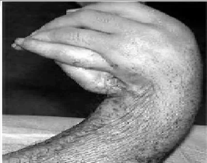
A 24-year-old man presents with a short forearm and a hand deformity. The patient is otherwise healthy with no other congenital defects. The clinical appearance of his forearm is shown (Slide). Your diagnosis is:
Explanation
Question 31
A 24-year-old man presents with a short forearm and a hand deformity. The patient is otherwise healthy with no other congenital defects. The clinical appearance of his forearm is shown (Slide). The patient has an elbow flexion contracture of 70° and desires lengthening. Which of the following statements is not true regarding lengthening:
Explanation

Question 32
A 24-year-old man presents with a short forearm and a hand deformity. The patient is otherwise healthy with no other congenital defects. The clinical appearance of his forearm is shown (Slide). The potential complications of lengthening are discussed, and the patient is advised against it. However, the elbow flexion contracture is corrected by gradual distraction. One year postoperatively, the patient has attained a 30° correction of the flexion deformity, which remains mobile. Now, he desires that his wrist deformity be corrected. The procedure of choice is:
Explanation
 Wrist arthrodesis is the best solution for this patient and his recurrent deformity because it provides a stable platform for grasp.C orrect Answer: Arthrodesis
Question 33
A 24-year-old man presents with a short forearm and a hand deformity. The patient is otherwise healthy with no other congenital defects. The clinical appearance of his forearm is shown (Slide). Although the patient has a thumb, it is in an abnormal position. Any attempt to make his thumb more functional will be influenced by:
Explanation
 The pattern of usage of the hand is established in the brain by 2 to 3 years of age. Although pollicization has been performed in adolescents, patients continue to prefer a scissor pinch. At 24 years of age, this pattern will be well established. The patient can be coaxed to use his thumb, but it will not be involuntary and automatic.C orrect Answer: Presence of a side-to-side finger grip
Question 34
A radial club hand is the result of an insult during which phase of the gestation period:
Explanation
 A radial club hand is the result of an insult during weeks 4 to 7 of gestation.
Question 35
A 15-day-old boy presents with deformity of the right hand. The boy was delivered prematurely and underwent an urgent arterial switch for transposition of great vessels. The patient is in stable condition. He has a radial club hand, and because the radial head cannot be palpated, total absence of radius is suspected. The thumb is absent and the index finger has camptodactyly. The forearm is short compared to the left side, and the patient flexes his elbow upon stimulation. Spontaneous finger motion is also present. A thorough physical examination is performed and a set of investigations is ordered. The results are as follows: complete blood count 10,000 mcu/L; platelet 254 254×103 mcu/L; neutophils 50%; Hb 14.2 mg/dL; lymphocytes 40%; Hct 45; and monocytes 10%. No renal abnormalities were noted on ultrasonogram of the abdomen. A radiograph of the spine is normal. Diagnosis is:
Explanation
Question 36
The principal abnormality associated with Holt-Oram syndrome is:
Explanation
Question 37
The hereditary pattern for Holt-Oram syndrome is:
Explanation
Question 38
A 15-day-old boy presents with deformity of the right hand. The boy was delivered prematurely and underwent an urgent arterial switch for transposition of great vessels. The patient is in stable condition. He has a radial club hand, and because the radial head cannot be palpated, total absence of radius is suspected. The thumb is absent and the index finger has camptodactyly. The forearm is short compared to the left side, and the patient flexes his elbow upon stimulation. Spontaneous finger motion is also present. A thorough physical examination is performed and a set of investigations is ordered. The results are as follows: complete blood count 10,000 mcu/L; 254×103 mcu/L; neutophils 50%; Hb 14.2 mg/dL; lymphocytes 40%; Hct 45; and monocytes 10%. No renal abnormalities were noted on ultrasonogram of the abdomen. A radiograph of the spine is normal. The next step in the management of the radial club hand is:
Explanation
Question 39
A 15-day-old boy presents with deformity of the right hand. The boy was delivered prematurely and underwent an urgent arterial switch for transposition of great vessels. The patient is in stable condition. He has a radial club hand, and because the radial head cannot be palpated, total absence of radius is suspected. The thumb is absent and the index finger has camptodactyly. The forearm is short compared to the left side, and the patient flexes his elbow upon stimulation. Spontaneous finger motion is also present. A thorough physical examination is performed and a set of investigations is ordered. The results are as follows: complete blood count 10,000 mcu/L; platelet 254×103 mcu/L; neutophils 50%; Hb 14.2 mg/dL; lymphocytes 40%; Hct 45; and monocytes 10%. No renal abnormalities were noted on ultrasonogram of the abdomen. A radiograph of the spine is normal. Centralization will be performed on the patient. All of the following statements are true about centralization except:
Explanation
Question 40
A 15-day-old boy presents with deformity of the right hand. The boy was delivered prematurely and underwent an urgent arterial switch for transposition of great vessels. The patient is in stable condition. He has a radial club hand, and because the radial head cannot be palpated, total absence of radius is suspected. The thumb is absent and the index finger has camptodactyly. The forearm is short compared to the left side, and the patient flexes his elbow upon stimulation. Spontaneous finger motion is also present. A thorough physical examination is performed and a set of investigations is ordered. The results are as follows: complete blood count 10,000 mcu/L; 254 103 mcu/L; neutophils 50%; Hb 14.2 mg/dL; lymphocytes 40%; Hct 45; and monocytes 10%. No renal abnormalities were noted on ultrasonogram of the abdomen. A radiograph of the spine is normal. When the patient is 10 years old, he is not satisfied with the length of his forearm and wishes to lengthen it. Which of the following is not a satisfactory recommendation:
Explanation
Question 41
Which of the following conditions is present in patients with radial club hand but not in patients with ulnar club hand:
Explanation
Question 42
All of the following developmental anomalies are associated with ulnar club hand except:
Explanation
Question 43
Which of the following syndromes is associated with ulnar club hand:
Explanation
Question 44
Which of the following areas is not involved in ulnar club hand:
Explanation
Question 45
All of the following are true statements regarding elbow involvement in ulnar club hand except:
Explanation
Question 46
All of the following statements are true regarding the carpal bones in patients with ulnar club hand except:
Explanation
Question 47
All of the following anomalies are present in patients with ulnar club hand except:
Explanation
Question 48
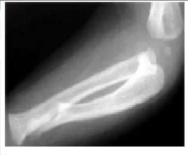
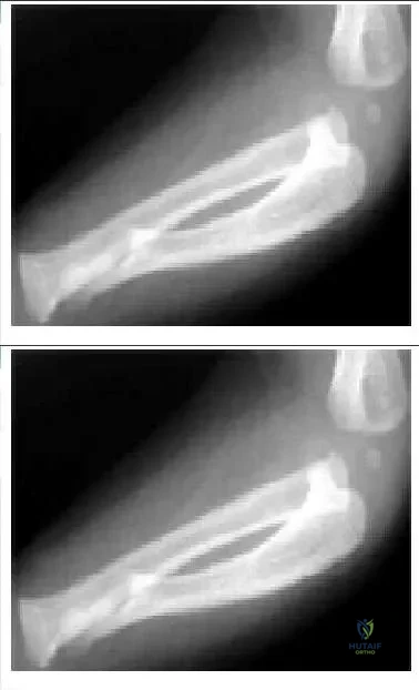
A 1-year-old boy was born full-term and pregnancy was uneventful. However, the parents noticed deformity of the patientâ s forearm, which progressed with growth. The parents consulted a pediatric orthopaedic surgeon 4 months prior and were advised to observe the growth. Multiple investigations in the form of two-dimensional echogram, abdomen ultrasonography, radiographs of the spine, and complete blood work did not reveal any abnormalities. No genetic or syndromic abnormality was reported. A radiograph taken 4 months prior is shown.
Explanation
The ulnar deficiency is longitudinal and the ulna is considered a postaxial bone. Ulnar agenesis means absence while radial club hand is a pre-axial longitudinal deficiency and cleft hand is a central deficiency.
Question 49
A 45-year-old man presents with marked lateral elbow pain. He says that the pain has been present for 3 weeks. He has no history of recent trauma. He is an avid tennis player, and he feels increased pain after playing tennis and when doing wrist extension exercises in the gym. His pain is maximally reproduced with resisted middle finger extension and with forearm supination with the elbow extended. Electromyography would confirm the diagnosis as:
Explanation
Question 50
A 45-year-old man presents with marked lateral elbow pain. He says that the pain has been present for 3 weeks. He has no history of recent trauma. He is an avid tennis player, and he feels increased pain after playing tennis and when doing wrist extension exercises in the gym. His pain is maximally reproduced with resisted middle finger extension and with forearm supination with the elbow extended. Which of the following is the appropriate initial treatment:
Explanation
Question 51
A 24-year-old motorcyclist sustains a traction injury to his right brachial plexus. Exam shows absent shoulder abduction, elbow flexion, and wrist extension. Sensation is absent in the C5, C6, and C7 dermatomes. Sensory nerve action potentials (SNAPs) for the median and radial nerves are normal in the right upper extremity. What does this indicate?
Explanation
Question 52
A 32-year-old woman presents with knee pain. Radiographs show an eccentric, lytic epiphyseal lesion in the distal femur. Biopsy reveals mononuclear cells and multinucleated giant cells.
What is the most appropriate definitive management?
Explanation
Question 53
A 4-month-old girl is placed in a Pavlik harness for developmental dysplasia of the hip. At a follow-up visit 2 weeks later, the parents report the infant has stopped kicking her left leg. On exam, she lacks active knee extension on the left. What is the most likely cause?
Explanation
Question 54
A 45-year-old male is brought to the ED after a crush injury. He is hemodynamically unstable with a blood pressure of 75/40 mmHg. Radiographs show an anteroposterior compression (APC) type III pelvic ring injury. A pelvic binder is applied. To maximize biomechanical efficacy, the binder should be centered over which anatomic landmark?
Explanation
Question 55
A 13-year-old gymnast presents with progressive lower back pain. Radiographs reveal a grade III L5-S1 isthmic spondylolisthesis. She has failed conservative management and reports radicular pain in the L5 distribution. What is the recommended surgical intervention?
Explanation
Question 56
A 65-year-old man presents with groin pain 8 years after a metal-on-metal total hip arthroplasty. Aspiration yields cloudy fluid with negative cultures.
Histology of the periprosthetic tissue is most likely to show:
Explanation
Question 57
During the surgical repair of a "terrible triad" injury of the elbow, which of the following sequences is the standard algorithm for reconstruction?
Explanation
Question 58
A patient presents with inability to actively flex the distal interphalangeal joint of the index finger and the interphalangeal joint of the thumb. To differentiate Anterior Interosseous Nerve (AIN) syndrome from flexor tendon rupture, which finding is pathognomonic for AIN syndrome?
Explanation
Question 59
A 13-year-old obese boy presents with a 3-week history of right groin pain and a limp. He walks with an externally rotated foot. AP and frog-leg lateral pelvis radiographs confirm a mild stable slipped capital femoral epiphysis (SCFE) on the right. What is the most appropriate management?
Explanation
Question 60
Bone morphogenetic proteins (BMPs) are used to enhance bone healing. Which of the following BMPs is an FDA-approved osteoinductive agent commonly used in anterior lumbar interbody fusion?
Explanation
Question 61
A 25-year-old overhead athlete presents with deep shoulder pain. MR arthrogram reveals a type II SLAP tear. During arthroscopy, a "peel-back" lesion is noted. This mechanism primarily involves which force applied to the biceps anchor during the late cocking phase of throwing?
Explanation
Question 62
A 70-year-old woman undergoes a right total knee arthroplasty. During the trial phase, the knee is stable in extension but opens symmetrically on the medial and lateral sides by 4 mm in flexion. What is the most appropriate surgical adjustment?
Explanation
Question 63
A 62-year-old man presents with dropping objects and a stiff gait. Exam shows hyperreflexia in the lower extremities and a positive Hoffmann sign.
T2-weighted MRI of the cervical spine is most likely to demonstrate which of the following?
Explanation
Question 64
A 25-year-old male presents with a complete C5-C6 root avulsion following a motorcycle accident 3 months ago. Clinical examination demonstrates absent elbow flexion and shoulder abduction, with preserved hand function. What is the most appropriate nerve transfer to restore active elbow flexion in this patient?
Explanation
Question 65
A 6-week-old infant is being treated with a Pavlik harness for developmental dysplasia of the hip. At the 2-week follow-up, the mother reports the infant has stopped kicking the affected leg. On examination, active knee extension is absent but ankle movements are preserved. What is the most likely cause of this complication?
Explanation
Question 66
An 8-year-old child sustains a displaced extension-type supracondylar fracture of the humerus. After closed reduction and percutaneous pinning, examination reveals an inability to flex the interphalangeal joint of the thumb and the distal interphalangeal joint of the index finger. Which nerve structure is most likely injured?
Explanation
Question 67
A 22-year-old man presents with wrist pain after a fall on an outstretched hand. Radiographs confirm a displaced fracture of the proximal pole of the scaphoid. The high risk of avascular necrosis and nonunion in this region is primarily due to the blood supply originating from branches of the:
Explanation
Question 68
A 45-year-old man presents with acute onset of severe lower back pain, bilateral sciatica, saddle anesthesia, and urinary retention following a heavy lifting episode. MRI reveals a massive L4-L5 central disc herniation. What is the most critical next step in management?
Explanation
Question 69
A 12-year-old boy presents with progressive knee pain and swelling. Radiographs show a destructive, permeative metaphyseal lesion in the distal femur with a "sunburst" periosteal reaction and Codman's triangle. Histological examination reveals malignant spindle cells producing osteoid matrix. What is the most likely diagnosis?
Explanation
Question 70
During a posterior approach for a total hip arthroplasty, the surgeon meticulously repairs the short external rotators and the posterior capsule to the greater trochanter. This specific closure is primarily performed to reduce the risk of which postoperative complication?
Explanation
Question 71
An 80-year-old female sustains a fall and complains of severe left hip pain. Radiographs reveal an intracapsular femoral neck fracture with complete displacement and no continuity of the trabecular lines. According to the Garden classification, this represents:
Explanation
Question 72
When comparing bone-patellar tendon-bone (BPTB) autograft to hamstring autograft for anterior cruciate ligament (ACL) reconstruction, which of the following is a recognized disadvantage of the BPTB graft?
Explanation
Question 73
A 55-year-old woman presents with advanced, neglected carpal tunnel syndrome, exhibiting severe thenar atrophy. Which specific intrinsic hand muscle is most classically atrophied and visible in this condition?
Explanation
Question 74
A 13-year-old overweight boy presents with a limp and right groin pain for 3 weeks. On physical examination, as the right hip is passively flexed, the limb obligatorily rotates externally. What is the most likely diagnosis?
Explanation
Question 75
A 28-year-old male sustains a closed comminuted tibial shaft fracture. Twelve hours post-injury, he develops severe pain out of proportion to the injury, pain with passive stretch of his toes, and a tense calf. What is the generally accepted threshold for intracompartmental pressure relative to diastolic blood pressure (Delta P) indicating the need for an emergent fasciotomy?
Explanation
Question 76
In the process of secondary bone healing, which physiological phase is characterized by the replacement of the cartilaginous soft callus with woven bone through endochondral ossification?
Explanation
Question 77
During a total knee arthroplasty, the surgeon performs gap assessment and notes that the knee is tight in flexion but perfectly balanced in extension. Which of the following surgical adjustments is most appropriate to specifically increase the flexion gap?
Explanation
Question 78
A 15-year-old gymnast presents with lower back pain exacerbated by spinal extension. Radiographs show a bilateral defect in the pars interarticularis of L5 with a 25% anterior translation of L5 over S1. Neurological examination is normal. What is the most appropriate initial management?
Explanation
Question 79
A 35-year-old man feels a sudden "pop" in his posterior ankle while playing basketball. He has a positive Thompson test on examination. Which specific clinical finding constitutes a positive Thompson test?
Explanation
Question 80
Articular cartilage derives its unique resilience and capacity to resist extreme compressive forces primarily from which of the following molecular interactions?
Explanation
Question 81
Review the clinical image
. A 40-year-old male arrives in the trauma bay in hemorrhagic shock following a high-energy crush injury. The AP radiograph demonstrates an "open book" pelvic fracture with wide symphyseal diastasis. What is the most critical initial orthopaedic intervention to control hemodynamics?
Explanation
Question 82
In the Ponseti method for the conservative management of idiopathic clubfoot, the sequence of deformity correction is strictly protocolized. Which of the following represents the correct order of correction?
Explanation
Question 83
A 60-year-old man presents with progressive deep pelvic pain. Radiographs reveal a large, lobulated lytic lesion in the ilium with "popcorn" stippled calcifications. Biopsy demonstrates a hyaline cartilage matrix with atypical chondrocytes. What is the mainstay of treatment for conventional low-grade chondrosarcoma of the pelvis?
Explanation
Question 84
A 4-month-old infant with obstetric brachial plexus palsy presents with an internal rotation contracture of the shoulder and complete absence of active biceps function. Wrist extension is also absent. What is the most appropriate next step in management?
Explanation
Question 85
A 25-year-old man suffered a C5-C6 root avulsion injury resulting in a permanent loss of elbow flexion. Hand and wrist functions remain entirely normal. A Steindler flexorplasty is planned to restore active elbow flexion. Which of the following anatomical structures is transferred during this procedure?
Explanation
Question 86
A 40-year-old male presents with sudden, severe, unprovoked right shoulder pain lasting for 2 weeks. As the pain subsides, he notices profound weakness in shoulder abduction and external rotation. EMG demonstrates denervation potentials in the supraspinatus and infraspinatus muscles. What is the most likely diagnosis?
Explanation
Question 87
During the clinical and electrodiagnostic evaluation of a patient with a traumatic closed brachial plexus injury, which of the following findings is most strongly indicative of a preganglionic root avulsion rather than a postganglionic lesion?
Explanation
Question 88
A 30-year-old man sustains a closed midshaft humeral fracture after a fall.
Upon initial clinical examination in the emergency department, he exhibits an inability to actively extend his wrist and metacarpophalangeal joints. Sensation is decreased over the dorsal first web space. What is the most appropriate initial management for this neurological deficit?
Explanation
Question 89
A newborn infant presents with a claw hand deformity, an absent grasp reflex, but a completely preserved Moro reflex in the upper arm and shoulder. Examination also reveals mild ptosis and miosis on the ipsilateral side. Which nerve roots of the brachial plexus are predominantly involved in this injury?
Explanation
Question 90
A 28-year-old female overhead athlete complains of vague pain, numbness, and tingling in her medial forearm and fourth and fifth digits. Symptoms are exacerbated by overhead activities. Provocative maneuvers such as the Roos stress test and Adson's test are positive. Which anatomical structure is most frequently responsible for this specific pattern of neural compression?
Explanation
Question 91
A 22-year-old patient with an upper trunk (C5-C6) brachial plexus injury is scheduled for an Oberlin transfer to restore elbow flexion, as his lower trunk function is fully intact. Which of the following best describes the standard surgical technique for an Oberlin transfer?
Explanation
Question 92
A 32-year-old professional volleyball player presents with an insidious onset of posterior shoulder pain and progressive weakness in external rotation. Clinical examination shows isolated atrophy of the infraspinatus muscle with a completely normal supraspinatus muscle bulk and strength. Where is the most likely site of nerve compression?
Explanation
Question 93
A 22-year-old male is evaluated 6 months after sustaining a massive upper extremity crush injury. He has intact shoulder abduction and elbow flexion but cannot actively extend his wrist or fingers.
EMG demonstrates absent motor units in the extensor carpi radialis brevis (ECRB) and extensor digitorum communis (EDC) with no signs of reinnervation. Tendon transfers are planned. Which of the following is the most standard and reliable tendon transfer to restore active wrist extension?
Explanation
None
