Orthopedic Prometric Exam Preparation MCQs - Part 10

Orthopedic Prometric Exam Preparation MCQs - Part 10
Comprehensive 100-Question Exam
00:00
Start Quiz
Question 1
Which digit is most commonly affected by macrodactyly:
Explanation
Question 2
Syndactyly is present in what percentage of patients with macrodactyly:
Explanation
Question 3
Macrodactyly that is present at birth is termed:
Explanation
Question 4
The most accepted theory for the cause of macrodactyly is:
Explanation
Question 5
Syndromes that may be associated with macrodactyly include:
Explanation
Question 6
Macrodactyly affects:
Explanation
Question 7
A 2-year-old child is brought to your office for evaluation of a "big hand." Upon examination, you notice that the child has mildly enlarged ring and small fingers. There is full range of motion without instability. After examination of the patient, you recommend:
Explanation
Question 8
A 2-year-old child is brought to your office for evaluation of a "big hand." Upon examination, you notice that the child has mildly enlarged ring and small fingers. There is full range of motion without instability. The childâ s parents inform you that they would like you to amputate the affected digits as soon as possible. You should:
Explanation
Question 9
You discover that a patient who you have been treating for macrodactyly has been followed by the Proteus Syndrome Foundation. Exhaustive work-up has been completed and radiographs of the hand reveal:
Explanation
Question 10
A 2-year-old child is brought to your office for evaluation of a "big hand." Upon examination, you notice that the child has mildly enlarged ring and small fingers. There is full range of motion without instability. After examination of the patient, you discuss the diagnosis of macrodactyly with the parents. The parents feel assured after your discussion of the disease process and your review of the radiographs. You should next see the patient:
Explanation
Question 11
Which of the following is not a complication of macrodactyly surgery:
Explanation
Question 12
Epiphysiodesis for macrodactyly should be performed at the following location:
Explanation
Question 13
When ruptured, which portion of the scapholunate ligament leads to scaphoid-lunate diastasis:
Explanation
Question 14
Which of the following radiographic views is not routinely used to diagnose scapholunate injury:
Explanation
Question 15
Which of the following treatments is not used for acute scapholunate ligament ruptures:
Explanation
Question 16
Which of the following is considered indicative of a scaphoid-lunate ligament tear on posteroanterior radiograph:
Explanation
Question 17
The Terry Thomas sign, which is considered indicative of scaphoid-lunate ligament rupture, is best described as:
Explanation
Question 18
The most important requirement for a diagnostic magnetic resonance image (MRI) study in cases of scaphoid-lunate ligament injury is:
Explanation
Question 19
In cases of subacute scaphoid-lunate ligament injury with no arthrosis, all of the following are acceptable options except:
Explanation
Question 20
Mallet finger injuries refer to:
Explanation
Question 21
In mallet finger injuries, the distal phalanx posture is:
Explanation
Question 22
Mallet finger injuries are typically:
Explanation
Question 23
Treatment of a type I mallet finger is typically closed. This involves:
Explanation
Question 24
Type I mallet finger injuries must be immobilized constantly for a minimum of:
Explanation
Question 25
The most common mallet finger injuries are:
Explanation
Question 26
On physical examination, a mallet finger assumes a:
Explanation
Question 27
The following mallet finger injuries always require tendon repair:
Explanation
Question 28
After placing a type I mallet finger in a splint at the initial visit, next follow- up should be:
Explanation
Question 29
The most common bone tumor of the upper extremity is:
Explanation
Question 30
The most common benign bone tumor of the hand is:
Explanation
Question 31
Osteochondromas are benign but can have a malignant transformation in which of the following cases:
Explanation
Question 32
The risk of malignant transformation in patients with multiple hereditary exostoses is:
Explanation
Question 33
Recurrence of osteochondroma is likely if:
Explanation
Question 34
Malignant transformation of osteochondroma commonly occurs to:
Explanation
Question 35
All of the following suggest a possibility of malignant transformation in multiple hereditary exostoses except:
Explanation
Question 36
Enchondromas are commonly involved in which of the following sites:
Explanation
Question 37
The most common forearm deformity in patients with hereditary multiple osteochondromatosis is:
Explanation
Question 38
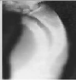
A 12-year-old boy is brought to the clinic by his concerned parents. The boyâ s forearm is bowed, and his parents are confused as to the possible diagnosis and treatment options. You notice that the right forearm of the child is bowed ulnarwards and is shorter compared to the left forearm. The pronosupination is markedly decreased on the right side but is also limited on the left side. The patient has a good grip, pinch, and grasp. He is neurologically intact as well. The parents say that they first noticed the deformity around 6 or 7 years ago, and the mother informs you that she had noticed a hard bump on the forearm. She has recently noticed another bump on his right leg. The child does not complain of pain and is using both of his hands quite well. The parents were informed by a previous physician that the child has Madelungâ s deformity and are concerned that the disease is now involving other areas of his body. You order a radiograph of the patientâ s forearm. The anteroposterior radiograph is shown (Slide). The next step is to order a:
Explanation
Question 39
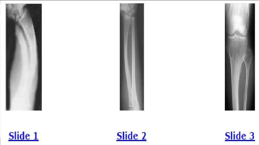
A 12-year-old boy is brought to the clinic by his concerned parents. The boyâ s forearm is bowed, and his parents are confused as to the possible diagnosis and treatment options. You notice that the right forearm of the child is bowed ulnarwards and is shorter compared to the left forearm. The pronosupination is markedly decreased on the right side but is also limited on the left side. The patient has a good grip, pinch, and grasp. He is neurologically intact as well. The parents say that they first noticed the deformity around 6 or 7 years ago, and the mother informs you that she had noticed a hard bump on the forearm. She has recently noticed another bump on his right leg. The child does not complain of pain and is using both of his hands quite well. The parents were informed by a previous physician that the child has Madelungs deformity and are concerned that the disease is now involving other areas of his body. An immediate appointment for magnetic resonance imagine (MRI) and computed tomography (C T) scan are not available, and a genetic evaluation has been carried out previously. As you await the report from the geneticist office, you decide to get a skeletal radiograph series on the patient. The radiograph of the opposite forearm (Slide 1) and right leg are shown (Slide 2). You order a radiograph of the forearm. The anteroposterior radiograph is shown (Slide 3). Your suspected diagnosis is:

Explanation
Question 40
A 12-year-old boy is brought to the clinic by his concerned parents. The boys forearm is bowed, and his parents are confused as to the possible diagnosis and treatment options. You notice that the right forearm of the child is bowed ulnarwards and is shorter compared to the left forearm. The pronosupination is markedly decreased on the right side but is also limited on the left side. The patient has a good grip, pinch, and grasp. He is neurologically intact as well. The parents say that they first noticed the deformity around 6 or 7 years ago, and the mother informs you that she had noticed a hard bump on the forearm. She has recently noticed another bump on his right leg. The child does not complain of pain and is using both of his hands quite well. The parents were informed by a previous physician that the child has Madelungs deformity and are concerned that the disease is now involving other areas of his body. You order a radiograph of the forearm. The anteroposterior radiograph is shown (Slide 1). The childâ s skeletal radiograph survey is also presented (Slide 2 and Slide 3). The genetic pattern seen in patients with this type of presentation is:
Explanation
Question 41
A 12-year-old boy is brought to the clinic by his concerned parents. The boy s forearm is bowed, and his parents are confused as to the possible diagnosis and treatment options. You notice that the right forearm of the child is bowed ulnarwards and is shorter compared to the left forearm. The pronosupination is markedly decreased on the right side but is also limited on the left side. The patient has a good grip, pinch, and grasp. He is neurologically intact as well. The parents say that they first noticed the deformity around 6 or 7 years ago, and the mother informs you that she had noticed a hard bump on the forearm. She has recently noticed another bump on his right leg. The child does not complain of pain and is using both of his hands quite well. The parents were informed by a previous physician that the child has Madelungs deformity and are concerned that the disease is now involving other areas of his body. You order a radiograph of the forearm. The anteroposterior radiograph is shown (Slide 1). The childâ s skeletal radiograph survey is also presented (Slide 2 and Slide 3). Which of the following areas is unlikely to be involved:
Explanation
Question 42
A 12-year-old boy is brought to the clinic by his concerned parents. The boyâ s forearm is bowed, and his parents are confused as to the possible diagnosis and treatment options. You notice that the right forearm of the child is bowed ulnarwards and is shorter compared to the left forearm. The pronosupination is markedly decreased on the right side but is also limited on the left side. The patient has a good grip, pinch, and grasp. He is neurologically intact as well. The parents say that they first noticed the deformity around 6 or 7 years ago, and the mother informs you that she had noticed a hard bump on the forearm. She has recently noticed another bump on his right leg. The child does not complain of pain and is using both of his hands quite well. The parents were informed by a previous physician that the child has Madelungâ s deformity and are concerned that the disease is now involving other areas of his body. You order a radiograph of the forearm. The anteroposterior radiograph is shown (Slide 1). The childâ s skeletal radiograph survey is also presented (Slide 2 and Slide 3). The chance of hand involvement in this child is:
Explanation
Question 43
A 12-year-old boy is brought to the clinic by his concerned parents. The boyâ s forearm is bowed, and his parents are confused as to the possible diagnosis and treatment options. You notice that the right forearm of the child is bowed ulnarwards and is shorter compared to the left forearm. The pronosupination is markedly decreased on the right side but is also limited on the left side. The patient has a good grip, pinch, and grasp. He is neurologically intact as well. The parents say that they first noticed the deformity around 6 or 7 years ago, and the mother informs you that she had noticed a hard bump on the forearm. She has recently noticed another bump on his right leg. The child does not complain of pain and is using both of his hands quite well. The parents were informed by a previous physician that the child has Madelungs deformity and are concerned that the disease is now involving other areas of his body. You order a radiograph of the forearm. The anteroposterior radiograph is shown (Slide 1). The childâ s skeletal radiograph survey is also presented (Slide 2 and Slide 3). The most likely complication in this child is:

Explanation
Question 44
A 12-year-old boy is brought to the clinic by his concerned parents. The boys forearm is bowed, and his parents are confused as to the possible diagnosis and treatment options. You notice that the right forearm of the child is bowed ulnarwards and is shorter compared to the left forearm. The pronosupination is markedly decreased on the right side but is also limited on the left side. The patient has a good grip, pinch, and grasp. He is neurologically intact as well. The parents say that they first noticed the deformity around 6 or 7 years ago, and the mother informs you that she had noticed a hard bump on the forearm. She has recently noticed another bump on his right leg. The child does not complain of pain and is using both of his hands quite well. The parents were informed by a previous physician that the child has Madelungs deformity and are concerned that the disease is now involving other areas of his body. You order a radiograph of the forearm. The anteroposterior radiograph is shown (Slide 1). The childâ s skeletal radiograph survey is also presented (Slide 2 and Slide 3). The difference between Madelungâ s deformity and this boyâ s condition is:

Explanation
Question 45
A 12-year-old boy is brought to the clinic by his concerned parents. The boys forearm is bowed, and his parents are confused as to the possible diagnosis and treatment options. You notice that the right forearm of the child is bowed ulnarwards and is shorter compared to the left forearm. The pronosupination is markedly decreased on the right side but is also limited on the left side. The patient has a good grip, pinch, and grasp. He is neurologically intact as well. The parents say that they first noticed the deformity around 6 or 7 years ago, and the mother informs you that she had noticed a hard bump on the forearm. She has recently noticed another bump on his right leg. The child does not complain of pain and is using both of his hands quite well. The parents were informed by a previous physician that the child has Madelungs deformity and are concerned that the disease is now involving other areas of his body. You order a radiograph of the forearm. The anteroposterior radiograph is shown (Slide 1). The childâ s skeletal radiograph survey is also presented (Slide 2 and Slide 3). All of the following are acceptable options, either alone or in combination, for management of this childâ s condition, except:

Explanation
Question 46
A 12-year-old boy is brought to the clinic by his concerned parents. The boys forearm is bowed, and his parents are confused as to the possible diagnosis and treatment options. You notice that the right forearm of the child is bowed ulnarwards and is shorter compared to the left forearm. The pronosupination is markedly decreased on the right side but is also limited on the left side. The patient has a good grip, pinch, and grasp. He is neurologically intact as well. The parents say that they first noticed the deformity around 6 or 7 years ago, and the mother informs you that she had noticed a hard bump on the forearm. She has recently noticed another bump on his right leg. The child does not complain of pain and is using both of his hands quite well. The parents were informed by a previous physician that the child has Madelungs deformity and are concerned that the disease is now involving other areas of his body. You order a radiograph of the forearm. The anteroposterior radiograph is shown (Slide 1). The childâ s skeletal radiograph survey is also presented (Slide 2 and Slide 3). Which of the following is not true regarding the possibility of malignant degeneration in this child:

Explanation
Question 47
Horners syndrome includes all of the following except:
Explanation
Question 48
Axonotmesis involves injury to the:
Explanation
Question 49
All of the following may be seen with preganglionic lesion except:
Explanation
Question 50
Weakness is not seen with root avulsion in the:
Explanation
Question 51
A 1-year-old child presents with simple syndactyly of the middle and ring fingers. What is the most appropriate timing and rationale for surgical release?
Explanation
Question 52
A 25-year-old male sustains a displaced basicervical femoral neck fracture. Biomechanically, which of the following is the most appropriate fixation method?
Explanation
Question 53
During a physical examination of a patient with a suspected anterior cruciate ligament (ACL) injury, a positive pivot shift test is elicited. This test primarily evaluates the competency of which functional bundle of the ACL?
Explanation
Question 54
Denosumab is often utilized in the management of unresectable Giant Cell Tumor of bone. What is the specific mechanism of action of this medication?
Explanation
Question 55
A 45-year-old man presents to the emergency department with acute saddle anesthesia, bilateral radiculopathy, and urinary retention secondary to a massive L4-L5 disc herniation. Current literature suggests that decompression within what time frame from the onset of symptoms provides the most significant improvement in urologic outcomes?
Explanation
Question 56
An infant with Developmental Dysplasia of the Hip (DDH) is being treated with a Pavlik harness. If the anterior straps are adjusted to place the hips in excessive hyperflexion (greater than 120 degrees), the child is at highest risk for developing which of the following complications?
Explanation
Question 57
The Martin-Gruber anastomosis is a well-described anatomical variant in the upper extremity. It involves the anomalous crossing of nerve fibers in the forearm from the:
Explanation
Question 58
A 35-year-old construction worker falls from a height and sustains a closed, highly comminuted, intra-articular calcaneus fracture. On examination in the ED, he has massive swelling and severe fracture blisters over the lateral hindfoot. What is the most appropriate initial management?
Explanation
Question 59
Which of the following transcription factors is considered the essential "master regulator" for the differentiation of mesenchymal stem cells into osteoblasts?
Explanation
Question 60
A 30-year-old male sustains an anteroposterior compression (APC) Type III pelvic ring injury following a high-speed motor vehicle collision. He is hemodynamically unstable. The primary source of life-threatening retroperitoneal hemorrhage in pelvic fractures is most commonly from:
Explanation
Question 61
A patient who underwent a metal-on-metal total hip arthroplasty three years ago presents with groin pain and swelling. MRI reveals a large pseudotumor. Histology shows an aseptic lymphocyte-dominated vasculitis-associated lesion (ALVAL). This reaction is classically described as which type of hypersensitivity?
Explanation
Question 62
A 24-year-old female sustains a Levine-Edwards Type IIA traumatic spondylolisthesis of the axis (Hangman's fracture). What is the mechanism of injury, and what is a critical consideration in her management?
Explanation
Question 63
In a child diagnosed with Legg-Calvé-Perthes disease (idiopathic avascular necrosis of the proximal femoral epiphysis), what is the single most significant independent prognostic factor for developing premature hip osteoarthritis in adulthood?
Explanation
Question 64
When evaluating a patient with recurrent anterior shoulder instability, what specifically defines an "off-track" Hill-Sachs lesion?
Explanation
Question 65
A 25-year-old male presents with knee pain. Radiographs show an eccentric, expansile, purely lytic lesion in the distal femoral epiphysis. Biopsy reveals multinucleated giant cells. Which genetic mutation is highly sensitive and specific for this tumor?
Explanation
Question 66
A newborn presents with radial longitudinal deficiency (radial club hand). If Holt-Oram syndrome is suspected as the underlying genetic etiology, what is the most common associated congenital cardiac anomaly?
Explanation
Question 67
In an APC-II (Anteroposterior Compression type II) pelvic ring injury, which of the following ligaments remains intact, thereby preventing complete vertical instability?
Explanation
Question 68
Which of the following clinical or pathologic findings represents the most significant adverse prognostic factor for overall survival in a 16-year-old patient diagnosed with high-grade intramedullary osteosarcoma of the distal femur?
Explanation
Question 69
A 65-year-old male presents with deteriorating fine motor skills, gait instability, and bilateral Hoffman's signs. MRI demonstrates cervical spondylotic myelopathy. Which specific MRI finding is considered the most reliable indicator of a poor postoperative neurological prognosis?
Explanation
Question 70
In modern total hip arthroplasty (THA), the selection of a ceramic-on-ceramic bearing surface is uniquely associated with which of the following postoperative complications?
Explanation
Question 71
During an anterior cruciate ligament (ACL) reconstruction using a bone-patellar tendon-bone autograft, a resident places the femoral tunnel too anteriorly. What classical clinical finding will be observed during intraoperative graft tensioning?
Explanation
Question 72
A 45-year-old male sustains a vertically oriented, displaced femoral neck fracture (Pauwels Type III). What is the primary biomechanical rationale for augmenting standard inverted-triangle cannulated screw fixation with a medially placed anti-glide plate?
Explanation
Question 73
A 4-month-old infant is undergoing closed reduction for developmental dysplasia of the hip (DDH). The intraoperative arthrogram demonstrates medial pooling of dye and a failure to achieve concentric reduction. Which anatomic structure most commonly blocks closed reduction extracapsularly?
Explanation
Question 74
During fracture healing via endochondral ossification, the production of Type X collagen in the fracture callus is primarily mediated by which of the following cell types?
Explanation
Question 75
A 25-year-old male falls on an outstretched hand and sustains a proximal pole scaphoid fracture. What is the major arterial supply to the scaphoid that makes this specific fracture pattern highly prone to avascular necrosis?
Explanation
Question 76
A 22-year-old football player sustains a high-energy midfoot injury. Radiographs show widening between the 1st and 2nd metatarsal bases.
Which ligamentous connection is anatomically disrupted in a classic Lisfranc injury?
Explanation
Question 77
A 13-year-old obese boy presents with left thigh pain and an obligatory external rotation of the hip during active flexion. A diagnosis of Slipped Capital Femoral Epiphysis (SCFE) is made. What is the most devastating complication associated with forceful closed reduction of a displaced SCFE?
Explanation
Question 78
A 30-year-old male with a closed tibial shaft fracture develops disproportionate pain out of proportion to the injury. He complains of severe paresthesias in the first dorsal web space of the foot. Which fascial compartment of the leg is most likely experiencing critically elevated tissue pressures?
Explanation
Question 79
Five years following a primary total knee arthroplasty, a patient presents with pain and progressive varus deformity. Radiographs reveal focal osteolysis around the medial tibial plateau without systemic signs of infection.
What is the predominant cell type mediating this periprosthetic osteolysis?
Explanation
Question 80
A 40-year-old falls from a height and sustains an L1 thoracolumbar burst fracture. Which of the following criteria most strongly mandates surgical stabilization rather than conservative management with a TLSO brace?
Explanation
Question 81
A 24-year-old overhead throwing athlete presents with posterior shoulder pain. Physical examination reveals a positive active compression test (O'Brien test) that elicits deep joint pain with the forearm pronated, which is relieved with the forearm in supination. What is the most likely associated finding on MR arthrography?
Explanation
Question 82
According to the Sunderland classification of peripheral nerve injury, which of the following histological descriptions correctly defines a fourth-degree nerve injury?
Explanation
Question 83
A newborn presents with an absent radius and an absent thumb. Which of the following tests is most appropriate to rule out a life-threatening associated condition?
Explanation
Question 84
A 12-year-old boy presents with a pathologic fracture through a radiolucent lesion in the proximal humerus.
The "fallen leaf" sign is seen. What is the most appropriate initial management after the fracture has healed?
Explanation
Question 85
According to the Wassel classification of thumb polydactyly, which type is the most common and involves duplication at the metacarpophalangeal joint?
Explanation
Question 86
In a patient with a displaced midshaft clavicle fracture, which of the following is considered an absolute indication for operative fixation?
Explanation
Question 87
A 35-year-old laborer sustains a severe laceration to his index finger requiring flexor tendon repair.
Which of the following pulley combinations is most critical to preserve or reconstruct to prevent biomechanical bowstringing?
Explanation
Question 88
A 65-year-old diabetic patient presents with severe back pain and elevated inflammatory markers. MRI confirms pyogenic spondylodiscitis at L3-L4. What is the most common causative organism for this condition?
Explanation
Question 89
Which of the following bearing surface combinations in total hip arthroplasty has the lowest volumetric wear rate but carries the highest risk of catastrophic brittle failure?
Explanation
Question 90
A 25-year-old athlete sustains a twisting knee injury. MRI reveals a full-thickness anterior cruciate ligament (ACL) tear and a displaced bucket-handle tear of the medial meniscus. What is the most appropriate management?
Explanation
Question 91
A 13-year-old obese boy presents with a 3-week history of left thigh pain and a limp.
On physical examination, his left hip obligatorily externally rotates when flexed. What is the most appropriate definitive treatment?

Explanation
Question 92
Which of the following bone tumors is characterized histologically by a proliferation of mononuclear cells and multinucleated giant cells, frequently harboring an H3F3A gene mutation?
Explanation
None
