العربية
Part of the Master Guide

AAOS Pediatric Orthopedic MCQs (Set 2): DDH, SCFE & Spinal Deformities | Board Review

Orthopedic Pediatrics 2026 MCQs: Board Review Questions & Answers (Part 4)

23 Apr 2026 95 min read 66 Views
Figure for Pediatrics 2004 MCQs - Part 4 - Question 76

Key Takeaway

Your ultimate guide to Orthopedic Pediatrics 2026 MCQs: Board Review Questions & Answers (Part 4) starts here. Top-rated Orthopedic Pediatrics 2026 MCQs bank. Practice with clinical case questions, orthopedic surgery board review, and evidence-based answers updated for 2026.

Orthopedic Pediatrics 2026 MCQs: Board Review Questions & Answers (Part 4)

Comprehensive 100-Question Exam


00:00

Start Quiz

Question 1

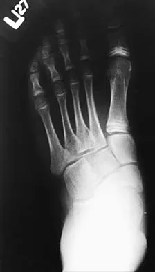
The mother of a 24-month-old girl reports that the child cannot rotate her right forearm. She also notes delayed development, with the child first walking at 18 months. The child has a five-word vocabulary and has not begun using simple phrases. Examination reveals that the right forearm is fixed in 80 degrees of pronation. The remainder of the examination of both upper extremities is otherwise normal. A radiograph is shown in Figure 41. Which of the following studies will best aid in diagnosis?


Explanation

The patient has classic radioulnar synostosis. Patients with this disorder frequently have duplication of sex chromosomes. Synostosis is often seen in females with 48-XXXX or 49-XXXXX in association with delayed development and mental retardation. In males, it can be associated with 48-XXXY or 49-XXXXY. Radioulnar synostosis is not usually associated with muscle disorders, congenital heart disease, or renal anomalies. MRI of the forearm can reveal other soft-tissue anomalies, but this information is not particularly helpful in planning therapy. Osteotomy is sometimes indicated to improve rotational position of the wrist, but this patient's rotation is quite functional for everyday tasks, and rotational osteotomy is not indicated.

Question 2

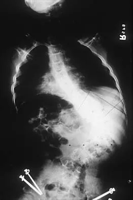
Figure 42 shows the radiograph of a patient with spinal muscular atrophy. Examination reveals good upper extremity function, and she can tie her shoes and propel a manual wheelchair. Posterior instrumentation and fusion may result in


Explanation

Spinal muscular atrophy is caused by an abnormal survival motor neuron gene that prevents apoptosis of the motor nerves. Spinal fusion results in better sitting balance, stabilized or improved pulmonary function, and high parental satisfaction, but it may result in at least temporary loss of upper extremity function. Bentley G, Haddad F, Bull TM, Seingry D: The treatment of scoliosis in muscular dystrophy using modified Luque and Harrington-Luque instrumentation. J Bone Joint Surg Br 2001;83:22-28. Furumasu J, Swank SM, Brown JC, Gilgoff I, Warath S, Zeller J: Functional activities in spinal muscular atrophy patients after spinal fusion. Spine 1989;14:771-775.

Question 3

A 2-week-old infant has had diminished movement of the right upper extremity since birth. Examination reveals weakness of shoulder abduction and external rotation, elbow flexion, and forearm supination. Both pupils are equally round and responsive to light. The remainder of the examination is normal. Radiographs of the upper limb show a healing middle-third clavicle fracture. Management should consist of


Explanation

The patient has a classic Erb's palsy with weakness of the muscles innervated by the fifth and sixth cervical roots. Horner syndrome, a poor prognostic indicator for recovery, is absent in this infant. All infants with brachial plexus birth palsies initially should be monitored for spontaneous recovery during the first 3 to 6 months of life. During this period of observation, glenohumeral motion, especially external rotation, should be maintained. Many infants will begin to show recovery within the first 6 to 8 weeks after birth and continue on to normal function. The timing of microsurgery is controversial. A recent study found that the outcome of microsurgical repair in patients who had no recovery of biceps function within 3 months after birth was similar compared to those who had recovery of biceps function between 3 and 6 months and no microsurgical repair. The author concluded that microsurgical repair was effective in improving function in those infants who had no evidence of recovery of biceps function within the first 6 months of life. Waters PM: Comparison of the natural history, the outcome of microsurgical repair, and the outcome of operative reconstruction in brachial plexus birth palsy. J Bone Joint Surg Am 1999;81:649-659.

Question 4

Progressive paralysis is most likely to be seen in association with what type of congenital vertebral abnormality?


Explanation

Anterior failure of formation results in a progressive kyphosis that may lead to cord compression and progressive neurologic deficit. Anterior failure of segmentation can also produce progressive kyphosis but usually is not severe enough to cause cord compression. Posterior failure of formation is seen in conditions such as myelomeningocele in which the neurologic deficit is generally stable. Lateral abnormalities and posterior failure of segmentation are rarely associated with progressive neurologic deficit. McMaster MJ, Singh H: Natural history of congenital kyphosis and kyphoscoliosis: A study of one hundred and twelve patients. J Bone Joint Surg Am 1999;81:1367-1383.

Question 5

A 9-year-old child has right groin pain after falling from a tree. Examination reveals that the right leg is held in external rotation, and there is significant pain with attempts at passive range of motion. Radiographs are shown in Figures 43a and 43b. Management should consist of


Explanation

43b The complications of femoral neck fractures in children include osteonecrosis, malunion, nonunion, and premature physeal closure. It is presumed that the risk of osteonecrosis is directly related to the amount of displacement at the time of injury and is not affected by the type of treatment. The risk of the other complications can be decreased depending on the type of treatment. Anatomic reduction by either closed or open methods can reduce the risk of malunion. The addition of internal fixation allows for maintenance of the reduction. In young children who cannot comply with a partial or non-weight-bearing status, the addition of a spica cast gives added protection. Canale ST: Fractures of the hip in children and adolescents. Orthop Clin North Am 1990;21:341-352.

Question 6

An 8-year-old boy with moderate factor VIII hemophilia played kickball earlier in the day and now reports progressively severe groin pain and is unable to walk. Examination reveals marked paresthesias over the medial aspect of the distal tibia. What is the most likely diagnosis?


Explanation

The iliacus muscle is a frequent site of hemorrhage in patients with severe or moderate hemophilia. In patients with moderate hemophilia, hemorrhage into the iliacus muscle often follows play or sporting events that include forceful contraction of the hip flexor muscles. An expanding iliacus hematoma compresses the adjacent femoral nerve, with one study reporting 60% complete femoral nerve palsy in hemophiliacs with an iliacus or iliopsoas hemorrhage. Femoral nerve compression typically includes paresthesias in the distribution of the terminal saphenous nerve branch. Hip joint hemarthrosis may occur, but this condition is not as frequent in hemophiliacs as muscle hemorrhage into the iliacus muscle. More importantly, a hip joint hemarthrosis is not associated with significant compression of the femoral nerve. Avulsion fractures of the anterior superior iliac spine typically occur during adolescence and are not associated with saphenous nerve paresthesias. Slipped capital femoral epiphysis does not have an increased association with hemophilia and usually occurs during the adolescent years. Greene WB: Diseases related to the hematopoietic system, in Morrissy RT, Weinstein SL (eds): Lovell and Winter's Pediatric Orthopaedics, ed 5. Philadelphia, PA, Lippincott Williams and Wilkins, 2001, pp 379-426.

Question 7

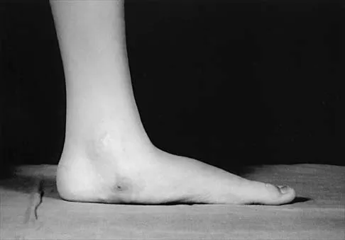
The patient shown in Figure 44 reports that her toes hurt when she walks. Management should consist of


Explanation

Brachymetatarsia is a congenital hypoplasia of one or more metatarsals. Shortening of the fourth metatarsal is the most common form of brachymetatarsia and is often bilateral. Taping and manipulative reduction attempts are ineffective, and extensor tenotomy and capsulotomy are not likely to sufficiently correct the deformity. Fusion or metatarsal lengthening result in complications and generally are not indicated. Shoe modifications, such as extra-depth or extra-wide shoes, generally will improve symptoms. If pressure and trauma persist in the older child, metatarsal lengthening or amputation may be indicated. Stevens PM: Toe deformities, in Drennan JC (ed): The Child's Foot and Ankle. New York, NY, Raven Press, 1992, p 195.

Question 8

Figures 45a and 45b show the AP and lateral radiographs of a 15-year old patient who is undergoing surgery to add 3 cm of length to the femur. Based on the radiographic findings, what is the next most appropriate step in management?


Explanation

45b Because the radiographs reveal poor regenerate bone, especially anteriorly and laterally, the first step in management is to slow the distraction rate. If this does not solve the problem, temporary reversal of the distraction, or "accordionization," can be used to induce a greater healing response. Maintaining the same distraction rate will further impair regenerate formation and delay healing. Bone grafting should be reserved as an option if decreasing the distraction rate or alternating a week of compression with a week of distraction fails to improve the callus formation. Repeat corticotomy is performed in patients with premature consolidation. Raney EM: Limb-length discrepancy, in Fitzgerald RH, Kaufer H, Malkani AL (eds): Orthopaedics. St Louis, MO, Mosby, 2002, pp 1519-1526.

Question 9

Examination of a 9-year-old boy reveals a right thoracic prominence on forward flexion. Neurologic examination is normal, and no other abnormalities are noted. AP radiographs reveal a 30-degree right thoracic curve. Initial management should consist of


Explanation

The patient has juvenile scoliosis. MRI has shown an association between juvenile scoliosis and intraspinal abnormalities, most often syringomyelia and Arnold-Chiari malformations. All juvenile curves greater than 20 degrees should be evaluated with MRI despite the absence of neurologic findings. Weinstein SL (ed): The Pediatric Spine: Principles and Practice, ed 1. New York, NY, Raven Press, 1994, pp 685-705 Nohria V, Oakes WJ: Chiari I malformation: A review of 43 patients. Pediatr Neurosurg 1990-91;16:222-227.

Question 10

A 13-year-old boy is comatose and has irregular breathing after being struck by a car while riding his bicycle. Auscultation suggests a pneumothorax on the right side and swelling about the right arm and leg. Initial management should consist of


Explanation

The first priority is to gain control of the airway with intubation. Following intubation, management should consist of ventilation and placement of a chest tube if needed, vascular access and circulatory stabilization, radiographs of the cervical spine and chest, and CT of the brain. American College of Surgeons Committee on Trauma. Advanced Trauma Life Support Course. Instructor's Manual. Chicago, IL, American College of Surgeons, 1984.

Question 11

Overgrowth of a limb in a patient with neurofibromatosis type 1 (NF1) is most likely associated with the presence of


Explanation

Plexiform neurofibromas are lesions found in patients with NF1. Clinical reports show the prevalence of plexiform neurofibroma to be 20% to 30% but increases to 40% when imaging studies are routinely obtained. The lesions are characterized by diffuse hypertrophy of the involved nerves but with preservation of the nerves' fascicular organization. The lesions may involve the dermis or may arise in the deeper structures. Palpation of a dermal lesion provokes an image of a "bag of worms." Plexiform neurofibromas may cause disfigurement and hyperpigmentation of the overlying skin. The lesions also can cause diffuse hypertrophy of the soft tissue and bone, with resultant changes ranging from a relatively minor limb-length discrepancy to gigantism of the entire extremity. Dural ectasia is frequently found in patients with NF1. Therefore, MRI should be obtained prior to planning spinal procedures in these patients; however, dural ectasia is not the cause of limb overgrowth. Lisch nodules are benign hamartomas of the iris. The lesions are uncommon during early childhood but are found in all adults with NF1. Juvenile xanthogranuloma has a low occurrence rate in patients with NF1; its presence is associated with juvenile chronic myeloid leukemia. Malignant peripheral nerve sheath tumors, formally called neurofibrosarcoma, result from malignant degeneration of a plexiform neurofibroma. This condition occurs in up to 4% of patients with NF1. Localized pain, an enlarging mass, or progressive neurologic symptoms suggest a malignant peripheral nerve sheath tumor in a patient with NF1. However, progressive neurologic symptoms also may occur with benign growth of a plexiform neurofibroma. Alman BA, Goldberg MJ: Syndromes of orthopaedic importance, in Morrissy RT, Weinstein SL (eds): Lovell and Winter's Pediatric Orthopaedics, ed 5. Philadelphia, PA, Lippincott Williams and Wilkins, 2001, pp 287-338.

Question 12

A 14-year-old girl with a right thoracic curve from T4 through L2 measuring 78 degrees is scheduled to undergo posterior spinal fusion for scoliosis. The surgical plan is to fuse from T3 through L2, using pedicle screws at L2 and about the apex at T8. What neural monitoring modality is most likely to identify a reversible neurologic deficit during surgery?


Explanation

Neural monitoring during scoliosis surgery was initially developed to avoid the devastating effects of spinal cord injury, particularly paraplegia. Monitoring in some form has become standard for this type of surgery. Somatosensory-evoked potentials in the lower extremities will detect many but not all neurologic difficulties with the spinal cord. Anterior spinal cord vascular disruption also can be detected by monitoring motor potentials. Electromyography following stimulation of lumbar pedicle screws can prevent nerve root injury that is the result of misplacement of the screws. This is best documented in the lumbar spine and has not been routinely used in the thoracic spine. The most common neural deficits following spinal surgery, however, are in the upper extremities because of the positioning of the patient in the prone position for long periods. In Schwartz and associates series of 500 patients, impending upper extremity neural injury was detected by somatosensory-evoked potentials in 18 (3.6%) patients. In contrast, lower extremity deficits were detected by combined motor- and sensory-evoked potentials in only 2 (0.4%) out of 500 patients in Padberg and associates series. Neural compression in the upper extremity can be easily detected by somatosensory-evoked potentials, and injury can be prevented by repositioning the patient. Padberg AM, Wilson-Holden TJ, Lenke LG, Bridwell KH: Somatosensory- and motor-evoked potential monitoring without wake-up test during idiopathic scoliosis surgery: An accepted standard of care. Spine 1998;23:1392-1400.

Question 13

Figure 46 shows the radiograph of an obese 12-year-old boy who has had left hip pain for the past 3 months. What is the best course of action?


Explanation

The patient has an obvious slipped capital femoral epiphysis of the left hip for which the recommended treatment is percutaneus pinning in situ. Development of a contralateral slip is less likely at this age; therefore, observation of the right hip is indicated because there is no general agreement regarding prophylactic fixation. Typically, there is no role for spica casting. Physical therapy is not indicated as a primary treatment, and reduction is contraindicated, as it has been associated with osteonecrosis. Loder RT, Aronsson DD, Greenfield ML: The epidemiology of bilateral slipped capital femoral epiphysis: A study of children in Michigan. J Bone Joint Surg Am 1993;75:1141-1147. Aronsson DD, Karol LA: Stable slipped capital femoral epiphysis: Evaluation and management. J Am Acad Orthop Surg 1996;4:173-181. Hurley JM, Betz RR, Loder RT, Davidson RS, Alburger PD, Steel HH: Slipped capital femoral epiphysis: The prevalence of late contralateral slip. J Bone Joint Surg Am 1996;78:226-230.

Question 14

A 7-year-old boy sustained a head contusion and small bowel injuries in a motor vehicle accident in which he was wearing a lap belt. He subsequently required a bowel resection. Six weeks after the accident, his parents note a painful mass in his lower back. His neurologic examination is normal. A radiograph and CT scans are shown in Figures 47a through 47c. Definitive management should now consist of


Explanation

47b 47c The posttraumatic lumbar kyphotic deformity will not remodel and is likely to worsen with time because the central line of gravity lies anterior to the deformity and the ligamentous disruption will not heal. The worsening deformity also puts the patient at some risk for future neurologic damage. Ebraheim NA, Savolain ER, Southworth SR, et al: Pediatric lumbar seat belt injuries. Orthopedics 1991;14:1010-1013.

Question 15

A 5-month-old girl with arthrogryposis has a limb-length discrepancy. Examination and radiographs reveal unilateral hip dislocation. Management should consist of


Explanation

In this age group of patients with arthrogryposis, open reduction through a medial approach is generally recommended. Open reduction through an anterior approach is reserved for patients in which a medial approach has failed or for older patients who require simultaneous femoral shortening and/or pelvic osteotomy. Closed treatment of unilateral hip dislocation in association with arthrogryposis is rarely successful. In bilateral hip dislocation associated with arthrogrypsis, the consensus is that the hips are best left unreduced because of the difficulty in obtaining excellent clinical and radiographic results bilaterally. Staheli LT, Chew DE, Elliot JS, Mosca VS: Management of hip dislocations in children with arthrogryposis. J Pediatr Orthop 1987;7:681-685. Szoke G, Staheli LT, Jaffe K, Hall JG: Medial-approach open reduction of hip dislocation in amyoplasia-type arthrogryposis. J Pediatr Orthop 1996;16:127-130.

Question 16

A 10-year-old boy who has had progressive low back and right buttock pain for the past 3 days is now unable to bear weight on the right side secondary to pain. He has a temperature of 101.3 degrees F (38.5 degrees C). Examination reveals full hip range of motion; but he reports pain on the right side with external rotation. Pain is elicited with compression of the iliac wings and with direct palpation of the right sacroiliac (SI) joint. An MRI scan of the pelvis shows no abscess, but there is inflammation of the SI joint. Management should consist of


Explanation

The clinical presentation and MRI findings are consistent with an acute infection of the SI joint. Bed rest and nonsteroidal anti-inflammatory drugs alone are insufficient to treat the problem. Staphylococcus aureus is the causative organism in most of these infections; therefore, unless there is an unusual factor in the history such as IV drug use, immune system compromise, or unusual travel, SI joint aspiration is unnecessary. It is often difficult to enter the SI joint, even under radiographic guidance. Management should consist of hospital admission and IV antibiotics. Blood cultures may be positive and should be obtained prior to starting antibiotics. Surgical fusion of the SI joint is not indicated. Morrissey RT: Bone and Joint Sepsis in Pediatric Orthopaedics. Philadelphia, PA, JB Lippincott, 1990. Beaupre A, Carroll N: The three syndromes of iliac osteomyelitis in children. J Bone Joint Surg Am 1979;61:1087-1092.

Question 17

Figures 48a and 48b show the elbow radiographs of a 5-year-old boy who fell from a tree after dinner. Examination reveals that he is unable to extend his wrist. Management should consist of immediate


Explanation

48b In the absence of vascular compromise, there has been no proven value to proceeding immediately to surgery, especially when the patient has a full stomach and runs a significant risk of perioperative aspiration. It would be more prudent to wait until the next morning with a surgical plan of closed reduction and pinning. Open reduction should be reserved for the unusual case of where closed treatment has not been successful. The implication that there may be a radial nerve injury associated with this fracture does not alter the treatment plan, and with a high level of certainty would be expected to resolve. Attempting closed reduction in the emergency department creates the opportunity for uncertain results and is not tolerated well by most patients. Skeletal traction, with its associated lengthy hospitalization and the technical difficulties associated with both the traction and radiographic evaluations, has fallen into disfavor for typical clinical situations. Iyengar SR, Hoffinger SA, Townsend DR: Early versus delayed reduction and pinning of type III displaced supracondylar fractures of the humerus in children: A comparative study. J Orthop Trauma 1999;13:51-55.

Question 18

In addition to the radiographic features seen in Figures 49a and 49b, this patient will most likely have which of the following findings?


Explanation

49b The radiographs show the characteristic features of osteopetrosis. The condition results from defective resorption of immature bone by osteoclasts. There are three distinct clinical forms: (1) infantile-malignant, which is autosomal recessive and fatal in the first few years of life if untreated; (2) intermediate autosomal recessive; and (3) autosomal dominant. These conditions do not follow a malignant course, and patients have normal life expectancy with orthopaedic problems and anemia. In the malignant form, the clinical features include frequent fractures, macrocephaly, progressive deafness and blindness, hepatosplenomegaly, and severe anemia beginning in early infancy or in utero. Deafness and blindness are generally thought to represent effects of pressure on nerves and usually occur later in life. The anemia is caused by encroachment of bone on marrow, resulting in obliteration, and the hepatosplenomegaly is caused by compensatory extramedullary hematopoiesis. Dental caries and abscesses, as well as osteomyelitis of the mandible, are also seen. Most patients have normal intelligence. Treatment of the malignant form includes high dose 1,25 dihydroxy vitamin D with a low-calcium diet to stimulate bone resorption, not because there are vitamin deficiencies. Bone marrow transplant has also been successful. Herring JA: Tachdjian's Pediatric Orthopedics, ed 4. Philadelphia, PA, WB Saunders, 2002, p 1550. Zaleske DJ: Metabolic and endocrine abnormalities, in Morrissy RT, Weinstein SL (eds): Lovell and Winter's Pediatric Orthopaedics, ed 5. Philadelphia, PA, Lippincott Williams and Wilkins, 2001, pp 212-214.

Question 19

A biopsy of the involved physis in a patient with slipped capital femoral epiphysis (SCFE) would most likely reveal


Explanation

Vascular invasion, histologically similar to granulation tissue, has been noted between the columns in the zone of hypertrophy, leading to the theory of microtrauma as an etiology. SCFE is also associated with conditions that increase the height of the zone of hypertrophy, including the adolescent growth spurt and endocrinopathies. The perichondral ring has been shown to decrease in thickness with age. Normal undulations in the growth plate also decrease during this time, possibly further destabilizing the physis. Abnormal accumulations of proteoglycan have been reported. Chung SM, Batterman SC, Brighton CT: Shear strength of the human femoral capital epiphyseal plate. J Bone Joint Surg Am 1976;58:94-103.

Question 20

A 12-year-old girl has a 4-cm limb-length discrepancy following a fracture of the left distal femur 2 years ago. Examination reveals 18 degrees of genu valgum on the involved side, with 7 degrees of genu valgum on the opposite side. Radiographs show that the left distal femoral growth plate is now closed; however, the tibial growth plate is still open. Her bone age matches her chronologic age. Management should consist of


Explanation

The patient has a projected limb-length discrepancy of 7 cm. This includes the 4 cm she already has, plus 3 cm expected growth of the uninvolved distal femur during the 3 years of growth she has remaining. She also has moderate limb deformity. Femoral lengthening is considered the treatment of choice because it can address both the limb-length discrepancy and the deformity. Epiphyseodesis will not result in limb-length equality at maturity, with only approximately 1.8 cm of equalization expected from this procedure. Use of closed femoral shortening of 7 cm runs the risk of weakening the quadriceps on the normal side and will leave the patient with a remaining residual valgus deformity. Tibial lengthening will leave the knees at different levels. A shoe lift can be prescribed as a temporary measure but is not a good long-term solution. Westh RN, Menelaus MB: A simple calculation for the timing of epiphyseal arrest: A further report. J Bone Joint Surg Br 1981;63:117-119. Sasso RC, Urquhart BA, Cain TE: Closed femoral shortening. J Pediatr Orthop 1993;13:51-56.

Question 21

The parents of a previously healthy 3-year-old child report that she refused to walk on awakening. Examination later in the day reveals that the patient can walk but with a noticeable limp. She has a temperature of 99.5 degrees F (37.5 degrees C). Range of motion measurements are shown in Figure 50. An AP pelvis radiograph is normal. Laboratory studies show a WBC count of 9,000/mm3 and an erythrocyte sedimentation rate of 10 mm/h. Management should consist of


Explanation

The patient has the typical history and presentation of transient synovitis of the hip, a condition that is more common in children age 2 to 5 years but which may affect children up to 12 years. The discomfort typically is noted on awakening, and the child will refuse to walk. Later in the day, the pain commonly improves and the child can walk but will have a limp. Mild to moderate restriction of hip abduction is the most sensitive range-of-motion restriction. The extent of the evaluation for transient synovitis depends on the intensity and duration of symptoms. Because she has been afebrile for the past 24 hours, observation is the management of choice. In the differential diagnosis of suspected transient synovitis, septic arthritis of the hip is the primary disorder to exclude. Osteomyelitis of the proximal femur also should be considered. In most patients, clinical examination will differentiate of these disorders to a reasonable certainty. Plain radiographs are normal in the early stage of an infectious process. Ultrasonography shows increased fluid in the hip joint in both transient synovitis and septic arthritis. MRI can differentiate the two conditions; however, this test would require general anesthesia and is not required in most patients in this age group. If a child with transient synovitis has a concurrent infectious process such as an upper respiratory tract infection or otitis media, the temperature will most likely be elevated. In this situation, a full evaluation for an infectious process and initiation of IV antibiotics should be considered. This would include radiographs, CBC count, erythrocyte sedimentation rate, blood cultures, aspiration of the hip joint, and IV antibiotics. Del Beccaro MA, Champoux AN, Bockers T, Mendelman PM: Septic arthritis versus transient synovitis of the hip: The value of screening laboratory tests. Annals Emerg Med 1992;21:1418-1422.

Question 22

A 12-year-old boy with hemophilia A and no known inhibitors sustains a tibia fracture and has pain with passive motion of the deep toe flexors. Appropriate management should consist of


Explanation

In a patient with hemophilia, factor replacement followed by assessment of compartment pressures is essential. If the patient has inhibitors, the problem is more difficult. Porcine factor has been helpful in patients with inhibitory antibodies. Recent evidence points to using activated factor VII and bypassing the intrinsic pathway. Desmopressin is an adjunct to therapy but is not as effective as factor VII. Dumontier C, Sautet A, Man M, Bennani M, Apoil A: Entrapment and compartment syndromes of the upper limb in haemophilia. J Hand Surg Br 1994;19:427-429. Carr ME Jr, Loughran TP, Cardea JA, Smith WK, Kuhn JG, Dottore MV: Successful use of recombinant factor VIIa for hemostasis during total knee replacement in a severe hemophiliac with high-titer factor VIII inhibitor. Int J Hematol 2002;75:95-99.

Question 23

A 10-year-old boy with spastic diplegic cerebral palsy walks in a crouched position with the hips and knees flexed. Maximum knee flexion is 15 degrees during early swing phase. Instrumented gait analysis shows quadriceps activity from terminal stance throughout swing phase. Treatment should consist of


Explanation

The rectus femoris muscle spans two joints and is active during running, sprinting, and walking at a fast pace during the preswing and early swing phase of gait. In these situations, the muscle helps to generate power to initiate hip flexion while absorbing or controlling the rate of knee flexion during early swing phase. Quadriceps activity, including the rectus femoris, is not normally needed when walking at a routine cadence. However, rectus femoris activity is commonly noted during preswing and the swing phase in patients with cerebral palsy, particularly those with diplegia. In an effort to initiate swing phase, the rectus femoris is "overactive." As a result, the knee flexion that commonly occurs at terminal stance and initial swing is restricted. Instead of achieving the normal 50 to 60 degrees of flexion during early swing, this patient's knee flexion is limited to 15 degrees. The goal of treatment is to retain rectus femoris activity for initiation of hip flexion but to diminish its restraint on knee flexion. Studies have shown that transfer of the distal rectus femoris tendon provides more flexion of the knee during the swing phase of gait than simply releasing the tendon. V-Y lengthening of the quadriceps tendon or a Z lengthening of the patellar tendon causes too much weakening of the quadriceps muscle and worsens the crouch deformity. In addition to transfer of the rectus femoris tendon, other procedures are often done concomitantly to obtain the best balance and realignment of hip-knee-ankle activity. Aiona MD: Guidelines for managing lower extremity problems in cerebral palsy, in Fitzgerald RH, Kaufer H, Malkani AL (eds): Orthopaedics. St Louis, MO, Mosby, 2002, pp 1534-1541. Chambers H, Laure A, Kaufman K, Cardelia M, Sutherland D: Prediction of outcome after rectus femoris surgery in cerebral palsy: The role of cocontraction of the rectus femoris and vastus lateralis. J Pediatr Orthop 1998;18:703-711.

Question 24

A 4-year-old boy with arthrogryposis has little active motion of his knees or elbows. Both elbows are in full extension with good triceps strength, but he is unable to bring his hand to his face or feed himself. Management should consist of


Explanation

Elbow release and triceps transfer to restore motion can be performed in children who are age 4 years and older. The ability to flex the elbow either actively or passively is of great assistance in activities of daily living. Van Heest A, Waters PM, Simmons BP: Surgical treatment of arthrogrypsosis of the elbow. J Hand Surg Am 1998;23:1063-1070.

Question 25

A 14-year-old competitive gymnast has had activity-related low back pain for the past month. Examination reveals no pain with forward flexion, but she has some discomfort when resuming an upright position. She also has pain with extension and lateral bending of the spine. The neurologic examination is normal. Popliteal angles measure 20 degrees. AP, lateral, and oblique views of the lumbar spine are negative. What is the next most appropriate step in management?


Explanation

Symptoms of activity-related low back pain, physical findings of pain with extension, lateral bending, and resuming an upright position, and relative hamstring tightness are consistent with spondylolysis. While the initial diagnostic work-up should include plain radiographs of the lumbosacral spine, the findings may be negative because it can take weeks or months for the characteristic changes to become apparent. SPECT has been a useful adjunct in the diagnosis of spondylolysis when plain radiographs are negative. Since the patient's pain is activity related and she is otherwise healthy, evaluation for infection is not indicated. Because the neurologic examination is normal, electromyography, nerve conduction velocity studies, and MRI are not indicated. CT can be used in those instances in which SPECT and bone scans are negative. Ciullo JV, Jackson DW: Pars interarticularis stress reaction, spondylolysis, and spondylolisthesis in gymnasts. Clin Sports Med 1985;4:95-110. Collier BD, Johnson RP, Carrera GF, et al: Painful spondylolysis or spondylolisthesis studied by radiography and single photon emission computed tomography. Radiology 1985;154:207-211. Jackson DW, Wiltse LL, Cirincione RT: Spondylolysis in the female gymnast. Clin Orthop 1976;117:68-73.

Question 26

A 6-year-old boy falls on an outstretched hand and sustains a supracondylar humerus fracture. A radiograph reveals a completely displaced extension-type (Gartland Type III) fracture.

On physical examination in the emergency department, an anterior interosseous nerve (AIN) palsy is diagnosed. Which of the following clinical findings is most likely present to confirm this diagnosis?


Explanation

Anterior interosseous nerve (AIN) palsy is the most common neurologic injury associated with extension-type supracondylar humerus fractures. The AIN is a pure motor branch of the median nerve that innervates the flexor pollicis longus (FPL), the flexor digitorum profundus (FDP) to the index and middle fingers, and the pronator quadratus. Clinically, it presents as an inability to make the 'A-OK' sign due to weakness in flexing the distal interphalangeal joint of the index finger and the interphalangeal joint of the thumb. It does not cause sensory deficits.

Question 27

A 6-year-old girl with spastic quadriplegic cerebral palsy (GMFCS level V) is evaluated for hip pain and difficulty with perineal hygiene. An anteroposterior pelvis radiograph reveals a migration percentage of 45% on the right hip and 40% on the left hip, with moderate acetabular dysplasia.

What is the most appropriate surgical management for this patient?


Explanation

In children with cerebral palsy, hip surveillance is critical. For significant hip subluxation (migration percentage > 40%) with acetabular dysplasia in a child of this age, soft tissue releases alone are inadequate and have an unacceptably high failure rate. Bony reconstruction with a varus derotational osteotomy (VDRO) of the femur combined with a volume-reducing pelvic osteotomy (e.g., Dega or San Diego osteotomy) is the gold standard. It provides durable coverage, reduces the head into the true acetabulum, and halts progressive subluxation.

Question 28

A 6-week-old female infant is undergoing treatment with a Pavlik harness for developmental dysplasia of the left hip. During the 2-week follow-up visit, the mother notes that the child has stopped kicking her left leg. On physical examination, the infant exhibits an absent patellar reflex on the left side and decreased active knee extension. Sensibility appears intact. What is the most appropriate next step in management?


Explanation

The clinical scenario describes a femoral nerve palsy, a known complication of Pavlik harness treatment resulting from excessive hip flexion (typically > 120 degrees), which compresses the femoral nerve against the rim of the pelvis. The standard and most appropriate management is to remove the harness or cease its use until quadriceps function returns (which usually takes a few days to a few weeks). Only after the nerve palsy has resolved should alternative bracing or modified harness treatment be attempted.

Question 29

A 4-year-old boy presents with a relapsed left idiopathic clubfoot. The deformity was initially treated successfully in infancy with the Ponseti method. The parents report he is now walking on the outside of his foot. Gait analysis demonstrates dynamic supination of the foot during the swing phase. Physical examination reveals an easily correctable deformity with completely passive plantigrade positioning. What is the most appropriate surgical treatment?


Explanation

Relapsed clubfoot frequently presents with dynamic supination due to muscle imbalance, predominantly overactivity of the anterior tibial tendon (ATT) overpowering the peroneal muscles. The definitive treatment for dynamic supination in a relapsed clubfoot is a transfer of the entire ATT to the lateral cuneiform. However, a strict prerequisite for this tendon transfer is that the foot must be completely passively correctable. Any residual fixed deformity must first be corrected by a short period of serial Ponseti casting before the tendon transfer is performed.

Question 30

An 8-year-old boy presents with a 3-month history of a painless limp and right hip stiffness. Radiographs demonstrate fragmentation of the capital femoral epiphysis. Measurements reveal that exactly 40% of the lateral pillar height is maintained.

According to the Herring Lateral Pillar Classification, what group does this patient fall into, and what is the typical outcome associated with surgical containment for his age and classification?


Explanation

The Herring Lateral Pillar Classification evaluates the height of the lateral pillar of the capital femoral epiphysis during the fragmentation stage of Legg-Calvé-Perthes disease. Group A retains 100% height, Group B retains >50% height, and Group C retains <50% height. This patient has 40% height, placing him in Group C. For children 8 years of age and older with Group C hips, the prognosis is generally poor (high likelihood of Stulberg III-V outcome). Large multicenter studies have demonstrated that surgical containment (e.g., femoral or pelvic osteotomy) does not significantly improve radiographic outcomes compared to non-operative treatment in this specific subgroup.

Question 31

A 14-year-old boy complains of vague midfoot pain and recurrent ankle sprains for the past 6 months. Physical examination reveals bilateral flatfeet with significantly decreased subtalar motion and peroneal spasticity. A lateral radiograph of the right foot demonstrates the 'anteater nose' sign. Which of the following statements regarding his condition is correct?


Explanation

The 'anteater nose' sign on a lateral radiograph is pathognomonic for a calcaneonavicular coalition, which represents an elongated anterior process of the calcaneus approaching the navicular. The best initial radiograph to visualize this specific coalition is the 45-degree internal oblique view. Talocalcaneal coalitions are typically visualized on Harris axial views and are associated with the 'C-sign' on lateral films. Calcaneonavicular coalitions ossify and become symptomatic earlier (ages 8-12) than talocalcaneal coalitions (ages 12-16). Initial management is non-operative (e.g., immobilization, orthotics).

Question 32

A 6-year-old girl with Osteogenesis Imperfecta (Sillence Type III) is referred to the orthopedic clinic after sustaining her fourth diaphyseal femur fracture in two years. She is currently undergoing cyclic intravenous bisphosphonate therapy.

Which of the following surgical interventions is the gold standard for managing recurrent diaphyseal femur fractures in this patient population?


Explanation

In patients with moderate to severe Osteogenesis Imperfecta (OI) exhibiting recurrent fractures or progressive long bone bowing, intramedullary rodding is the standard of care to straighten the bone, prevent recurrent fractures, and provide internal support. Telescopic rods, such as the Fassier-Duval rod, are the gold standard for growing children because they elongate with the child's growth. This prevents the bone from 'growing off' the end of the rod, a common complication with static rods (like TENs) that leads to fractures adjacent to the implant. Plate fixation is contraindicated due to poor bone stock and the high risk of stress risers.

Question 33

A 13-year-old female competitive gymnast presents with progressive low back pain and bilateral radicular symptoms radiating to the posterior thighs. Examination reveals a palpable 'step-off' at the lumbosacral junction, a crouched gait, and severe hamstring tightness. Standing lateral radiographs reveal a Meyerding Grade III isthmic spondylolisthesis at L5-S1 with a slip angle of 45 degrees. What is the most appropriate definitive management?


Explanation

The patient has a high-grade (Meyerding Grade III, >50% slip) isthmic spondylolisthesis with a high slip angle, clinical deformity, and neurologic symptoms. High-grade slips with radicular symptoms and sagittal imbalance in adolescents mandate surgical intervention. The contemporary standard involves posterior spinal decompression (to relieve the L5 roots), partial or complete reduction of the slip (to restore sagittal balance), and instrumented fusion (L5-S1 or L4-S1 depending on the specific anatomy). Pars repair is reserved for low-grade slips (Grade I) without disc degeneration. In situ fusion in the setting of a high slip angle carries an unacceptable risk of pseudarthrosis and progressive deformity.

Question 34

A newborn male is evaluated in the nursery for a significant right upper extremity deformity. The forearm is shortened and bowed volarly, and the hand is in fixed radial deviation. Radiographs confirm an absent radius and an absent thumb. Before addressing the orthopedic deformity, which of the following systemic evaluations is most critical?


Explanation

Radial longitudinal deficiency (radial clubhand) is highly associated with several systemic syndromes. The most critical and common associations include VACTERL association (Vertebral anomalies, Anal atresia, Cardiac defects, Tracheoesophageal fistula, Renal anomalies, Limb defects), Holt-Oram syndrome (cardiac septal defects), and TAR syndrome (Thrombocytopenia-Absent Radius). Therefore, a thorough systemic workup is mandatory prior to any surgical intervention. An echocardiogram and a renal ultrasound are paramount to rule out potentially life-threatening congenital heart and renal anomalies.

Question 35

A 2-year-old boy presents with a 3-day history of right knee swelling, a mild limp, and a low-grade fever (37.9°C). Initial laboratory work reveals a WBC count of 11,000/mm3, an ESR of 30 mm/hr, and a CRP of 2.5 mg/dL. Synovial fluid aspiration yields 65,000 WBCs/mm3 with 80% neutrophils. Gram stain is negative, and routine aerobic solid cultures show no growth at 48 hours. Which of the following pathogens is the most likely culprit, and what specific diagnostic technique is required to identify it?


Explanation

Kingella kingae is a leading cause of pediatric osteoarticular infections, particularly in children under 4 years of age. Clinically, it frequently presents with milder systemic symptoms (low-grade fever, mildly elevated ESR/CRP) compared to infections caused by Staphylococcus aureus. K. kingae is famously fastidious and rarely grows on routine solid media. Diagnosis is significantly enhanced by directly inoculating the synovial fluid into aerobic BACTEC blood culture vials or by utilizing nucleic acid amplification tests (PCR).

Question 36

A 13-year-old obese male presents with an inability to bear weight on the right leg after a minor fall. He reports having had a mild, aching knee pain for 2 months prior to the fall. Radiographs reveal a severe posterior and inferior displacement of the proximal femoral epiphysis. If an open reduction and internal fixation via a surgical dislocation approach (modified Dunn procedure) is planned, preservation of which of the following vessels is most critical to prevent osteonecrosis of the femoral head?


Explanation

The deep branch of the medial circumflex femoral artery (MCFA) is the primary blood supply to the femoral head in adolescents. During a modified Dunn procedure for an unstable slipped capital femoral epiphysis (SCFE), meticulous protection of the external rotator muscles and the retinacular vessels arising from the deep branch of the MCFA is paramount to minimize the high risk of avascular necrosis.

Question 37

A 3-year-old girl is evaluated for bilateral bowing of the lower extremities. Her BMI is above the 95th percentile. Standing radiographs demonstrate a metaphyseal-diaphyseal angle (Drennan's angle) of 18 degrees bilaterally, with prominent medial beaking of the proximal tibia. What is the most appropriate initial management?


Explanation

This patient has infantile Blount disease. A metaphyseal-diaphyseal angle > 16 degrees strongly suggests true Blount disease rather than physiologic bowing. In a child under 3 to 4 years of age with Langenskiöld stage I or II, nonoperative management with a daytime Knee-Ankle-Foot Orthosis (KAFO) is the initial treatment of choice. Surgical intervention is typically reserved for those who fail bracing or present at an older age with advanced stages.

Question 38

A 6-week-old female is being treated with a Pavlik harness for a dislocated left hip. At the 2-week follow-up, an ultrasound confirms the hip remains dislocated. The examiner also notes that the infant has decreased spontaneous extension of the left knee, though she vigorously kicks the right leg. What is the most likely cause of this new clinical finding?


Explanation

Femoral nerve palsy is a well-documented complication of the Pavlik harness, typically caused by hyperflexion of the hip which compresses the nerve against the inguinal ligament. It presents as decreased active knee extension. When this occurs, or if the hip fails to reduce after 3-4 weeks of harness wear, the harness must be discontinued to prevent further nerve damage and 'Pavlik harness disease' (excoriation, dysplasia, or AVN).

Question 39

A 6-year-old boy falls from the monkey bars and sustains a completely displaced, extension-type supracondylar humerus fracture. On arrival, his hand is pink but the radial pulse is absent. Capillary refill is brisk (< 2 seconds). Following closed reduction and percutaneous pinning, the hand remains pink with brisk capillary refill, but the radial pulse remains unpalpable. What is the next best step in management?


Explanation

The management of a 'pink, pulseless' hand following a well-reduced and pinned supracondylar humerus fracture is observation. Because perfusion is clinically adequate (pink color, brisk capillary refill), the collateral circulation is sufficient to sustain the limb. Vascular exploration is indicated only if the hand becomes white and pulseless (ischemia) after reduction, which suggests brachial artery entrapment or severe intimal injury without adequate collateral flow.

Question 40

An 8-year-old boy presents with a limp and right hip pain of several months' duration. Radiographs demonstrate sclerosis and early fragmentation of the proximal femoral epiphysis.

Which of the following is considered the most significant prognostic factor for the development of early osteoarthritis in patients with this condition?


Explanation

Age at clinical onset is the single most important prognostic factor in Legg-Calvé-Perthes disease. Children who present at an older age (typically defined as > 8 years) have less remaining growth potential for femoral head remodeling, leading to an increased risk of permanent aspherical deformity (coxa magna) and subsequent early-onset osteoarthritis. A Gage sign is a radiographic 'head at risk' sign, but age remains the primary prognostic determinant.

Question 41

A 7-year-old with spastic quadriplegic cerebral palsy (GMFCS Level V) presents for routine hip surveillance. An AP pelvis radiograph demonstrates a migration percentage (Reimer's index) of 45% on the right and 15% on the left. Clinically, the right hip can be abducted to 20 degrees. What is the most appropriate management for the right hip?


Explanation

In children with cerebral palsy, a migration percentage > 40% combined with restricted abduction typically indicates progressive hip subluxation that is no longer amenable to soft-tissue releases alone. Bony reconstruction with a varus derotational osteotomy (VDRO) of the proximal femur, frequently accompanied by a pelvic osteotomy (e.g., Dega or San Diego), is the standard of care to restore joint congruency and prevent painful dislocation.

Question 42

A 3-month-old infant is undergoing serial casting for idiopathic congenital talipes equinovarus using the Ponseti method. During the casting process, the physician attempts to correct the equinus deformity by dorsiflexing the foot before the heel varus and forefoot adductus are fully corrected. This technical error is most likely to result in which of the following iatrogenic deformities?


Explanation

In the Ponseti method, attempting to dorsiflex the foot before the calcaneocuboid joint is unlocked (which requires full correction of cavus, adductus, and varus) will cause a breach at the midtarsal joint rather than true ankle dorsiflexion. This midfoot break results in an iatrogenic rocker-bottom foot. The equinus deformity must be addressed last, usually requiring a percutaneous Achilles tenotomy.

Question 43

A 4-year-old girl is evaluated for recurrent fractures following minimal trauma. Clinical examination reveals blue sclerae and evidence of dentinogenesis imperfecta. Genetic testing confirms a mutation in COL1A1. Which of the following mechanisms best describes the action of the medical therapy most commonly used to decrease fracture burden in this patient?


Explanation

The patient has Osteogenesis Imperfecta (OI), characterized by defective type I collagen. The mainstay of medical management to reduce fracture burden is the use of bisphosphonates (e.g., pamidronate or zoledronic acid). Bisphosphonates act by inducing osteoclast apoptosis and inhibiting osteoclast-mediated bone resorption, thereby increasing cortical thickness and trabecular bone density, though they do not correct the underlying collagen defect.

Question 44

A newborn is evaluated for a shortened right lower extremity. Physical examination reveals an anteromedial bowing of the tibia, a dimple over the anterior mid-tibia, and an absent lateral ray (3-ray foot).

Which of the following is the most common associated skeletal anomaly found in the ipsilateral limb of patients with this specific condition?


Explanation

The clinical presentation is classic for fibular hemimelia, which includes anteromedial tibial bowing, an anterior dimple, and absent lateral rays of the foot. Fibular hemimelia is highly associated with congenital femoral deficiency (formerly proximal focal femoral deficiency), as well as cruciate ligament deficiency and ball-and-socket ankle geometry. Pseudarthrosis of the tibia is typically associated with anterolateral bowing and neurofibromatosis type 1.

Question 45

A 14-year-old male presents with rigid, painful flatfeet and a history of recurrent ankle sprains. Examination shows significantly restricted subtalar motion and peroneal spasticity. A CT scan confirms a middle facet talocalcaneal coalition involving 60% of the posterior facet area, accompanied by early degenerative changes in the talonavicular joint. What is the most appropriate definitive surgical intervention?


Explanation

Resection of a talocalcaneal coalition is generally contraindicated if the coalition involves > 50% of the posterior facet or if there is evidence of advanced degenerative arthritis in adjacent joints. Because this patient has extensive facet involvement (>50%) and concomitant talonavicular arthritis, resection alone is destined to fail. A triple arthrodesis (fusion of the talocalcaneal, talonavicular, and calcaneocuboid joints) is the treatment of choice to address both the rigid deformity and the arthritic changes.

Question 46

A 9-year-old boy presents with left knee pain and an obligatory external rotation of the left lower extremity with hip flexion. Radiographs demonstrate a severe, chronic slipped capital femoral epiphysis (SCFE) on the left. The patient's medical history is significant for chronic kidney disease and secondary hyperparathyroidism. Following in situ pinning of the left hip, what is the most appropriate management for the asymptomatic right hip?


Explanation

The patient has a known endocrinopathy/metabolic disorder (renal osteodystrophy with secondary hyperparathyroidism), which places him at an exceptionally high risk for developing a contralateral slipped capital femoral epiphysis (SCFE). Prophylactic prophylactic in situ percutaneous pinning of the contralateral hip is strongly indicated in patients with underlying endocrinopathies, radiation therapy history, and in very young patients (typically less than 10 years old) presenting with a unilateral SCFE. Observation is appropriate for older, idiopathic cases, but not for this high-risk patient.

Question 47

A 6-week-old female infant is undergoing treatment with a Pavlik harness for a dislocated right hip (developmental dysplasia of the hip). During a follow-up visit after 1 week, the parents report that the infant is no longer extending her right knee spontaneously. On examination, the knee extension is absent, and the patellar reflex is diminished. This complication is most likely the result of which of the following positioning errors in the harness?


Explanation

The clinical presentation describes a femoral nerve palsy, which is the most common nerve palsy associated with the use of a Pavlik harness. It is caused by excessive hyperflexion of the hip. The appropriate management is to temporarily remove the harness or adjust the anterior straps to decrease the amount of flexion until the nerve palsy resolves. In contrast, excessive hip abduction in the harness places the hip at high risk for avascular necrosis (AVN) of the femoral head.

Question 48

An 18-month-old girl presents with bilateral genu varum. Standing full-length lower extremity radiographs reveal a metaphyseal-diaphyseal angle (Drennan's angle) of 18 degrees bilaterally, along with beaking of the medial proximal tibial metaphysis. What is the most appropriate initial management?


Explanation

The patient's clinical and radiographic presentation is consistent with infantile Blount's disease (tibia vara). A metaphyseal-diaphyseal angle (Drennan's angle) greater than 16 degrees has a high predictive value for true infantile Blount's disease rather than physiologic bowing. For a child under the age of 3 with Blount's disease (typically stages I and II), the standard initial treatment is bracing with Knee-Ankle-Foot Orthoses (KAFOs) worn during weight-bearing. Surgical intervention (osteotomy or guided growth) is reserved for older children, those who fail bracing, or those presenting with more advanced Langenskiold stages.

Question 49

A 7-year-old boy with spastic quadriplegic cerebral palsy (GMFCS Level IV) is evaluated during a routine hip surveillance clinic. Anteroposterior pelvis radiographs demonstrate a migration percentage of 55% in the right hip, with breaking of Shenton's line. The hip is reducible on physical examination. What is the most appropriate surgical intervention?


Explanation

In children with cerebral palsy, a migration percentage (Reimer's index) greater than 40-50% indicates significant hip subluxation that is unlikely to respond to soft-tissue releases alone. Bony reconstruction is indicated to provide a stable, concentric reduction and prevent progression to a painful, dislocated hip. The standard reconstructive procedure involves a varus derotational osteotomy (VDRO) of the proximal femur to correct coxa valga and excessive anteversion, combined with a pelvic osteotomy (e.g., Dega or San Diego) to correct the acetabular dysplasia. Soft-tissue releases alone are reserved for migration percentages less than 30-40% in younger children (typically <4-5 years old).

Question 50

An orthopaedic surgeon is treating a 2-week-old infant with idiopathic congenital clubfoot using the Ponseti method. When applying the first cast to correct the cavus deformity, which specific manipulative maneuver is required?


Explanation

In the Ponseti method of clubfoot casting, the deformities are corrected in a specific sequence (CAVE: Cavus, Adductus, Varus, Equinus). The cavus deformity is driven by a pronated forefoot relative to the hindfoot. To correct the cavus, the first metatarsal must be elevated, which effectively supinates the forefoot to align it properly with the hindfoot. Once the forefoot is supinated and the cavus is corrected in the first cast, subsequent casts will focus on abducting the foot around the fixed talar head.

Question 51

A 6-year-old boy sustains a Gartland type III extension-type supracondylar humerus fracture. On initial evaluation in the emergency department, the hand is pink and warm with brisk capillary refill, but the radial pulse is not palpable. After emergent closed reduction and percutaneous pinning in the operating room, the fracture is anatomically aligned, but the radial pulse remains absent. The hand remains warm and pink. What is the most appropriate next step in management?


Explanation

The management of a 'pulseless, pink hand' following a supracondylar humerus fracture involves urgent closed reduction and percutaneous pinning (CRPP) to restore anatomy and potentially release a kinked or entrapped brachial artery. If the hand remains pink and well-perfused (capillary refill <2 seconds) after anatomic reduction and stabilization, despite an absent palpable pulse, the standard of care is observation and continuous monitoring. Many of these hands have adequate collateral circulation. Open exploration of the brachial artery is indicated if the hand becomes or remains 'pulseless and white' (ischemic) after reduction.

Question 52

An 8-year-old boy is diagnosed with Legg-Calvé-Perthes disease of the right hip. Which of the following radiographic findings is considered the most significant poor prognostic indicator, predictive of a poor long-term outcome (Stulberg class IV or V)?


Explanation

In Legg-Calvé-Perthes disease, lateral subluxation of the femoral head out of the acetabulum leads to hinge abduction, where the extruded, enlarged femoral head impinges on the lateral acetabular margin during abduction. This causes severe mechanical damage, failure of the head to remodel concentrically, and a poor long-term outcome (Stulberg IV or V). An intact lateral pillar (>50% height, Herring A or B) is a good prognostic sign. The crescent sign and metaphyseal cysts are diagnostic and part of the staging but are not the primary drivers of mechanical failure compared to loss of containment and lateral subluxation.

Question 53

A healthy 3-year-old boy weighing 15 kg sustains an isolated, closed, spiral fracture of the middle third of the right femur after falling off a trampoline. There is 2 cm of shortening and no neurovascular deficits. What is the standard of care for definitive management?


Explanation

For pediatric femur fractures, treatment algorithms are primarily based on age and weight. In children aged 6 months to 4-5 years old (and typically weighing less than 20 kg) with an isolated femur fracture and acceptable shortening (<2-3 cm), early spica casting is the standard of care and provides excellent outcomes. Flexible intramedullary nails are indicated for children aged 5 to 11 years. Rigid locked antegrade nailing is contraindicated in young children due to the risk of avascular necrosis of the femoral head and growth arrest from piriformis fossa entry.

Question 54

A 14-year-old boy presents with chronic, vague hindfoot pain and a history of recurrent ankle sprains. Physical examination reveals bilateral rigid, flat feet with markedly decreased subtalar motion and peroneal spasticity. A 45-degree internal oblique radiograph of the foot demonstrates an elongated anterior process of the calcaneus bridging to the navicular, often referred to as the 'anteater nose' sign. What is the most likely diagnosis?


Explanation

The clinical presentation of a rigid flatfoot with peroneal spasticity in an adolescent is characteristic of a tarsal coalition. The 'anteater nose' sign on a 45-degree internal oblique radiograph is pathognomonic for a calcaneonavicular coalition. A talocalcaneal coalition typically presents with the 'C-sign' on a lateral radiograph and is best visualized on a coronal CT scan. Vertical talus presents at birth with a rigid rocker-bottom foot.

Question 55

A 5-year-old girl with Osteogenesis Imperfecta (Type III) has a history of recurrent long bone fractures and progressive bowing deformities. She is receiving cyclical intravenous pamidronate therapy. This pharmacological agent decreases the incidence of fractures primarily through which of the following cellular mechanisms?


Explanation

Pamidronate is a nitrogen-containing bisphosphonate widely used in the medical management of moderate to severe Osteogenesis Imperfecta. These agents work by being internalized by osteoclasts during bone resorption. Inside the osteoclast, they inhibit the enzyme farnesyl pyrophosphate (FPP) synthase in the mevalonate pathway. This disruption prevents the prenylation of small GTPase proteins essential for osteoclast function and survival, ultimately leading to osteoclast apoptosis and decreased bone turnover. They do not correct the underlying Type I collagen defect.

Question 56

A 12-year-old obese boy presents with a 4-week history of left groin pain and a new inability to bear weight on the left leg for the past 48 hours. On examination, his left lower extremity is externally rotated, and attempts at hip flexion result in obligate external rotation. Radiographs demonstrate a left slipped capital femoral epiphysis (SCFE). He undergoes in situ percutaneous pinning. Which of the following factors presents the highest risk for the development of avascular necrosis (AVN) of the femoral head in this patient?


Explanation

The inability to bear weight with or without crutches characterizes an unstable SCFE according to the Loder classification. Unstable SCFE is the single most significant risk factor for the development of avascular necrosis (AVN), carrying an AVN rate of up to 47%, compared to nearly 0% in stable SCFE. Age, duration of prodromal symptoms, degree of slip, and use of single versus double screw fixation do not predict AVN as strongly as the stability of the slip.

Question 57

A 6-year-old boy falls from the monkey bars and sustains a widely displaced, extension-type supracondylar humerus fracture.

On presentation in the emergency department, his hand is pink and warm, but the radial pulse is not palpable. He is taken emergently to the operating room. After successful closed reduction and percutaneous pinning, the hand remains pink with brisk capillary refill, but the radial pulse is still absent on Doppler ultrasound. What is the most appropriate next step in management?


Explanation

The management of a 'pulseless, pink hand' after a supracondylar humerus fracture reduction is close clinical observation. As long as the hand remains well-perfused (warm, pink, capillary refill < 2 seconds), adequate collateral circulation is present. Vascular exploration is indicated only if the hand becomes dysvascular (white, cold, prolonged capillary refill) after reduction, or if there is a loss of a previously palpable pulse following reduction.

Question 58

A 4-year-old boy with a history of idiopathic clubfoot, initially treated successfully with the Ponseti method, presents to the clinic with his parents who report he is 'walking on the outside of his foot'. Gait analysis reveals a dynamic supination deformity during the swing phase. Physical examination shows his ankle passively dorsiflexes to 15 degrees beyond neutral with the knee extended. What is the most appropriate surgical management for this relapse?


Explanation

Dynamic supination during the swing phase in a patient with a relapsed Ponseti-treated clubfoot is typically caused by an overpowering tibialis anterior muscle. The gold standard treatment, provided there is adequate passive ankle dorsiflexion, is a full transfer of the tibialis anterior tendon to the lateral cuneiform. A split transfer (SPLATT) is generally reserved for patients with spasticity (e.g., cerebral palsy) to avoid overcorrection. Repeat Achilles tenotomy is not indicated since passive dorsiflexion is well preserved (15 degrees).

Question 59

A 13-year-old girl complains of frequent right ankle sprains and midfoot pain that worsens with activity. On examination, she has a rigid pes planovalgus foot and significantly limited subtalar motion. Radiographs reveal an elongated anterior process of the calcaneus (the 'anteater nose' sign).

Initial nonoperative management with a short leg cast fails to alleviate her symptoms. What is the most appropriate surgical intervention?


Explanation

The 'anteater nose' sign on the lateral radiograph is pathognomonic for a calcaneonavicular coalition. If nonoperative management (casting, orthotics) fails, the treatment of choice for a calcaneonavicular coalition in an adolescent without severe degenerative joint disease is surgical resection of the coalition with the interposition of tissue (most commonly the extensor digitorum brevis muscle or fat) to prevent recurrence.

Question 60

A 7-year-old child with spastic quadriplegic cerebral palsy is evaluated for progressive bilateral hip subluxation. His Gross Motor Function Classification System (GMFCS) level is V. Anteroposterior pelvis radiographs show a Reimers migration percentage of 55% bilaterally. There are no degenerative changes of the femoral head or acetabulum. What is the most appropriate treatment to achieve durable hip stability?


Explanation

In a child with cerebral palsy and a hip migration percentage > 50%, soft tissue releases alone (e.g., adductor/iliopsoas tenotomies) are highly likely to fail. Bony reconstruction with a varus derotational osteotomy (VDRO) of the proximal femur, typically combined with a pelvic osteotomy (e.g., Dega or San Diego), is the standard of care to achieve stable, concentric reduction. Salvage procedures like the Castle procedure are reserved for painful, chronically dislocated hips with severe articular damage.

Question 61

A 6-month-old girl with a dislocated left hip is brought to the operating room for a closed reduction and spica casting. An intraoperative arthrogram is performed to assess the adequacy of reduction.

On the arthrogram, the surgeon identifies a radiolucent block traversing the inferior aspect of the acetabulum, preventing concentric seating of the femoral head. Which of the following anatomical structures is most likely causing this specific block to reduction?


Explanation

The transverse acetabular ligament is located at the inferior aspect of the acetabulum and, when hypertrophied or contracted, presents as an inferior block to reduction on an arthrogram in developmental dysplasia of the hip (DDH). The inverted limbus presents as a superior/lateral block. The ligamentum teres and pulvinar present as medial blocks within the cotyloid fossa. The iliopsoas tendon causes an hourglass constriction of the joint capsule extra-articularly.

Question 62

A 2.5-year-old girl is evaluated for severe bilateral bowing of her legs. She is at the 95th percentile for weight. Standing radiographs show a metaphyseal-diaphyseal angle of 18 degrees bilaterally, with prominent medial metaphyseal beaking of the proximal tibias. What is the most appropriate initial management?


Explanation

The patient has infantile Blount disease, indicated by her age, obesity, metaphyseal beaking, and a metaphyseal-diaphyseal angle > 16 degrees (which distinguishes it from physiologic bowing). For infantile Blount disease in children under the age of 3, the initial treatment of choice is bracing with Knee-Ankle-Foot Orthoses (KAFOs). Surgical osteotomy is reserved for children who fail bracing, present after age 4, or have advanced Langenskiöld stage (III or greater).

Question 63

A 9-year-old boy presents with a 6-month history of a painless snapping sensation in his lateral right knee. He recently began experiencing pain and an inability to fully extend the knee. MRI reveals a discoid lateral meniscus. During arthroscopy, the meniscus is found to lack the normal posterior coronary ligament attachments and is hypermobile. Which specific variant of discoid meniscus does this presentation describe?


Explanation

The Wrisberg variant of the discoid lateral meniscus is characterized by an absence of the normal posterior capsular attachments (the meniscotibial or coronary ligaments). The meniscus is stabilized only by the meniscofemoral ligament of Wrisberg. This lack of posterior attachment allows the meniscus to hyper-translate or 'slip' anteriorly into the joint, causing the classic 'snapping knee' syndrome.

Question 64

A 4-year-old child with a known history of recurrent fractures and blue sclerae is being treated with intravenous bisphosphonate therapy (pamidronate).

Radiographs of the long bones demonstrate multiple dense horizontal bands ('zebra lines') in the metaphyses. What is the primary mechanism by which this medication produces these radiographic findings?


Explanation

The patient has Osteogenesis Imperfecta (OI). Bisphosphonates, such as pamidronate, act by inhibiting osteoclast activity and inducing osteoclast apoptosis, thereby decreasing bone resorption. When administered cyclically, the periods of osteoclast inhibition leave behind bands of unresorbed calcified cartilage in the growing metaphyses, appearing as parallel radiodense lines known as 'zebra lines'.

Question 65

An 8-year-old boy is diagnosed with Legg-Calvé-Perthes disease of the right hip. Anteroposterior and frog-leg lateral radiographs taken during the fragmentation stage demonstrate a >50% depression of the lateral pillar height of the capital femoral epiphysis. According to the Herring lateral pillar classification, what is his classification and the expected clinical prognosis?


Explanation

The Herring lateral pillar classification evaluates the height of the lateral portion of the femoral head during the fragmentation stage. A loss of >50% of the lateral pillar height is classified as Herring Group C. Patients with Group C disease, particularly those 8 years of age or older at the onset of symptoms, generally have a poor prognosis characterized by an aspherical femoral head, coxa magna, and early onset of degenerative joint disease, regardless of the treatment applied.

Question 66

A 7-year-old child with spastic quadriplegic cerebral palsy (GMFCS level IV) presents with progressive bilateral hip displacement. Recent radiographs show a migration percentage of 55% bilaterally with increased femoral anteversion and coxa valga, but no significant degenerative joint changes. She has painful hips and difficulty with perineal care. What is the most appropriate surgical management?


Explanation

For children with cerebral palsy (particularly GMFCS levels III-V) presenting with a migration percentage greater than 40-50%, bony reconstruction is the gold standard. A Varus Derotational Osteotomy (VDRO) corrects the abnormal femoral anteversion and coxa valga. A concomitant pelvic osteotomy (such as a Dega or San Diego procedure) is typically required to address the associated acetabular dysplasia and provide adequate anterolateral coverage. Soft tissue release alone (Option A) is inadequate for a migration percentage > 40%. Proximal femoral resection (Option C) is a salvage procedure reserved for painful, chronic, non-reconstructable, dislocated hips.

Question 67

A 30-month-old girl is evaluated for bilateral severe bowing of her legs. She has a BMI in the 95th percentile and achieved independent ambulation at 10 months of age. Radiographs reveal a varus deformity centered at the proximal tibia with metaphyseal beaking. Langenskiöld stage II changes are present.

What is the most appropriate initial management?


Explanation

This patient has infantile Blount disease (tibia vara), characterized by a varus deformity at the proximal tibia, early walking, obesity, and metaphyseal beaking. For a child under 3 years of age with Langenskiöld stage I or II disease, a trial of bracing with a Knee-Ankle-Foot Orthosis (KAFO) is the recommended first-line treatment. Surgical intervention (proximal tibial osteotomy) is indicated if bracing fails, if the child is over 4 years of age at presentation, or if they present with Langenskiöld stage III or higher.

Question 68

A 5-year-old boy sustains a widely displaced, extension-type supracondylar humerus fracture. On presentation, his hand is pink and warm, but the radial pulse is absent.

The patient undergoes emergent closed reduction and percutaneous pinning. Postoperatively, the hand remains well-perfused with brisk capillary refill (under 2 seconds) and normal oxygen saturation, but the radial pulse remains absent. What is the most appropriate next step in management?


Explanation

The management of a 'pink, pulseless' hand following closed reduction and percutaneous pinning of a pediatric supracondylar humerus fracture is close observation. Assuming the hand has excellent perfusion (warm, brisk capillary refill, good pulse oximetry waveform), the collateral circulation is sufficient to maintain viability. Vascular exploration is indicated for a 'white, pulseless' (dysvascular) hand that does not improve after fracture reduction.

Question 69

A 13-year-old boy with a BMI of 38 presents to the emergency department unable to bear weight on his left leg after stumbling on a step. Radiographs confirm a severe, displaced slipped capital femoral epiphysis (SCFE). He is unable to walk even with crutches. Which of the following complications is most specifically associated with this patient's acute presentation compared to a patient who is able to bear weight?


Explanation

This patient has an unstable SCFE, defined by the Loder classification as the inability to bear weight, even with crutches. Unstable SCFE carries a significantly higher risk of avascular necrosis (AVN), ranging from 20% to 50%, compared to stable SCFE, where the risk of AVN is near 0%. The precarious blood supply to the capital femoral epiphysis (mainly the lateral epiphyseal vessels) is acutely disrupted or kinked in an unstable slip.

Question 70

A 3-year-old girl is evaluated for a painless limp and leg length discrepancy. Examination reveals a positive Galeazzi sign and asymmetric thigh folds. Anteroposterior pelvis radiograph shows a dislocated right hip with significant acetabular dysplasia, a false acetabulum, and increased femoral anteversion. What is the most appropriate definitive surgical intervention?


Explanation

In a child older than 2 to 3 years presenting with a late, untreated Developmental Dysplasia of the Hip (DDH), the hip is typically high-riding with adaptive changes including significant acetabular dysplasia and increased femoral anteversion. Comprehensive surgical reconstruction is required. This involves an open reduction to clear obstacles to reduction, a femoral shortening and varus derotational osteotomy (VDRO) to reduce pressure on the joint (reducing AVN risk) and correct version, and a pelvic osteotomy (e.g., Pemberton, Dega, or Salter) to address the acetabular dysplasia.

Question 71

A 4-year-old boy who was successfully treated for an idiopathic right clubfoot as an infant using the Ponseti method now presents with a relapsed deformity. His parents note recurrent intoeing and that he walks on the lateral border of the foot. Physical examination shows dynamic supination of the foot during the swing phase of gait and fixed equinus of 10 degrees. What is the most appropriate treatment strategy?


Explanation

Relapse in Ponseti-treated clubfoot often presents with dynamic supination and recurrent equinus. For a child over 2.5 to 3 years of age, the standard of care for a symptomatic relapse involving dynamic supination is repeat serial casting to correct any fixed deformities (restoring passive dorsiflexion and abduction), followed by a transfer of the anterior tibial tendon (ATTT) to the third cuneiform. This balances the foot dynamically and prevents further relapse. Extensive posteromedial release (Option C) is largely historic and leads to a stiff, painful foot.

Question 72

A 3-year-old boy presents with a noticeable spinal curvature. Standing full-spine radiographs demonstrate a unilateral unsegmented bar spanning from T8 to T10, with a contralateral fully segmented hemivertebra at T9.

What is the expected natural history of this specific spinal anomaly if left untreated?


Explanation

This patient has congenital scoliosis. The combination of a unilateral unsegmented bar with a contralateral fully segmented hemivertebra at the same level represents the most aggressive form of congenital scoliosis. Because there is absent growth on the concavity (the bar) and excessive growth on the convexity (the hemivertebra), this deformity has a relentless natural history, typically progressing at a rapid rate of 5 to 10 degrees per year. Early surgical intervention (such as hemivertebra excision and short segment fusion) is indicated.

Question 73

A 6-year-old boy with Osteogenesis Imperfecta (OI) type III presents with progressive anterolateral bowing of bilateral femurs, causing pain and difficulty with ambulation. He has a history of multiple low-energy fractures. What is the preferred surgical intervention to address the severe long bone deformities and minimize the risk of recurrent fractures?


Explanation

The standard of care for addressing severe long bone deformity in patients with severe Osteogenesis Imperfecta (Type III) is the Sofield-Millar procedure. This involves making multiple osteotomies to straighten the bone, followed by internal fixation using an intramedullary device. In growing children, telescopic intramedullary rods (e.g., Fassier-Duval rods) are preferred because they elongate with bone growth, providing continuous internal splinting and significantly reducing the risk of recurrent deformity and fracture. Plating (Option C) creates stress risers at the ends of the plates in osteopenic bone.

Question 74

A 9-year-old boy is diagnosed with Legg-Calvé-Perthes disease (LCPD) of the right hip. Radiographs in the fragmentation stage demonstrate collapse of more than 50% of the lateral pillar of the femoral head (Herring Lateral Pillar Class C). Which of the following factors in this patient is most strongly associated with a poor radiographic and functional outcome at skeletal maturity?


Explanation

In Legg-Calvé-Perthes disease, the two most important prognostic factors are the age of the patient at the onset of symptoms and the extent of epiphyseal involvement/collapse (specifically lateral pillar height). An age of onset older than 8 years is universally associated with a poorer prognosis because there is less remaining growth potential for the femoral head to remodel before skeletal maturity. Lateral pillar class B or C also indicates a poor prognosis, but among the choices provided, age > 8 years is the primary unmodifiable demographic risk factor dictating a poor outcome.

Question 75

A 13-year-old boy with Duchenne Muscular Dystrophy (DMD) presents for evaluation. He lost the ability to ambulate 18 months ago and is wheelchair-dependent. Examination reveals poor sitting balance and a progressive spinal deformity. Radiographs demonstrate a neuromuscular scoliosis of 45 degrees extending from T4 to the pelvis. His current forced vital capacity (FVC) is 40% of predicted. What is the most appropriate management for his spinal deformity?


Explanation

In patients with Duchenne Muscular Dystrophy, scoliosis is virtually inevitable once they become wheelchair-bound and it progresses rapidly. Bracing is contraindicated as it does not halt progression and worsens restrictive lung disease. Surgical stabilization is recommended early (curves >20-30 degrees) before the patient's pulmonary function declines to a prohibitively dangerous level. The standard surgical procedure is a posterior spinal fusion from the upper thoracic spine down to the pelvis to correct pelvic obliquity, maintain sitting balance, and improve quality of life. An FVC >30-35% is generally considered adequate to proceed with surgery.

Question 76

A 6-year-old boy is brought to the emergency department after falling from monkey bars. Radiographs demonstrate a completely displaced extension-type supracondylar humerus fracture. On examination, the child's hand is pink and warm, but the radial pulse is absent. He is taken to the operating room for closed reduction and percutaneous pinning. Following anatomic reduction and secure pinning, the hand remains pink, warm, and well-perfused with brisk capillary refill, but the radial pulse remains absent. What is the most appropriate next step in management?


Explanation

In extension-type supracondylar humerus fractures, a pink, pulseless hand following a satisfactory closed reduction and percutaneous pinning is typically managed with observation. The collateral circulation is sufficient to maintain viability of the limb, and the radial pulse usually returns within a few days to weeks. Vascular exploration is indicated if the hand becomes white, cold, and poorly perfused (white and pulseless) after reduction.

Question 77

A 7-year-old child with spastic diplegic cerebral palsy undergoes comprehensive 3D gait analysis. The kinematic data reveals a characteristic stiff-knee gait with significantly decreased peak knee flexion during the swing phase. Spasticity and overactivity of which of the following muscles is primarily responsible for this gait abnormality?


Explanation

Stiff-knee gait in cerebral palsy is characterized by reduced peak knee flexion during the swing phase, which impairs foot clearance. This is classically caused by spasticity or prolonged firing (overactivity) of the rectus femoris muscle during the swing phase, preventing the knee from flexing adequately. Treatment often involves a distal rectus femoris transfer to the sartorius or gracilis to augment knee flexion.

Question 78

A 6-week-old infant with developmental dysplasia of the hip is being treated with a Pavlik harness. During a routine follow-up visit at 2 weeks post-application, the mother notes that the child is no longer actively kicking the affected leg. Examination reveals decreased active extension of the knee on the affected side, though sensory responses appear intact. Which of the following is the most appropriate next step in management?


Explanation

The clinical scenario describes a femoral nerve palsy, a known complication of Pavlik harness treatment caused by excessive hip flexion. The femoral nerve becomes compressed against the rim of the pelvis or inguinal ligament. The most appropriate immediate management is to decrease the amount of hip flexion by adjusting (loosening) the anterior straps, or if the palsy is profound, temporarily discontinuing the harness until spontaneous nerve recovery occurs.

Question 79

A 13-year-old obese boy presents with severe right groin pain after a minor slip. He is unable to bear weight on the affected limb, even with the assistance of crutches. Radiographs demonstrate a right slipped capital femoral epiphysis (SCFE) with a 45-degree slip angle. According to the Loder classification, what specific clinical factor in this patient is associated with the highest risk of developing avascular necrosis (AVN) of the femoral head?


Explanation

The Loder classification defines an unstable SCFE as one in which the patient cannot bear weight, even with crutches, regardless of the duration of symptoms. Unstable slips have a significantly higher rate of avascular necrosis (up to 47%), whereas stable slips have a very low rate of AVN (<10%). While slip angle determines severity, the functional instability (inability to bear weight) is the most critical prognostic risk factor for AVN.

Question 80

An 8-year-old boy presents with a painless limp of 3 months duration. Radiographs show sclerosis and fragmentation of the femoral head consistent with Legg-Calvé-Perthes disease.

According to the Herring lateral pillar classification, which of the following radiographic findings determines the poorest prognosis for subsequent femoral head sphericity?


Explanation

The Herring lateral pillar classification is highly prognostic in Legg-Calvé-Perthes disease, evaluated during the fragmentation stage. Group A has no lateral pillar involvement. Group B maintains >50% of the lateral pillar height. Group C has <50% of lateral pillar height maintained. Group C (>50% loss of height) has the poorest prognosis and highest likelihood of developing an aspherical incongruent joint. Age >8 years at onset is also a poor prognostic indicator.

Question 81

A 3.5-year-old boy who was successfully treated in infancy for bilateral idiopathic clubfoot using the Ponseti method presents with a recurrent deformity. On physical examination, he demonstrates dynamic supination of the foot during the swing phase of gait. Passive range of motion demonstrates that the foot is fully correctable without fixed equinus or cavus. What is the most appropriate next step in management?


Explanation

Dynamic supination in a previously treated clubfoot that remains passively correctable is a common sign of relapse in toddlers. It is driven by the strong pull of the tibialis anterior muscle acting without adequate antagonism. The definitive treatment for this specific dynamic deformity in a child over 2.5 to 3 years old is a full tibialis anterior tendon transfer (TATT) to the lateral cuneiform. If residual rigid equinus or cavus were present, a brief period of serial casting would precede the transfer.

Question 82

A 4-year-old otherwise healthy boy sustains an isolated midshaft transverse femur fracture after a fall from a trampoline. He weighs 16 kg (35 lbs). Shortening is measured at 1.5 cm on initial radiographs. What is the most appropriate definitive management for this patient?


Explanation

For children aged 6 months to 5 years with isolated diaphyseal femur fractures and less than 2 cm of shortening, early immediate hip spica casting is the gold standard of treatment. Flexible intramedullary nails are typically indicated for children aged 5 to 11 years or those weighing over 20 kg. Rigid IM nailing is contraindicated in this age group due to the risk of trochanteric arrest or avascular necrosis. Pavlik harnesses are reserved for infants <6 months old.

Question 83

A 2-year-old girl is evaluated for multiple recurrent fractures after minimal trauma. On clinical examination, she has blue sclerae and evidence of dentinogenesis imperfecta. Genetic testing confirms a mutation in the COL1A1 gene. The pathophysiologic basis of her condition primarily involves a quantitative or qualitative defect in the synthesis of which of the following?


Explanation

Osteogenesis imperfecta (OI) is primarily caused by autosomal dominant mutations in the COL1A1 or COL1A2 genes, which encode the alpha-1 and alpha-2 chains of type I collagen. Type I collagen is the major structural protein of bone, dentin, and sclerae, which explains the clinical triad of brittle bones, dentinogenesis imperfecta, and blue sclerae. FGFR3 is associated with achondroplasia, Type II collagen with spondyloepiphyseal dysplasia, and COMP with pseudoachondroplasia.

Question 84

A 4-year-old boy with a BMI in the 99th percentile presents with progressively worsening severe bowing of his left leg. Radiographs demonstrate an abrupt angulation at the proximal medial tibial metaphysis with a metaphyseal-diaphyseal angle (Drennan angle) of 18 degrees.

The radiographic appearance is consistent with a Langenskiöld stage III lesion. Observation over the last year has shown clear progression. What is the most appropriate surgical management?


Explanation

The patient has infantile Blount disease (tibia vara) that is progressive. A metaphyseal-diaphyseal angle >16 degrees is highly predictive of progression. Once a child is older than 3 to 4 years and presents with an advanced Langenskiöld stage (Stage III or higher), bracing is generally ineffective. The standard of care is a proximal tibial valgus-derotation osteotomy to realign the mechanical axis, unload the medial physis, and prevent permanent physeal bar formation.

Question 85

A 14-year-old boy presents with recurrent right ankle sprains and deep, aching midfoot pain that worsens with sporting activities. On physical examination, he has pes planus, a rigid subtalar joint, and valgus heel alignment. When asked to stand on his tiptoes, his right heel fails to invert. Lateral radiographs reveal an elongated anterior process of the calcaneus (the 'anteater nose' sign). Which of the following plain radiographic views is most specifically diagnostic for confirming the suspected condition?


Explanation

The clinical presentation of a rigid flatfoot with absent subtalar motion and an 'anteater nose sign' on a lateral radiograph is classic for a calcaneonavicular coalition. The best initial radiographic view to definitively visualize and confirm a calcaneonavicular coalition is the 45-degree internal oblique radiograph of the foot. Talocalcaneal coalitions, on the other hand, are better evaluated initially with Harris axial views or a CT scan (often indicated by the C-sign on a lateral X-ray).

Question 86

A 6-week-old female is currently being treated with a Pavlik harness for a dislocated left hip. During a follow-up visit after 2 weeks of treatment, the mother reports that the infant is not kicking her left leg as vigorously as the right. Physical examination reveals decreased active extension of the left knee, while ankle and toe movements remain symmetric and normal. What is the most likely cause of this physical finding?


Explanation

Femoral nerve palsy is a known complication of the Pavlik harness, typically resulting from hyperflexion of the hip. It presents as decreased active knee extension (quadriceps weakness). The appropriate management involves temporarily loosening the anterior straps to reduce hip flexion or discontinuing the harness until nerve function fully recovers, which is usually spontaneous.

Question 87

A 12-year-old obese boy presents to the emergency department with acute left hip pain and an inability to bear weight after a minor fall 2 days ago. Radiographs demonstrate a severe, displaced slipped capital femoral epiphysis (SCFE) on the left side. He is completely unable to bear weight even with crutches. According to the Loder classification, this specific presentation is associated with a significantly increased risk of which of the following complications?


Explanation

The Loder classification categorizes SCFE into stable (patient can bear weight) and unstable (patient cannot bear weight, even with crutches). Unstable SCFE has a substantially higher risk of avascular necrosis (AVN), historically reported to be as high as 47%, compared to nearly 0% in stable SCFE.

Question 88

A 6-year-old boy falls from the monkey bars and sustains a widely displaced, extension-type supracondylar humerus fracture. Upon initial presentation, his hand is pink and warm, with a capillary refill of less than 2 seconds, but the radial pulse is not palpable. He undergoes immediate closed reduction and percutaneous pinning. Post-operatively in the recovery room, his hand remains pink and warm, but the radial pulse is still absent. What is the most appropriate next step in management?


Explanation

The patient has a 'pink, pulseless' hand after reduction and pinning of a supracondylar fracture. The standard of care in this scenario is careful observation and monitoring. Collateral circulation in the pediatric elbow is generally sufficient to perfuse the hand even if the brachial artery is in spasm or injured. Vascular exploration is indicated if the hand becomes pale, cold, and poorly perfused (a 'white, pulseless' hand) after closed reduction.

Question 89

A 9-year-old boy with spastic diplegic cerebral palsy is evaluated for a worsening crouch gait. He has a history of prior bilateral Achilles tendon lengthenings performed at age 5. Physical exam reveals bilateral knee flexion contractures of 15 degrees. Which of the following findings is most likely to be seen on three-dimensional computerized gait analysis?


Explanation

Crouch gait in spastic diplegia is often characterized by excessive ankle dorsiflexion (frequently iatrogenic secondary to over-lengthening of the Achilles tendon), coupled with excessive knee and hip flexion during the stance phase. The lack of the normal plantarflexion-knee extension couple allows the tibia to fall forward into excessive dorsiflexion, which biomechanically forces the knee into increased flexion (crouch).

Question 90

A 3-year-old girl is evaluated for severe bilateral bowlegs. She has a BMI in the 98th percentile. Standing radiographs show an abrupt varus angulation at the proximal tibial metaphysis with a metaphyseal-diaphyseal angle (MDA) of 18 degrees, consistent with Langenskiöld stage II.

What is the most appropriate initial management for this patient?


Explanation

The patient presents with infantile Blount disease, as evidenced by her age, obesity, severe varus, and a metaphyseal-diaphyseal angle (MDA) greater than 16 degrees. For children younger than 4 years with early-stage infantile Blount disease (Langenskiöld stage I or II), initial management is conservative with knee-ankle-foot orthoses (KAFOs). Surgery (proximal tibial osteotomy) is reserved for children older than 4 years, advanced Langenskiöld stages, or failure of bracing.

Question 91

A 4-year-old boy treated with the Ponseti method for idiopathic right clubfoot presents with relapsed deformity. The parents report he has been compliant with the bracing protocol. Examination reveals dynamic supination of the foot during the swing phase of gait. Passive range of motion shows completely correctable hindfoot varus and forefoot adduction. Which of the following is the most appropriate surgical intervention?


Explanation

Dynamic supination during the swing phase in a relapsed clubfoot treated previously with Ponseti casting is typically caused by a strong anterior tibial muscle combined with weak everters. The standard of care for this specific dynamic relapse is a full transfer of the anterior tibial tendon (TATT) to the lateral cuneiform (or cuboid). This procedure successfully rebalances the foot to prevent recurrent supination.

Question 92

A 13-year-old boy presents with severe ankle pain after an external rotation injury while skateboarding. Radiographs reveal a Salter-Harris III intra-articular avulsion fracture of the anterolateral aspect of the distal tibial epiphysis (Tillaux fracture). The fracture pattern observed is directly related to the normal physiological pattern of physeal closure in the distal tibia. In what sequence does the distal tibial physis normally close?


Explanation

The distal tibial physis typically closes over an 18-month period, beginning centrally, then proceeding medially, and finally closing laterally. Because the anterolateral physis is the last to close, it remains susceptible to avulsion forces from the anterior inferior tibiofibular ligament (AITFL) during an external rotation injury, leading to the classic Tillaux fracture in adolescents.

Question 93

A 14-year-old boy complains of worsening distal femur pain for 3 months, predominantly occurring at night and unassociated with activity. Radiographs demonstrate a mixed lytic and sclerotic lesion in the distal femoral metaphysis with a distinct 'sunburst' periosteal reaction.

In addition to securing a core needle biopsy, understanding the genetic basis is crucial. Which of the following genetic alterations is most strongly associated with the pathogenesis of this patient's likely diagnosis?


Explanation

The clinical presentation and radiographic findings (sunburst periosteal reaction, mixed lytic/sclerotic lesion in the metaphysis) are classic for osteosarcoma. Osteosarcoma pathogenesis is highly associated with mutations in the RB1 (retinoblastoma) and TP53 (Li-Fraumeni syndrome) tumor suppressor genes. Option A is characteristic of Ewing sarcoma; Option B represents multiple hereditary exostoses; Option D is synovial sarcoma; and Option E is associated with fibrous dysplasia (McCune-Albright syndrome).

Question 94

A 12-year-old boy presents with frequent ankle sprains and chronic vague foot pain. Examination reveals rigid, flat feet bilaterally with severely restricted subtalar motion. The peroneal tendons are noted to be spastic. Lateral radiographs reveal an elongated anterior process of the calcaneus (the 'anteater nose' sign).

Which of the following represents the most appropriate initial management for this condition?


Explanation

The patient has a calcaneonavicular tarsal coalition, strongly indicated by the rigid flatfoot, peroneal spasticity, and the radiographic 'anteater nose' sign. Initial conservative management for symptomatic tarsal coalition usually involves rigid immobilization to reduce joint inflammation and relieve muscle spasm. A short-leg walking cast for 4-6 weeks is the most effective initial non-operative treatment.

Question 95

An 8-year-old boy presents with a 4-month history of a painless limp. Examination shows restricted hip abduction and internal rotation. AP and frog-leg lateral pelvis radiographs show sclerosis, flattening, and fragmentation of the right femoral head.

Which of the following clinical or radiographic factors indicates the worst prognosis for this patient?


Explanation

In Legg-Calvé-Perthes disease, a poor prognosis is associated with older age at onset (typically >8 years), extensive epiphyseal involvement, loss of lateral pillar height (Herring B/C or C), and signs of a 'head at risk'. Loss of containment with lateral subluxation (extrusion) is a severe 'head at risk' sign that subjects the softened femoral head to deforming mechanical forces against the lateral acetabular rim, leading to permanent aspherical deformity and early osteoarthritis.

Question 96

A 12-year-old obese boy presents with sudden inability to bear weight on the left leg after a minor fall. He had been experiencing vague left thigh pain for 3 weeks prior to the fall. On physical examination, the left hip is held in external rotation and he is entirely unable to ambulate even with crutches. Radiographs show a severe posterior and inferior displacement of the proximal femoral epiphysis. He undergoes urgent in-situ pinning with a single cannulated screw. Based on his presentation, which of the following complications is he at the highest risk for developing compared to a patient who is able to bear weight?


Explanation

The patient has an unstable Slipped Capital Femoral Epiphysis (SCFE). The Loder classification dictates that an unstable SCFE is defined by the inability to bear weight, even with assistive devices. The distinction is critical because unstable SCFE has a significantly higher risk of avascular necrosis (AVN), reported to be between 10% and 47%, compared to nearly 0% in stable SCFE. Chondrolysis is typically associated with unrecognized pin penetration into the joint space. While femoroacetabular impingement is a known long-term sequela of SCFE deformity, AVN is the specific, devastating complication most closely linked to the unstable nature of the slip.

Question 97

A 5-year-old girl falls from monkey bars and sustains a Gartland type III supracondylar humerus fracture

. On presentation to the emergency department, her hand is pink and well-perfused with a capillary refill of 2 seconds, but the radial pulse is not palpable. She undergoes emergent closed reduction and percutaneous pinning. In the recovery room, her hand remains pink and warm, but the radial pulse remains absent on palpation and Doppler ultrasound. What is the most appropriate next step in management?


Explanation

The management of the 'pulseless, pink hand' following a pediatric supracondylar humerus fracture is observation. If the hand remains well-perfused (pink, warm, capillary refill < 2 seconds) after closed reduction and pinning, the collateral circulation is sufficient, and arterial exploration or advanced imaging is not indicated. The brachial artery may be in spasm or have a small intimal tear that will often resolve or remodel without ischemic consequence. Arterial exploration is strictly indicated if the hand is 'white and pulseless' (ischemic) after reduction and pinning.

Question 98

A 4-week-old female infant is being treated with a Pavlik harness for developmental dysplasia of the left hip (DDH). At her 1-week follow-up visit, the mother reports that the infant is no longer kicking her left leg. On examination, the hip remains successfully reduced, but there is absent active extension of the left knee. Sensation to a light pinprick on the anterior thigh appears diminished compared to the contralateral side. What is the most appropriate management of this complication?


Explanation

The infant has developed a femoral nerve palsy, a known complication of Pavlik harness treatment. It is primarily caused by excessive hyperflexion of the hip, which compresses the femoral nerve against the inguinal ligament. The standard of care when a femoral nerve palsy is identified is to discontinue the Pavlik harness immediately to prevent permanent nerve damage. Once the harness is removed, motor function typically recovers over days to a few weeks. After full recovery of quadriceps function, an alternative treatment method, such as a rigid abduction orthosis or careful reinstitution of the harness with less flexion, can be considered.

Question 99

A 3-year-old boy presents with a history of 4 low-energy fractures. Physical examination reveals blue sclerae, joint hyperlaxity, and mild dentinogenesis imperfecta. Genetic testing confirms a mutation in the COL1A1 gene. The patient is initiated on intravenous pamidronate therapy. Which of the following best describes the expected radiographic changes following treatment and the specific cellular mechanism of this medication?


Explanation

The patient has Osteogenesis Imperfecta (OI). Bisphosphonates, such as pamidronate, are the medical treatment of choice for moderate to severe OI. They are antiresorptive agents that act by inhibiting osteoclast activity and inducing osteoclast apoptosis. Because the normal resorption of calcified cartilage at the physis is inhibited during cyclical IV bisphosphonate therapy, horizontal sclerotic lines form parallel to the growth plate. These are known as 'zebra lines' or dense metaphyseal bands. Bisphosphonates do not directly stimulate osteoblasts or down-regulate collagen synthesis.

Question 100

A 7-year-old boy with spastic quadriplegic cerebral palsy (GMFCS Level IV) is evaluated for bilateral hip pain that interferes with seating and perineal hygiene. An anteroposterior pelvic radiograph demonstrates a right hip Reimers migration percentage of 55%, a broken Shenton's line, and an acetabular index of 35 degrees. The left hip shows a migration percentage of 20% with normal acetabular parameters. What is the most appropriate surgical management for the right hip?


Explanation

In children with cerebral palsy, the management of hip displacement depends on the Reimers migration percentage (MP) and the presence of acetabular dysplasia. An MP > 50% combined with a high acetabular index (> 25-30 degrees) indicates significant subluxation and bony dysplasia. Soft tissue releases alone are indicated for early displacement (MP < 30%) in younger children. A VDRO alone is appropriate for moderate displacement (MP 30-40%) without severe acetabular dysplasia. For an MP of 55% with an acetabular index of 35 degrees, a combined procedure (proximal femoral VDRO and a volume-reducing pelvic osteotomy, such as a Dega or San Diego osteotomy) is required to stabilize the hip and correct the dysplasia. A salvage procedure (Castle) is reserved for severe, painful, un-reconstructable dislocated hips in older, non-ambulatory patients.

None

Clinic OS
Medically Verified Content by
Prof. Clinic OS
Consultant Orthopedic & Spine Surgeon
Chapter Index