
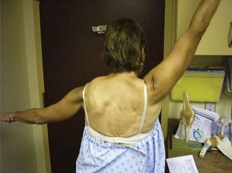
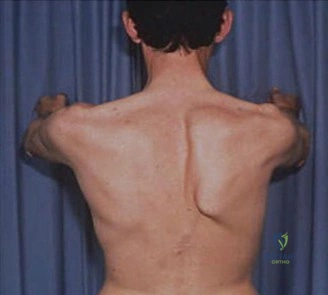
A 32-year-old amateur weightlifter presents with an 8-month history of right shoulder pain and "clicking" during overhead movements. He describes a feeling of instability. On examination, he demonstrates a prominent scapular medial border during a wall push-up test. How do you approach the clinical classification and immediate diagnostic workup?
Candidate: I suspect long thoracic nerve palsy. I would classify the winging as medial and confirm it with the wall push-up test. I need to order an EMG and nerve conduction study to look for signs of denervation in the serratus anterior. I would also order an MRI of the brachial plexus to rule out compression from masses or fibrous bands.
Failing to distinguish between medial and lateral winging, or jumping straight to "surgical options." A poor candidate ignores the importance of ruling out other nerve palsies (like the spinal accessory nerve) and fails to mention the crucial timeline (8 months), which directly influences the surgical vs. conservative decision pathway.
Start by categorizing the winging: "This is medial scapular winging, characteristic of long thoracic nerve (LTN) palsy affecting the serratus anterior."
1. Differential Diagnosis: Differentiate from lateral winging (spinal accessory nerve/trapezius) and subtle winging (dorsal scapular nerve/rhomboids).
2. Workup: Gold standard is EMG/NCS to differentiate neurapraxia from neurotmesis and assess reinnervation.
3. Imaging: MRI of the shoulder/brachial plexus is mandatory to rule out compressive masses (e.g., cysts/tumors) or anatomical anomalies, and to assess for fatty infiltration/atrophy of the serratus anterior muscle, which dictates the surgical salvage potential.
The patient has failed 12 months of intensive physical therapy. EMG shows persistent denervation in the serratus anterior. You decide to proceed with surgical intervention. Discuss your choice of technique given the anatomy.
Candidate: Given the chronicity of 12 months and failed conservative management, I would offer a nerve transfer. I would use the thoracodorsal nerve to reinnervate the long thoracic nerve. This is preferred over a muscle transfer if the muscle is not yet severely atrophied.
Forgetting to discuss the viability of the muscle. Candidates often fail to mention that if the serratus anterior is irreversibly atrophied (>18-24 months), a nerve transfer is futile. They also frequently miss the importance of "tension-free" coaptation and the role of the thoracodorsal nerve donor site morbidity.
Structure the answer by Reconstructive Hierarchy:
1. Nerve Transfer: Thoracodorsal nerve to LTN is the choice for chronic but potentially recoverable serratus anterior (typically 6-18 months). Mention using a branch of the thoracodorsal nerve to ensure minimal donor site morbidity for the latissimus dorsi.
2. Technical Pearl: Emphasize the need for microsurgical, tension-free epineurial repair.
3. Salvage/Late stage: If the muscle is fibrotic or the timeframe exceeds 24 months, shift the plan to a Pectoralis Major Transfer (Sahrmann procedure), which acts as a dynamic mechanical tether rather than a reinnervation strategy.
You have performed a thoracodorsal to long thoracic nerve transfer. Describe the intraoperative view and the key steps in ensuring a successful coaptation.
Candidate: I would use the lateral approach, identifying the long thoracic nerve on the chest wall. I'd then locate the thoracodorsal nerve branches near the latissimus. I would perform the nerve transfer using microsutures.
Ignoring the "free drape" position. Without the arm draped freely, it is impossible to maintain the necessary tension-free repair. Candidates often fail to mention using the operating microscope or loupes, which are mandatory for fascicular alignment.
1. Positioning: Lateral decubitus with the arm draped free to allow limb manipulation and eliminate tension on the nerve coaptation.
2. Exposure: Longitudinal incision along the mid-axillary/posterior-axillary line.
3. Dissection: Trace the LTN proximal to the serratus digitations and the thoracodorsal nerve branch to the latissimus.
4. Coaptation: Use the operating microscope. Perform a tension-free, end-to-end epineurial repair using 9-0 or 10-0 monofilament sutures. Ensure no nerve twisting occurs to allow proper axonal regeneration across the coaptation site.
Detailed Chapters & Topics
Dive deeper into specialized chapters regarding shoulder-and-elbow-cases-scapular-winging