Suprascapular Nerve Entrapment: Surgical Anatomy, Diagnosis, and Management
Key Takeaway
Suprascapular Nerve Entrapment (SSNE) is a complex cause of shoulder pain and dysfunction, often overlooked. It results from nerve compression, commonly at the suprascapular or spinoglenoid notches. Diagnosis involves integrating clinical signs like deep pain, weakness, and atrophy with electrodiagnostic studies and advanced imaging for precise localization and effective management.
A 26-year-old volleyball player presents with a 4-month history of deep, aching posterior shoulder pain and a feeling of "heaviness" in the arm. He notes increasing difficulty with overhead serving. On physical examination, you note subtle atrophy in the supraspinatus and infraspinatus fossae. What is your immediate diagnostic work-up to investigate for potential suprascapular nerve entrapment?
Candidate: I would start with an MRI of the shoulder to look for cysts or masses. I'd also order EMG/NCS to confirm nerve involvement and help localize the lesion. Finally, I'd perform a clinical examination, including the nerve stretch test.
The candidate fails to distinguish between the two anatomical sites (suprascapular vs. spinoglenoid). They also miss the importance of advanced imaging specific to nerve pathology (MRN) and fail to mention that EMG changes take time to manifest, potentially yielding a false negative in early stages.
I would structure the work-up as follows: 1) Clinical correlation: Look for supraspinatus AND infraspinatus atrophy (proximal) vs. isolated infraspinatus (distal). Perform provocative testing (cross-body adduction/nerve stretch). 2) Imaging: MRI/MRN to visualize the course of the nerve and identify space-occupying lesions like paralabral cysts. Crucially, I would specifically look for a posterior labral tear as the source of a spinoglenoid cyst. 3) Electrodiagnostics: EMG/NCS to assess for denervation and confirm the level of entrapment, while acknowledging that findings depend on the duration of symptoms.
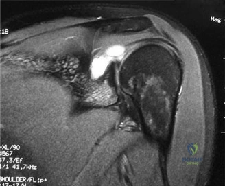
During the assessment, your imaging reveals a large ganglion cyst at the spinoglenoid notch. Please describe the relationship of the nerve to this anatomy and the critical surgical considerations when approaching this lesion.
Candidate: The suprascapular nerve enters the spinoglenoid notch beneath the inferior transverse scapular ligament. The cyst compresses the nerve here. I would decompress the cyst and release the ligament.
The candidate ignores the etiology of the cyst. If you only decompress the cyst without addressing the labral tear (the source of the fluid), you have a high risk of recurrence. They also fail to mention the importance of nerve visualization to avoid iatrogenic injury.
The suprascapular nerve passes through the spinoglenoid notch beneath the inferior transverse scapular ligament. Crucially, I must address the source of the ganglion. Most spinoglenoid cysts originate from a posterior labral tear. The Gold Standard is an arthroscopic approach: first, address the intra-articular labral pathology to stop the "one-way valve" effect producing the cyst, then perform a safe arthroscopic decompression of the cyst and release the spinoglenoid ligament, ensuring constant visualization of the nerve to avoid iatrogenic injury.
Consider the anatomical relationship at the suprascapular notch. Contrast this with the spinoglenoid notch, and explain why the surgical approach for decompression differs.
Candidate: At the suprascapular notch, the nerve goes under the superior transverse scapular ligament, but the artery goes over it. This makes it a tighter, more dangerous area to operate on compared to the spinoglenoid notch.
Insufficient detail regarding the "Army-Navy" mnemonic (Artery over, Nerve under) and failing to discuss why open vs arthroscopic preference shifts based on location (ossification of ligament vs. simple cysts).
Anatomically, at the suprascapular notch, the "Artery goes Over and Nerve goes Under" (the superior transverse scapular ligament). Because this site is often affected by bony spurs or ossification of the ligament, an open approach is frequently preferred for direct visualization and safe management of the vessels. Conversely, the spinoglenoid notch is typically more amenable to arthroscopic intervention, as it is usually compressed by cysts related to the glenohumeral joint, allowing for simultaneous intra-articular work.
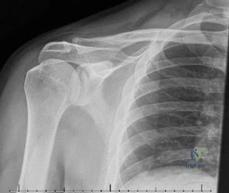
Look at this radiograph/image. If you decide to proceed to surgery, what are the key risks you would discuss during the informed consent process?
Candidate: I would warn the patient about infection, bleeding, and nerve damage. Also, that the pain might not go away completely and they might need a long time for the muscle strength to return.
The candidate provides a generic list. A high-scoring candidate must specifically address the "chronicity" aspect: if there is already fatty atrophy on the MRI, they must manage the patient's expectations that motor function might NOT recover.
In addition to standard surgical risks (infection, hematoma, scarring), I would specifically discuss: 1) Iatrogenic nerve injury: The potential for permanent deficit during neurolysis. 2) Incomplete recovery: Particularly the difference between pain relief (often good) and motor recovery (often guarded if there is pre-existing fatty atrophy). 3) Recurrence: The risk of cyst reformation if the labral pathology is not fully addressed. 4) Timeline: Emphasizing that nerve regeneration is slow, potentially taking 6-18 months.
