Periprosthetic Femoral Fracture with Aseptic Stem Loosening: A Comprehensive Case Study
Key Takeaway
Periprosthetic femoral fractures with aseptic stem loosening are diagnosed through clinical presentation of acute pain and inability to bear weight post-trauma. Imaging, including AP/lateral X-rays and CT scans, is crucial to identify fracture patterns, confirm stem instability (e.g., lucency, subsidence), and assess bone loss for surgical planning.
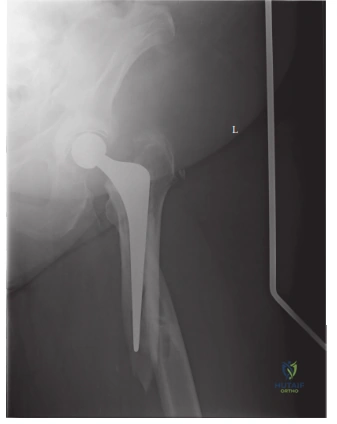
You are in the emergency department assessing a 78-year-old female who sustained a fall. She is 10 years post-op from a left Total Hip Arthroplasty. She presents with inability to weight-bear and a classic shortened, externally rotated deformity. Looking at the radiograph provided, how would you classify this injury, and what are the three critical factors you must address to guide your management?

Candidate: I would classify this as a Vancouver B2 periprosthetic femoral fracture. The stem is loose, evidenced by subsidence and radiolucent lines, but the bone stock appears sufficient for distal fixation. The three factors are: implant stability, quality of bone stock, and the level/geometry of the fracture.
Candidates often fail to provide the specific classification or simply call it a "fracture around the stem." A major failing is neglecting to mention the stability of the implant; assuming it is stable leads to a recommendation for ORIF, which is a catastrophic error for a loose B2 stem.
State clearly: "This is a Vancouver B2 periprosthetic fracture." The three pillars for management are: 1) Implant Stability: Determining if the current stem is fixed or loose (this dictates ORIF vs. Revision). 2) Bone Stock: Evaluating the remaining proximal and diaphyseal bone to ensure adequate purchase for a revision implant. 3) Infection Status: Ruling out occult PJI, which would change the management to a two-stage revision.
During your planning for the revision of this B2 fracture, why might you consider using an Extended Trochanteric Osteotomy (ETO), and what are the specific technical risks associated with this step?
Candidate: I would use an ETO to allow safe removal of the loose stem and any residual cement, and to provide direct visualization of the canal to prevent iatrogenic perforation. The risks include non-union of the trochanteric fragment and the potential for proximal migration or abductor deficiency.
Failing to mention the vascular supply to the greater trochanter or neglecting to address how you secure the fragment (cabling/tension bands) at the end of the case. Also, missing the risk of "pedestal" formation if the osteotomy is poorly executed.
Explain that an ETO is "the key to the canal." It allows for: 1) Safe extraction of well-fixed distal cement or stems without cortical breach. 2) Accurate entry point for reaming. Technical Risks: Include non-union of the osteotomy site (requiring secure tension band/cable fixation), and the risk of compromising the blood supply to the femur. It is critical to perform the osteotomy in a controlled, subvastus fashion to preserve the soft-tissue sleeve and vascularity.
Look at this image of the surgical site. You have decided on a long, fluted, tapered titanium stem. Why choose this specific implant design over a cemented stem in this scenario?
Candidate: I would choose a long, fluted, tapered stem because it provides excellent primary fixation in the diaphysis. The flutes provide rotational stability and the tapered geometry provides axial stability. Cementing would be very difficult given the fracture and the likely poor interdigitation in the compromised medullary canal.
Suggesting a cement-in-cement revision without justifying why it would be superior to a fluted stem, or failing to acknowledge the risk of cement extrusion through the fracture site causing thermal necrosis or vascular injury.
The "Wagner-style" fluted tapered stem is the gold standard for B2/B3 fractures. 1) Biomechanical Advantage: Provides immediate "interference fit" stability in the intact diaphysis distal to the fracture. 2) Rotational Control: Longitudinal flutes engage the endosteal cortex, which is vital in a fractured femoral shaft. 3) Avoidance of Cement: Cementing is contraindicated in long-gap fractures due to the risk of soft-tissue extravasation and the difficulty of obtaining a dry, contained field necessary for bone cement mantle integrity.
