Pediatric Osteomyelitis: Epidemiology, Surgical Anatomy, and Clinical Approach

Key Takeaway
Pediatric osteomyelitis is a serious bone infection in children, often affecting long bone metaphyses. *Staphylococcus aureus* is the leading pathogen. Unique pediatric skeletal anatomy, such as transphyseal vessels in infants and the thick periosteum, profoundly impacts infection spread. Prompt and precise evaluation is critical to mitigate long-term morbidity and preserve growth.
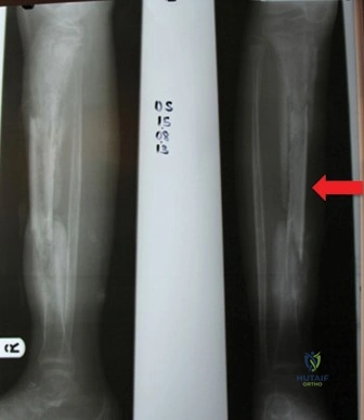
A 4-year-old child presents with a 3-day history of refusing to bear weight on the left leg and local tenderness over the distal femur. A temperature of 38.5°C is recorded. You are presented with the following radiograph.
What is your immediate differential diagnosis, and how would you structure your further assessment?
Candidate: I would consider acute hematogenous osteomyelitis as the primary diagnosis. My differential would include septic arthritis, trauma, or a malignancy like Ewing sarcoma. I would perform a thorough clinical exam, order blood tests including inflammatory markers (ESR, CRP), blood cultures, and obtain an MRI of the limb.
The candidate jumps straight to "MRI" without acknowledging the importance of systemic screening or the age-specific etiology. Failing to mention "septic arthritis" as a critical rule-out (especially in children where the metaphysis is intracapsular) is a major red flag for examiners.
Start with a structured approach: "I would approach this child with a high index of suspicion for acute hematogenous osteomyelitis." Define the differential based on the "Septic Hip/Knee/Bone" workup: (1) Septic arthritis, (2) Osteomyelitis, (3) Trauma/Occult fracture, (4) Malignancy, (5) Transient synovitis. Mention specific age-related pathogens (*Kingella kingae* in this age group). State: "I would obtain a CBC, ESR, CRP, and blood cultures immediately. I would order an MRI—the gold standard—to localize the infection and check for subperiosteal abscesses or physeal involvement. I would also include ultrasound if I suspect joint effusion."
The child's inflammatory markers are significantly elevated. An MRI confirms a large metaphyseal subperiosteal abscess. You proceed to surgical debridement. Explain your surgical technique and key safety considerations regarding the physis.
Candidate: I would perform a longitudinal incision over the abscess, drain the purulence, and perform a limited cortical fenestration or drill holes. I would be very careful to avoid the growth plate to prevent arrest.
Vague technique description. Failing to mention sending tissue for formal culture (aerobic/anaerobic/fungal), failing to discuss the importance of "meticulous debridement" while preserving the "structural integrity" of the bone, or ignoring the need for post-operative drain management.
Structure the answer: (1) Exposure through an internervous plane to minimize soft tissue trauma. (2) Subperiosteal abscess drainage and culture collection. (3) Cortical decompression via "multiple small drill holes" or a "cortical window" if the abscess is large, emphasizing that these must be "distal to the physis" to protect growth. (4) Copious irrigation (3-6L) with pulsatile lavage. (5) Multidisciplinary collaboration regarding IV antibiotics. (6) Protecting the physis by avoiding aggressive curettage near the growth cartilage, acknowledging the risk of iatrogenic physeal arrest.
