Beyond Arthroscopy: Surgical Hip Dislocation for Complex Instability
Key Takeaway
Learn more about Beyond Arthroscopy: Surgical Hip Dislocation for Complex Instability and how to manage it. **Surgical hip dislocation** is a comprehensive treatment strategy employed for complex hip pathologies like multifactorial dysplasia, high femoral anteversion, and joint instability following previous interventions. This approach facilitates extensive reconstructive procedures such as labrum reconstruction, subtrochanteric derotation, and periacetabular osteotomy. It aims to address underlying mechanical issues, restore hip function, and alleviate persistent symptoms.
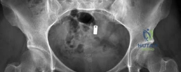
You are reviewing a 32-year-old female patient who presents with chronic, debilitating hip pain. She underwent an arthroscopic labral debridement 18 months ago, which provided no relief. Her clinical examination reveals a positive impingement test and a distinct, reproducible apprehension sign in extension and external rotation. Her AP pelvis radiograph is shown below.
How do you interpret this clinical and radiographic presentation, and what is the underlying biomechanical failure?
Candidate: The patient has symptomatic hip dysplasia and has failed previous arthroscopic management. The labral debridement has likely compromised the fluid seal of the joint, leading to microinstability. The apprehension sign suggests the joint is failing to contain the femoral head during extension and external rotation.
Failing to identify the "iatrogenic" nature of the instability or focusing only on the dysplasia. A weak candidate ignores the significance of the labral resection and the potential role of femoral version abnormalities in the patient's biomechanical failure.
Acknowledge this as a "failed arthroscopy in the setting of structural instability." Describe the pathology as an "iatrogenic loss of the fluid seal" in a patient with borderline/frank dysplasia. Emphasize that the labrum was a compensatory stabilizer; its resection in a dysplastic hip leads to anterolateral microinstability, articular cartilage shear, and chronic pain. Mention the need to investigate for concomitant femoral version abnormalities (torsion) which, if present, exacerbate this anterior uncoverage.
You have decided to proceed with a Surgical Hip Dislocation (SHD). A critical component of this procedure is the safe exposure of the hip joint while maintaining the blood supply to the femoral head. Can you describe the surgical anatomy of the femoral head's blood supply and how the Ganz approach specifically protects it?
Candidate: The blood supply is dominated by the medial circumflex femoral artery (MFCA). The Ganz approach uses a trochanteric flip osteotomy. By keeping the short external rotators attached to the posterior femoral neck, you protect the retinacular vessels that run along the posterosuperior aspect of the neck.
Vague explanations of "the arteries at the back." A senior candidate must specifically mention the MFCA course (between obturator externus and quadratus femoris) and the critical importance of keeping the external rotators intact to prevent tension on the retinacular vessels.
Detail the path of the MFCA: it arises from the profunda femoris, passes between the pectineus and iliopsoas, then between the obturator externus and quadratus femoris. The deep branch crosses the posterior obturator externus tendon to enter the trochanteric fossa. The Ganz "digastric" osteotomy keeps the external rotators (obturator internus/externus, gemelli, piriformis) attached to the distal femur, which acts as a "vascular tether" protection, preventing disruption of the posterosuperior retinacular vessels during dislocation.
During your work-up, you calculate the patient's femoral version on CT, revealing 38° of anteversion. How does this finding modify your surgical plan for the SHD?
Candidate: High femoral anteversion at 38° means the femoral head is functionally uncovered anteriorly. I would need to perform a derotational femoral osteotomy (DFO) in addition to the SHD and potential labral reconstruction to restore normal biomechanical orientation.
Focusing only on the acetabulum (PAO). Examiners are looking for the realization that "three-dimensional deformities require three-dimensional solutions." Missing the need for a DFO in the face of excessive anteversion is a significant oversight for a senior candidate.
State clearly that excessive anteversion drives the femoral head anteriorly, exacerbating the instability caused by the dysplastic acetabulum. The treatment of choice is an SHD (to perform labral reconstruction and intra-articular work) combined with a subtrochanteric derotational femoral osteotomy (DFO) to reduce anteversion to 10–15°. Emphasize that failing to correct the torsion would leave the patient with persistent microinstability despite perfect acetabular coverage.
