Bernese Periacetabular Osteotomy: Your Definitive Case Title Bernese
Key Takeaway
Your ultimate guide to Bernese Periacetabular Osteotomy: Your Definitive Case Title Bernese starts here. A 21-year-old female with severe acetabular dysplasia, lateral femoral head migration, and labral tear, experienced chronic groin and trochanteric pain despite two years of non-surgical treatment. The chosen intervention for this **case title bernese** periacetabular osteotomy involved surgically reorienting the acetabulum to correct the hip instability and alleviate pain.
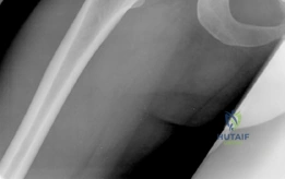
You are presented with this 21-year-old female patient complaining of chronic, activity-related groin pain. Describe your systematic radiographic approach to evaluating this hip, and identify the key findings visible in the provided image.
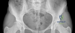
Candidate: I would look at the AP pelvis to check for hip dysplasia. The patient has a shallow socket, the Shenton's line is broken, and there is lateral uncoverage. The LCEA looks reduced, likely confirming DDH.
Candidates often jump straight to "dysplasia" without a structured radiographic approach. They fail to mention the Tönnis angle, the specific measurement of the LCEA, or checking the posterior wall/acetabular version, which are essential for surgical planning.
I would perform a systematic assessment of the AP pelvis: 1. Coverage: Measure the LCEA (Wiberg); values <20° confirm dysplasia. 2. Acetabular Inclination: Evaluate the Tönnis angle to assess acetabular steepness. 3. Stability: Assess the Shenton’s line; a disruption confirms proximal/lateral migration. 4. Version: Screen for signs of retroversion (crossover, ischial spine, and posterior wall signs). 5. Joint Integrity: Grade the Tönnis arthritic change (0-3). The image shows clear lateral uncoverage and a broken Shenton’s line, classic for developmental dysplasia.
The patient has failed 24 months of conservative management. You are considering a Bernese Periacetabular Osteotomy (PAO). Explain the clinical relevance of this specific radiographic view in your preoperative planning.
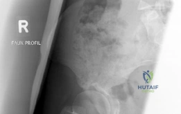
Candidate: This is an abduction view. It shows how the acetabulum covers the femoral head when moved. It's used to see if the PAO will work by showing if the joint space becomes more even.
Failure to mention the concept of "joint congruency." A candidate who misses the "hinge abduction" sign—where the joint space narrows medially—risks recommending a PAO in a patient with pre-existing arthrosis who will not benefit from joint preservation.
This is the AP Abduction view, the "litmus test" for joint preservation. It simulates the rotation of the acetabular fragment during a PAO. If the hip achieves congruency—where the joint space is restored—it confirms the joint is salvageable. If the joint remains incongruent or demonstrates "hinging" (medial joint space narrowing), it indicates the cartilage is too damaged, and a PAO is contraindicated as it would accelerate arthrosis.
During the Bernese PAO, you must perform four osteotomies. Identify the most technically demanding cut and explain the risks associated with it, particularly regarding the neurovascular structures.
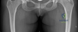
Candidate: The retroacetabular cut is the hardest. You have to be careful not to hit the sciatic nerve behind the hip or enter the joint capsule. You need to angle the osteotome correctly.
Candidates often provide vague anatomical warnings. Examiners want to hear specific technical strategies: e.g., keeping the hip in flexion/internal rotation to relax the sciatic nerve and utilizing specific osteotome angles.
The retroacetabular osteotomy is the most challenging. The primary risk is injuring the sciatic nerve posteriorly or iatrogenically breaching the hip joint superiorly. Technical pearls: 1. Keep the hip in slight flexion and internal rotation to relax the sciatic nerve and move it away from the osteotomy path. 2. Use tactile feedback and fluoroscopic guidance for the osteotome angle (approx 15° distal/10° medial). 3. Protect the obturator neurovascular bundle using a blunt retractor during the pubic osteotomy.
