Periacetabular Osteotomy: End Hip Pain, Improve Flexion and Internal Motion
Key Takeaway
Looking for accurate information on Periacetabular Osteotomy: End Hip Pain, Improve Flexion and Internal Motion? A 28-year-old female presented with over one year of right hip anterior groin pain, exacerbated by activities involving **flexion and internal** rotation. Clinical examination revealed positive specific tests, and radiological imaging confirmed bilateral symptomatic mild hip dysplasia, characterized by femoral head undercoverage. Treatment involved staged bilateral periacetabular osteotomies (PAOs) to correct the dysplasia.
A 28-year-old female presents with insidious onset of right groin pain, worsening with prolonged standing. On examination, she has a positive impingement sign and mild abductor fatigue. Her AP pelvis radiograph is provided below. How would you interpret these findings, and what is the primary biomechanical rationale for the procedure indicated?
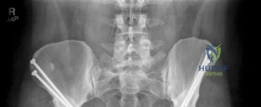
Candidate: The patient has developmental dysplasia of the hip (DDH). The radiographs show a shallow acetabulum with poor lateral femoral head coverage. The PAO is indicated to reorient the socket. By lateralizing the acetabular rim and medializing the joint center, we decrease the joint reaction forces and improve the abductor moment arm, which prevents further cartilage wear.
Failing to mention the "static overload" vs. "dynamic instability" mechanism. Candidates often omit the crucial point regarding the preservation of the posterior column, which is what distinguishes the Ganz PAO from older osteotomies (like the Salter or Steel) that compromise the pelvic ring/birth canal.
The patient exhibits radiographic features of DDH: increased Tonnis angle and decreased lateral CE angle. The PAO addresses the pathophysiology of increased contact stress (due to reduced surface area) and high resultant joint reaction forces (due to a lateralized center of rotation). By performing the Ganz osteotomy, we achieve multiplanar reorientation while preserving the posterior column. This maintains pelvic ring stability, allows for early mobilization, and avoids distorting the birth canal—a key consideration for a patient of childbearing age.
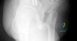
During the intrapelvic dissection, you are preparing to perform the ischial osteotomy. What are the specific neurovascular structures at risk in this region, and how do you mitigate the risk of injury to the sciatic nerve?
Candidate: The sciatic nerve is at risk posteriorly, as are the obturator nerve and vessels medially. To protect the sciatic nerve, I must keep the osteotome strictly anterior to the posterior cortex of the posterior column and use fluoroscopic guidance, specifically an obturator oblique view, to ensure the cut is complete but does not exit the posterior bone.
Ignoring the "corona mortis" when discussing the pelvic dissection. Examiners look for candidates who acknowledge the vascular risks anteriorly (external iliac/obturator) as well as the sciatic nerve posteriorly.
The ischial osteotomy is the most hazardous step. The sciatic nerve lies just posterior to the quadrilateral plate. Risk mitigation includes using a specialized curved Ganz osteotome, direct subperiosteal dissection along the quadrilateral plate to identify the safe corridor, and continuous fluoroscopic confirmation (obturator oblique/false profile views). I must also mention the 'corona mortis'—an anastomosis between the external iliac and obturator systems—which must be identified and ligated during the pubic dissection to avoid significant hemorrhage.
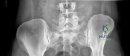
The image below shows a clinical scenario where the osteotomies have been completed. What is the significance of the "Tonnis angle" and "Lateral Center Edge Angle" in your postoperative assessment, and how do you know if you have overcorrected?
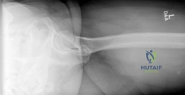
Candidate: The goal is to normalize the lateral center edge angle to 25–35 degrees and reduce the Tonnis angle to less than 10 degrees. Overcorrection is a major concern because it causes iatrogenic pincer-type impingement, which restricts ROM and causes labral pain.
Failing to mention the intraoperative range of motion (ROM) check. Radiographic parameters are static; the functional impingement test under fluoroscopy is the definitive way to rule out overcorrection before finalizing fixation.
The radiographic targets are an LCEA of 25–35° and a Tonnis angle < 10°. However, these are guidance markers. The most critical 'Gold Standard' step is the intraoperative ROM check: if the hip encounters hard impingement in flexion and internal rotation, it indicates over-coverage (iatrogenic pincer). This must be corrected by re-mobilizing the fragment or, if necessary, performing rim-trimming before final fixation to ensure long-term joint health.
