Conservative Head Reduction Osteotomy: Restoring Young Hip Function
Key Takeaway
Learn more about Conservative Head Reduction Osteotomy: Restoring Young Hip Function and how to manage it. **Head reduction osteotomy** is a surgical procedure for complex hip deformities, often chosen for conditions like Perthes disease causing pain and limited motion. This technique involves reducing the femoral head, lengthening the femoral neck, and advancing the greater trochanter. Its goal is to improve hip mechanics, alleviate impingement, and restore function in cases with significant anatomical irregularities like head flattening or extrusion.
A 22-year-old patient presents with chronic, activity-related groin pain and a persistent limp. You review their imaging. Describe the pathophysiology of the condition shown in this radiograph and the mechanical consequence of the deformity.
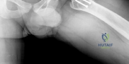
Candidate: The radiograph shows a deformed femoral head, likely secondary to Perthes disease, known as coxa magna. This causes femoroacetabular impingement because the head is too large for the socket. During abduction, the head hinges against the lateral acetabular rim, which causes pain and eventual cartilage wear.
Failing to use the specific biomechanical term "hinge abduction." A poor candidate describes the shape but fails to explain the kinematic failure—specifically how the abnormal head geometry levers the medial articular surface away from the weight-bearing acetabulum, leading to eccentric loading.
Identify the diagnosis as sequelae of pediatric hip disease resulting in 'Coxa Magna.' Explain the biomechanics: The aspherical/enlarged head leads to 'hinge abduction,' where the lateral prominence levers the femoral head out of the acetabulum during abduction. This results in non-concentric articulation, loss of articular cartilage contact, and rapid development of secondary osteoarthritis.
You have decided to proceed with a Conservative Head Reduction Osteotomy. The most critical step is the surgical approach to ensure the viability of the femoral head. Describe the anatomy of the vascular supply at risk and how the Ganz surgical dislocation protects it.
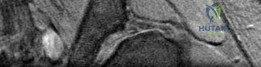
Candidate: The main blood supply is the medial femoral circumflex artery (MFCA). It travels along the obturator externus muscle. The Ganz approach uses a trochanteric flip osteotomy, which keeps the muscles attached to the greater trochanter, allowing us to safely dislocate the hip without cutting the retinacular vessels that run on the posterior-superior neck.
Omitting the critical role of the "short external rotators" and the "obturator externus." Simply saying "the MFCA" is insufficient; the examiner wants to hear that you understand the relationship between the surgical interval (piriformis/gluteus minimus) and the preservation of the vascular leash.
The primary supply is the deep branch of the MFCA. It runs posterior to the obturator externus and anterior to the quadratus femoris. The Ganz surgical dislocation protects this by performing a trochanteric flip osteotomy, which leaves the short external rotators intact. This maintains the retinacular vessels, which enter the femoral head at the posterosuperior aspect of the neck, completely undisturbed during the 360-degree joint visualization.
What are the primary complications of this procedure, and how do you counsel a patient regarding the risks of revision to total hip arthroplasty (THA)?
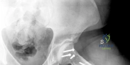
Candidate: The biggest risk is iatrogenic AVN from damaging the blood supply. Other risks include non-union of the trochanteric osteotomy, heterotopic ossification, and hardware failure. I would explain to the patient that this is a joint-preservation procedure, not a cure, and it aims to delay, rather than eliminate, the need for a future THA.
Providing a generic list of surgical complications. The FRCS examiner expects you to rank them by clinical significance (e.g., AVN is the "catastrophic" complication, while heterotopic ossification is a "frequent" complication) and mention prophylaxis like Indomethacin for HO.
Structure the answer into 'Vascular,' 'Mechanical,' and 'Biological' complications. Highlight iatrogenic AVN (2-5%) as the most significant, followed by trochanteric non-union (3-8%) and heterotopic ossification (10-20%). Regarding counseling: frame it as a 'bridge' procedure to delay THA. Explicitly state that if AVN or structural collapse occurs, the threshold for conversion to THA is lowered, and note the potential technical challenges of revision due to prior hardware and altered anatomy.
