Part of the Master Guide

Revision THA Acetabulum: Overcoming Paprosky IIIA & Metallosis

Acetabular Revision: Solving Challenges Post Two-Stage Arthroplasty

17 Apr 2026 14 min read 146 Views
Illustration of title acetabular revision - Dr. Mohammed Hutaif

Key Takeaway

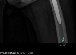
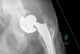
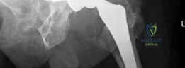
For anyone wondering about Acetabular Revision: Solving Challenges Post Two-Stage Arthroplasty, A **title acetabular revision** is a complex procedure addressing issues like septic loosening, dislocation, and significant osteolysis in total hip arthroplasty. For a 71-year-old male with a dislocated left THA due to infection and periacetabular bone loss, treatment involved a two-stage exchange followed by revision with a press-fit hemispherical cup and screw fixation.

Introduction and Epidemiology

Two-stage exchange arthroplasty remains the gold standard for the treatment of chronic periprosthetic joint infection of the hip. While highly effective, a subset of patients will present for subsequent acetabular revision due to persistent or recurrent infection, aseptic loosening, component malposition, or progressive bone loss following the reimplantation stage. Addressing these challenges requires a sophisticated understanding of complex acetabular reconstruction. The increasing incidence of primary total hip arthroplasty coupled with an aging population and higher rates of comorbidities contributes to a rising burden of periprosthetic joint infection, subsequently increasing the demand for complex revision surgeries.

Image

Periprosthetic joint infection is a devastating complication, with reported incidences ranging from 0.5% to 2% for primary total hip arthroplasty and significantly higher, up to 10% or more, for revision procedures. Following a successful two-stage exchange, approximately 5% to 10% of patients may still experience treatment failure, necessitating further intervention. This failure can stem from a variety of causes, including inadequate debridement during the first stage, biofilm persistence, suboptimal local antibiotic delivery, or the development of aseptic mechanical complications post-reimplantation.

The "hostile acetabulum" encountered in these scenarios is often the result of cumulative bone loss from the initial infection, the mechanical wear of an articulating antibiotic spacer, and the subsequent reimplantation and eventual failure of the revision components. Persistent deep infection is a primary concern, but mechanical failure of the acetabular component due to underlying bone deficiency, improper component selection, or technical errors during reimplantation also mandates revision. The decision-making process for these complex cases is multifactorial, requiring a comprehensive diagnostic approach and a tailored surgical strategy to restore pelvic biomechanics and eradicate infection.

Surgical Anatomy and Biomechanics

A thorough understanding of acetabular anatomy and biomechanics is paramount for successful acetabular revision surgery. The acetabulum is a complex structure formed by the confluence of the ilium, ischium, and pubis, articulating with the femoral head. Its integrity is crucial for load bearing and hip joint stability.

Key Anatomical Considerations

The structural foundation of the acetabulum relies on the pelvic columns. The anterior iliopectineal column and posterior ilioischial column provide the primary structural support. Defects in these columns significantly compromise acetabular stability and dictate specific reconstructive strategies. The acetabular quadrants superior, anterior, posterior, and inferior help delineate bone loss patterns and identify safe zones for hardware fixation.

Image

The Paprosky classification system is widely utilized to categorize acetabular bone defects based on the amount of remaining host bone, specifically the integrity of the rim and columns.
* Type I: Minimal bone loss, intact rim and columns.
* Type II: Significant cavitary defects, intact columns.
* IIA: Superior migration, intact ischium.
* IIB: Medial migration, intact ischium.
* IIC: Global cavitary defect.
* Type III: Extensive bone loss involving rim and columns.
* IIIA: Greater than 50% host bone contact for an uncemented cup, often superior-posterior column defect.
* IIIB: Less than 50% host bone contact for an uncemented cup, often extensive column and medial wall involvement.
* Type IV: Pelvic discontinuity, representing a complete separation of the superior and inferior halves of the acetabulum.

Image

Proximity to critical neurovascular structures dictates the surgical approach and the limits of safe dissection. The superior gluteal nerve and artery, exiting superior to the piriformis, are vulnerable during superior dissection, especially with revision via posterior or modified lateral approaches. The sciatic nerve courses posterior to the acetabulum and is highly susceptible to injury during the posterior approach, component insertion, or limb lengthening. The femoral nerve and vessels, located anterior to the acetabulum, are at risk during anterior approaches, aberrant retractor placement over the anterior rim, or when placing anterior column screws outside of the designated safe zones. Additionally, the corona mortis, an anastomosis between the obturator and external iliac vascular systems, lies on the posterior aspect of the superior pubic ramus and can cause life-threatening hemorrhage if violated during anterior column exposure or screw placement.

Biomechanical Objectives

The primary biomechanical goals in acetabular revision are the restoration of the anatomic center of rotation, achieving initial rigid mechanical stability of the implant, and facilitating long-term biologic fixation. Superior and lateral displacement of the hip center increases joint reactive forces and decreases the abductor moment arm, leading to accelerated wear, instability, and an altered gait pattern. Re-establishing the center of rotation inferiorly and medially to its native position optimizes abductor mechanics and minimizes sheer forces on the reconstructive construct.

Indications and Contraindications

The decision to proceed with a subsequent revision after a failed two-stage arthroplasty requires careful patient selection and a thorough risk-benefit analysis. The distinction between septic failure and aseptic failure is the primary driver of the algorithmic approach.

Diagnostic Criteria for Intervention

Diagnosis of recurrent or persistent periprosthetic joint infection relies on the International Consensus Meeting criteria, utilizing serum markers such as Erythrocyte Sedimentation Rate and C-Reactive Protein, alongside synovial fluid analysis for cell count, neutrophil percentage, and alpha-defensin. Aseptic loosening is diagnosed radiographically via progressive radiolucent lines, component migration, or hardware failure in the absence of infectious markers.

Clinical Scenario Operative Indications Non Operative Indications
Aseptic Loosening Progressive component migration, severe pain, impending catastrophic bone failure, Paprosky III or IV defects. Asymptomatic stable radiolucencies, medically unfit for major surgery, acceptable functional status.
Recurrent Infection Acute or chronic recurrent periprosthetic joint infection, sinus tract formation, systemic sepsis originating from the joint. Terminal illness, absolute medical contraindications to anesthesia, suppressive antibiotic therapy success in a stable implant.
Instability Recurrent dislocations due to component malposition, severe abductor deficiency requiring constrained or dual mobility constructs. Single dislocation event successfully managed closed, non-compliance with postoperative precautions.
Pelvic Discontinuity Documented separation of the hemipelvis with symptomatic instability or progressive deformity. Asymptomatic fibrous non-union in low-demand, elderly patients with high surgical risk.

Contraindications to complex acetabular reconstruction include active, untreated systemic infections, severe peripheral vascular disease precluding adequate wound healing, and profound medical comorbidities that yield an unacceptable perioperative mortality risk. In cases of recurrent, intractable infection with massive bone loss where reconstruction is impossible, salvage procedures such as resection arthroplasty or hip disarticulation must be considered.

Pre Operative Planning and Patient Positioning

Meticulous preoperative planning is the cornerstone of success in complex acetabular revision. The "hostile acetabulum" presents unpredictable bone defects that require a comprehensive armamentarium of reconstructive options available in the operating room.

Image

Advanced Imaging and Templating

Standard anteroposterior pelvis and cross-table lateral radiographs are mandatory. Judet views (iliac and obturator obliques) are critical for assessing the integrity of the anterior and posterior columns. However, plain radiography often underestimates the extent of osteolysis and bone loss.

High-resolution Computed Tomography with Metal Artifact Reduction Sequence is essential for three-dimensional evaluation of the remaining bone stock. Computed Tomography allows for the definitive diagnosis of pelvic discontinuity, the assessment of the medial wall, and the quantification of ischial and iliac bone available for fixation.

Image

Digital templating is performed to estimate the size of the required acetabular component, the necessity for porous metal augments, and the potential need for a cup-cage construct or a custom triflange acetabular component. In cases of severe Paprosky IIIB defects or pelvic discontinuity, three-dimensional printed pelvic models can be generated from the Computed Tomography data. These models allow for tactile preoperative planning, trial of implants, and the manufacturing of patient-specific custom implants.

Patient Positioning and Preparation

Patients are typically positioned in the lateral decubitus position. Rigid pelvic fixation using peg boards or specialized pelvic positioners is critical, as the application of significant force is often required during component extraction and impaction. The surgeon must ensure the pelvis is perfectly vertical to allow for accurate intraoperative assessment of component version and inclination. The entire hemipelvis, flank, and lower extremity should be prepped and draped free to allow for extensile approaches and assessment of leg length and offset.

Detailed Surgical Approach and Technique

The surgical execution of an acetabular revision post two-stage arthroplasty is technically demanding. The presence of dense scar tissue, altered anatomy, and compromised bone stock requires strict adherence to anatomical principles and meticulous dissection.

Surgical Exposure

The posterior approach is the workhorse for acetabular revision. The incision is often an extension of previous surgical scars. Deep dissection involves identifying and protecting the sciatic nerve, which may be encased in scar tissue or displaced by heterotopic ossification. Neurolysis of the sciatic nerve is frequently necessary to prevent traction injury during the procedure.

Image

In cases of a well-fixed femoral stem that impedes acetabular exposure, or when simultaneous femoral revision is required, an Extended Trochanteric Osteotomy is highly recommended. The Extended Trochanteric Osteotomy provides unparalleled, extensile exposure to the acetabulum, facilitates safe extraction of the femoral component, and protects the abductor mechanism.

Component Extraction and Debridement

Removal of the existing acetabular component must be performed with extreme care to preserve all remaining host bone. Explant systems utilizing curved gouges and specialized extraction tools are preferred over aggressive levering. Once the component is removed, meticulous debridement of all fibrous tissue, cement mantles, and necrotic bone is performed.

Image

If recurrent infection is suspected, multiple tissue samples (minimum of five) must be sent for aerobic, anaerobic, fungal, and mycobacterial cultures, as well as permanent histology. A radical synovectomy is completed until bleeding, healthy host bone is encountered.

Acetabular Preparation and Defect Classification

Following debridement, the acetabulum is sequentially reamed to bleeding subchondral bone. Reaming should be directed medially and inferiorly to recreate the true anatomic center of rotation. During this phase, the surgeon must dynamically assess the remaining bone stock and definitively classify the defect according to the Paprosky system, as intraoperative findings frequently dictate a more severe defect than appreciated on preoperative imaging.

Image

Reconstructive Strategies Based on Bone Loss

The reconstruction technique is directly dictated by the Paprosky classification and the presence or absence of pelvic discontinuity.

Paprosky Type I and II Defects:
These defects can typically be managed with a hemispherical, highly porous metal (tantalum or titanium foam) multi-hole acetabular component. The porous metal provides a high coefficient of friction for initial scratch fit and an optimal scaffold for biologic ingrowth. Particulate allograft may be used to fill contained cavitary defects prior to cup insertion. Multiple screws are placed into the safe zones of the ilium and ischium to maximize initial stability.

Paprosky Type IIIA Defects:
These defects are characterized by superior and lateral bone loss but retain greater than 50% host bone contact. Reconstruction often involves a "jumbo cup" (typically greater than 62mm in males and 58mm in females) combined with highly porous metal augments.

Image

The augment is trialed and fixed to the superior ilium using screws, effectively converting the uncontained defect into a contained one. The hemispherical cup is then impacted and cemented into the augment, or a modular system is utilized where the cup and augment are mechanically linked.

Paprosky Type IIIB Defects:
With less than 50% host bone contact, a hemispherical cup alone will fail to achieve biologic fixation. These massive defects require structural support spanning from the ilium to the ischium. A cup-cage construct is a highly effective solution.

Image

First, a highly porous multi-hole cup is placed into the defect and secured with screws. Next, an ilioischial cage is contoured and placed over the cup, spanning the defect and fixed to the ilium and ischium. Finally, a polyethylene liner is cemented into the cage. This construct unloads the porous cup, allowing for biologic ingrowth while the cage provides immediate mechanical stability.

Pelvic Discontinuity (Type IV):
Pelvic discontinuity represents the most formidable challenge in acetabular revision. The inferior and superior halves of the pelvis move independently, precluding standard fixation. Treatment depends on the biologic potential of the discontinuity.
1. Distraction Osteogenesis: For mobile discontinuities with adequate remaining bone, a highly porous customized or jumbo cup is impacted into the defect, effectively distracting the superior and inferior segments. This tension provides immediate stability, and the porous metal bridges the gap to allow for simultaneous biologic fixation and healing of the discontinuity.
2. Cup Cage Constructs: As described above, spanning the discontinuity with a cage while utilizing a porous cup for ingrowth.
3. Custom Triflange Acetabular Components: For chronic, massive defects where standard implants cannot achieve fixation, a custom triflange is utilized.

Image

Manufactured from preoperative Computed Tomography scans, these implants feature flanges that screw directly into the ilium, ischium, and pubis, providing rigid immediate fixation.

Complications and Management

The complication profile for acetabular revision following a failed two-stage arthroplasty is significantly higher than that of primary arthroplasty. Surgeons must be prepared to manage intraoperative and postoperative adverse events.

Instability and Dislocation

Dislocation is the most common postoperative complication, driven by compromised abductor musculature, altered offset, and the necessity for extensile approaches.

Image

To mitigate this risk, the utilization of dual mobility articulations has become a standard of care in complex revisions. Dual mobility constructs increase the jump distance and the effective head size, drastically reducing dislocation rates. In cases of profound abductor deficiency or complete paralysis, a constrained liner may be necessary, though this transfers significant stress to the acetabular bone-implant interface and increases the risk of aseptic loosening.

Recurrent Periprosthetic Joint Infection

Given the history of a prior two-stage exchange, the risk of recurrent infection is a paramount concern. Persistent biofilms, inadequate initial debridement, or the introduction of new pathogens can lead to failure.

Image

Management of recurrent infection in the setting of massive hardware (e.g., custom triflange or cup-cage) is extraordinarily difficult. If the components are well-fixed, a thorough Debridement, Antibiotics, and Implant Retention strategy may be attempted, often followed by lifelong chronic antibiotic suppression. If components are loose, a repeat multi-stage exchange or salvage procedures such as a Girdlestone resection arthroplasty must be considered.

Summary of Complications

Complication Estimated Incidence Prevention and Salvage Strategies
Dislocation 10% - 25% Optimize offset/version; utilize dual mobility or constrained liners; postoperative abduction bracing.
Recurrent Infection 5% - 15% Meticulous debridement; local antibiotic delivery (calcium sulfate beads); chronic antibiotic suppression; salvage resection.
Sciatic Nerve Injury 2% - 8% Direct visualization and neurolysis; intraoperative neuromonitoring; avoid excessive limb lengthening.
Aseptic Loosening 5% - 10% Achieve rigid initial fixation; utilize highly porous metals for ingrowth; correct underlying pelvic discontinuity.
Vascular Injury < 1% Strict adherence to safe zones for screw placement; careful anterior retractor placement; vascular surgery standby for high-risk medial wall defects.

Post Operative Rehabilitation Protocols

Rehabilitation following complex acetabular revision must be highly individualized, balancing the need for early mobilization to prevent medical complications with the necessity of protecting the mechanical construct to allow for biologic ingrowth.

For standard revisions utilizing hemispherical cups in Paprosky I or II defects, patients may be allowed immediate weight-bearing as tolerated. However, for complex reconstructions involving massive augments, cup-cage constructs, custom triflanges, or the management of pelvic discontinuity, strict weight-bearing restrictions are mandatory. These patients are typically restricted to toe-touch weight-bearing or non-weight-bearing on the operative extremity for 6 to 12 weeks.

Standard posterior hip precautions are enforced. In patients identified intraoperatively as high risk for dislocation, an abduction brace may be utilized for the first 6 weeks. Aggressive deep vein thrombosis prophylaxis is required, balancing the risk of venous thromboembolism against the risk of postoperative hematoma formation, which can serve as a nidus for recurrent infection.

Summary of Key Literature and Guidelines

The management of acetabular defects in the revision setting has evolved significantly, driven by advancements in highly porous metals and custom manufacturing capabilities.

Image

The foundational work by Paprosky et al. remains the standard for classifying bone defects and guiding initial reconstructive algorithms. The advent of trabecular metal (tantalum) and highly porous titanium has revolutionized the treatment of severe bone loss. Studies by Sporer and Paprosky have demonstrated excellent mid-to-long-term survivorship of highly porous cups and augments in Paprosky IIIA and IIIB defects, shifting the paradigm away from structural allografts, which historically suffered from high rates of late resorption and collapse.

For the management of pelvic discontinuity, the distraction technique utilizing highly porous jumbo cups has shown high rates of biologic fixation and healing of the discontinuity in appropriately selected patients. In cases requiring cup-cage constructs, literature from the Mayo Clinic (Berry et al.) demonstrates reliable mechanical stability and prevention of catastrophic failure in patients with massive uncontained defects.

Guidelines from the International Consensus Meeting on Periprosthetic Joint Infection provide the definitive diagnostic criteria for identifying recurrent infection prior to revision surgery. The consensus emphasizes the necessity of a multidisciplinary approach, combining orthopedic surgical expertise with infectious disease specialists to optimize systemic and local antibiotic therapies, thereby maximizing the probability of a successful, durable reconstruction following a failed two-stage arthroplasty.


Dr. Mohammed Hutaif
Medically Verified Content by
Prof. Dr. Mohammed Hutaif
Consultant Orthopedic & Spine Surgeon
Chapter Index