العربية
Part of the Master Guide

AAOS & ABOS Basic Science MCQs (Set 3): Bone Biology, Biomechanics & Anatomy Review

Anatomy Board Review MCQs (Set 4): Peripheral Nerve & Musculoskeletal Systems | AAOS ABOS OITE

23 Apr 2026 55 min read 105 Views
Anatomy 2005 MCQs - Part 4

Key Takeaway

This high-yield question set for anatomy board review (Set 4) rigorously assesses understanding of peripheral nerve anatomy, including innervation patterns, plexuses, and common entrapments. It also challenges knowledge of detailed musculoskeletal bone and joint structures, crucial for orthopedic residents preparing for ABOS, AAOS, and OITE exams.

Anatomy Board Review MCQs (Set 4): Peripheral Nerve & Musculoskeletal Systems | AAOS ABOS OITE

Comprehensive 100-Question Exam


00:00

Start Quiz

Question 1

Portions of which of the following normal structures help compose the spiral cord seen in Dupuytren's contracture?


Explanation

The normal fascial components that become diseased and compose the spiral cord include the pretendinous band, spiral band, lateral digital sheet, and Grayson's ligament. Cleland's ligament lies dorsal and is not involved with spiral cord formation. The intrinsic muscle and tendon, the natatory ligament, and the intermetacarpal ligament are well proximal to the digit and are not involved in spiral cord formation. McGrouther D: Dupuytren's contracture, in Green DP, Hotchkiss RN, Pederson WC (eds): Green's Operative Hand Surgery, ed 4. New York, NY, Churchill Livingstone, 1999, vol 1, pp 565-569.

Question 2

Figure 43 shows an arthroscopic view of the posteromedial compartment of a patient's left knee using a 70-degree arthroscope placed through the intercondylar notch. The arrow is pointing to what structure?


Explanation

Passing the 70-degree arthroscope through the intercondylar notch provides excellent visualization of the posteromedial corner of the knee. This view should be part of every knee arthroscopy because these structures are often not well visualized from the anterior portals. If this view is omitted, tears of the peripheral posterior horn of the medial meniscus can be overlooked. The arrow points to the peripheral aspect of the posterior horn of the medial meniscus. With an intact medial meniscus, the medial tibial plateau should not be seen from this view. The semimembranosus and gastrocnemius tendons are extra-articular and not visualized. Miller MD: Basic arthroscopic principles, in DeLee JC, Drez D Jr, Miller MD (eds): Orthopaedic Sports Medicine, ed 2. Philadelphia, PA, Saunders, 2003, pp 224-237.


Question 3

Which of the following statements best describes the location of the nerve that is at risk in a direct posterior approach to the Achilles tendon?


Explanation

The sural nerve lies lateral to the Achilles tendon at the level of the foot but follows an oblique course proximally to lie directly over the tendon as it heads to the popliteal fossa. It is at risk with any proximal dissection from a direct posterior approach and in particular with procedures done at the musculotendinous junction. The nerve crosses over the lateral border of the Achilles tendon at an average of 9.8 cm above its insertion. Webb J, Moorjani N, Radford M: Anatomy of the sural nerve and its relation to the Achilles tendon. Foot Ankle Int 2000;21:475-477.

Question 4

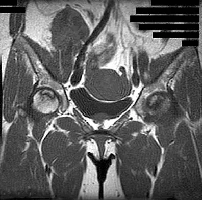
A 46-year-old woman has bilateral groin pain, with more severe pain on the left side than on the right side. Figures 44a and 44b show a radiograph and a T1-weighted MRI scan. What is the most likely diagnosis?


Explanation

The radiograph reveals bilateral patchy sclerosis of the femoral heads without evidence of collapse. The MRI scan shows bilateral head involvement with a common serpentine-like low-intensity signal of the superior femoral head that is common to osteonecrosis. Changes confined to the femoral head effectively exclude rheumatoid arthritis and osteoarthritis. Resnick D (ed): Diagnosis of Bone and Joint Disorders, ed 4. Philadelphia, PA, WB Saunders, 2002, vol 4, pp 3160-3162.


Question 5

Figures 45a through 45c show the radiograph, CT scan, and MRI scan of a 15-year-old boy who has lateral ankle pain. What is the most likely diagnosis?


Explanation

The elongated anterior process of the calcaneus reaching distally toward the navicular is an abnormal finding. Instead of viewing the rounded, blunt distal anterior process of the calcaneus, a bridge extends to the navicular, albeit incomplete. These findings are consistent with a fibrous coalition. CT can reveal a stress fracture of the calcaneus, arthritis of the subtalar joint with subchondral cysts, or an os peroneal bone disruption in the peroneus longus, but those entities are not shown here. The plantar fascia is intact. Richardson EG: Sesamoids and accessory bones of the foot, in Coughlin MJ, Mann RA (eds): Surgery of the Foot and Ankle, ed 7. St Louis, MO, Mosby, 1999, pp 702-732.


Question 6

A 5-year-old girl has had a low-grade fever, right hip and buttock pain, and a right-sided limp for the past 5 days. Examination shows diffuse tenderness and extreme pain on range of motion of the hip. Laboratory studies show a peripheral WBC count of 13,500/mm3 and an erythrocyte sedimentation rate of 55 mm/h. A radiograph is shown in Figure 46a, and an axial postgadolinium T1-weighted MRI scan with fat suppression and an axial T2-weighted fast spin echo MRI scan are shown in Figures 46b and 46c. What is the most likely diagnosis?


Explanation

MRI findings of acute osteomyelitis include a decrease in the normally high signal intensity of bone marrow on T1-weighted imaging; however, a postgadolinium T1-weighted image with fat suppression will show osteomyelitis as a bright marrow signal compared to the surrounding fat. Osteomyelitis is also brighter than normal fat on T2-weighted imaging. There is no rim-enhancing lesion suggesting an abscess, although myositis is seen in the obturator internus and short external rotators. The clinical scenario and imaging studies do not support the diagnosis of septic hip, eosinophilic granuloma, or pelvic fracture. Dormans JP, Drummond DS: Pediatric hematogenous osteomyelitis: New trends in presentation, diagnosis and treatment. J Am Acad Orthop Surg 1994;2:333-341.


Question 7

The palmar cutaneous branch of the median nerve (PCBMN) originates from the


Explanation

The PCBMN originates from the median nerve proper between 3 and 21 cm proximal to the wrist with moderate variation. It virtually always originates from the radial side of the nerve and travels distally with the median nerve, radial to the palmaris longus, and ulnar to the flexor carpi radialis. Hobbs RA, Magnussen PA, Tonkin MA: Palmar cutaneous branch of the median nerve. J Hand Surg Am 1990;15:38-43.

Question 8

The blood supply to the anterior cruciate ligament is primarily derived from what artery?


Explanation

Microvascular studies have shown that the majority of the blood supply to the cruciate ligaments comes from the middle geniculate artery, although there is collateral flow through the other geniculates and from bone. Arnoczky SP: Blood supply to the anterior cruciate ligament and supporting structures. Orthop Clin North Am 1985;16:15-28.

Question 9

Figures 47a and 47b show the CT scans of a patient who reports persistent pain in the sinus tarsi following a fall. The avulsion fracture fragment remains attached to what ligament?


Explanation

The bifurcate ligament bifurcates to connect the dorsal aspect of the anterior process of the calcaneus to both the cuboid and the navicular. Inversion injuries on the side of the foot can result in avulsion fractures (arrow) of the anterior process of the calcaneus. Sarrafian SK: Anatomy of the Foot and Ankle: Descriptive, Topographic, Functional, ed 2. New York, NY, Lippincott, 1993, p 192.


Question 10

Figure 48 shows an MRI scan of the knee. The arrow is pointing to what structure?


Explanation

The arrow points to the biceps femoris, which is inserted onto the fibula. The biceps femoris lies at the posterolateral aspect of the thigh. The semimembranosus and the semitendinous lie at the posterior medial aspect of the thigh. Gray H: Anatomy of the Human Body. Philadelphia, PA, Lea and Febiger, 1918, 2000.


Question 11

In Figure 49, line AB connects the anterior arch of C1 to the posterior margin of the foramen magnum. Line CD connects the anterior margin of the foramen magnum to the posterior arch of C1. What is the normal ratio of displacement from CD to AB (Power's ratio)?


Explanation

The ratio of displacement from CD to AB normally equals 1.0. If the ratio is greater than 1.0, an anterior atlanto-occipital dislocation may exist. Ratios slightly less than 1.0 are normal except in posterior dislocations, fractures of the odontoid process or ring of the atlas, or congenital abnormalities of the foramen magnum. In these conditions, the ratio may approach 0.7. Powers B, Miller MD, Kramer RS, et al: Traumatic anterior atlanto-occipital dislocation. Neurosurgery 1979;4:12-17.


Question 12

Figure 50 shows the AP radiograph of an asymptomatic 82-year-old woman who underwent total hip arthroplasty 16 years ago. What is the most likely diagnosis?


Explanation

Pelvic osteolysis in the presence of a well-fixed porous-coated socket is a recognized complication in total hip arthroplasty. The radiograph shows large lytic lesions superiorly adjacent to an acetabular screw and inferiorly extending into the ischium. It also reveals eccentricity of the femoral head with respect to the acetabular component, consistent with polyethylene wear. Koval KJ (ed): Orthopaedic Knowledge Update 7. Rosemont, IL, American Academy of Orthopaedic Surgeons, 2002, p 440.


Question 13

The MRI findings shown in Figure 51 would most likely create which of the following signs and symptoms?


Explanation

The MRI scan shows a far lateral disk herniation. With the L4-5 disk, a far lateral herniation abuts the left L4 nerve root. The findings would be consistent with those of a left L4 radiculopathy and would include pain or a sensory deficit on the anteromedial aspect of the knee, diminished patellar tendon reflex, and quadriceps weakness, perhaps making it difficult to walk up and down stairs. Fardin DF, Garfin SR (eds): Orthopaedic Knowledge Update: Spine 2. Rosemont, IL, American Academy of Orthopaedic Surgeons, 2002, p 329.


Question 14

Figure 52 shows the MRI scan of a 28-year-old baseball pitcher. Examination will most likely reveal which of the following findings?


Explanation

A ganglion cyst compressing the suprascapular nerve results in poorly localized pain in the shoulder girdle. Sensation is intact, with weakness of external rotation and abduction. Supraspinatus and infraspinatus atrophy is often noted when viewed from behind. These cysts are typically associated with labral tears. Deltoid weakness is associated with an axillary nerve injury, and scapular winging results from injury to the long thoracic nerve. Piatt BE, Hawkins RJ, Fritz RC, et al: Clinical evaluation and treatment of spinoglenoid notch ganglion cysts. J Shoulder Elbow Surg 2002;11:600-604.


Question 15

A 23-year-old man has had heel pain and fullness for the past several months. He reports that initially the pain was present only with activity, but more recently the pain has become constant. Figures 53a through 53d show a radiograph, a bone scan, and T2-weighted and gadolinium MRI scans. What is the most likely diagnosis?


Explanation

The imaging studies reveal an expansile lesion with the classic soap bubble appearance that involves most of the calcaneus. The bone scan reveals a very active lesion with intense uptake, and the MRI scans show the classic, loculated appearance of the lesion with multiple fluid-fluid levels. While it is important to rule out telangiectatic osteosarcoma, the most likely diagnosis is an aneurysmal bone cyst. While giant cell tumor might have a similar appearance, the multiple fluid levels in a expansile lesion strongly favor an aneurysmal bone cyst. Parsons TW: Benign bone tumors, in Fitzgerald RH, Kaufer H, Malkani AL (eds): Orthopaedics. St Louis, MO, Mosby, 2002, pp 1027-1035.


Question 16

A 77-year-old woman who underwent total knee arthroplasty 16 years ago now reports pain, swelling, and notable crepitation with range of motion. AP, lateral, and Merchant radiographs are shown in Figures 54a through 54c. What is the most likely diagnosis?


Explanation

The Merchant radiograph shows a lateral patellar shift with total polyethylene failure, resulting in a metal-on-metal bearing. This problem is associated with metal-backed patellar components. Component fixation appears solid, and no osteolysis is evident. Poss R (ed): Orthopaedic Knowledge Update 3. Park Ridge, IL, American Academy of Orthopaedic Surgeons, 1990, pp 590-593. Leopold SS, Berger RA, Patterson L, et al: Serum titanium level for diagnosis of a failed metal-backed patellar component. J Arthroplasty 2000;15:938-943.


Question 17

A 65-year-old woman who works as a florist has had pain in her right elbow for the past 6 months after lifting a flowerpot. MRI scans are shown in Figures 55a and 55b. The area of increased signal intensity seen in Figure 55b most likely represents which of the following findings?


Explanation

The MRI scans reveal a chronic distal biceps tendinitis. The T1-weighted scan shows the anatomic detail of the biceps tendon, and the T2-weighted scan shows increased signal caused by edema surrounding the tendon. The T1-weighted scan is not consistent with an antecubital lipoma. The chronicity of the lesion makes hematoma unlikely. An aneurysm usually appears with blood flow through the region and is dark on T1- and T2-weighted scans. Morrey BF: Tendon injuries about the elbow, in Morrey BF (ed): The Elbow and Its Disorders, ed 2. Philadelphia, PA, WB Saunders, 1993, pp 492-504.


Question 18

The carpal tunnel canal is narrowest (smallest cross-sectional area) at what level?


Explanation

The carpal tunnel canal has an hourglass shape in the coronal plane and is narrowest at the level of the hook of the hamate. Cobb TK, Dalley BK, Posteraro RH, et al: Anatomy of the flexor retinaculum. J Hand Surg Am 1993;18:91-99.

Question 19

When harvesting iliac crest bone graft during a posterior spinal decompression and fusion, injury to which of the following nerves may result in painful neuromas or numbness over the skin of the buttocks?


Explanation

The superior cluneal nerves (L1, L2, and L3) are at greatest risk when harvesting iliac crest bone graft during a posterior decompression and fusion. The nerves pierce the lumbodorsal fascia and cross the posterior iliac crest beginning at 8 cm lateral to the posterior superior iliac spine. The ilioinguinal and iliohypogastric nerves innervate anterior structures, and the lateral femoral cutaneous nerve lies in proximity to the anterior superior iliac spine and is at risk with anterior iliac crest bone graft harvesting. The superior gluteal nerve courses through the sciatic notch and supplies motor branches to the gluteus medius, minimus, and tensor fascia lata muscles. An HS: Principles and Techniques of Spine Surgery. Baltimore, MD, Williams and Wilkins, 1998, pp 770-773.

Question 20

The injury seen in the CT scan shown in Figure 56 is related to or associated with injury to which of the following structures?


Explanation

The right syndesmosis appears disrupted on the CT scan when compared to the normal left side. CT can be helpful in determining injury to the syndesmosis, especially with occult clinical findings. Ebraheim NA, Lu J, Yang H, et al: The fibular incisure of the tibia on CT scan: A cadaver study. Foot Ankle Int 1998;19:318-321. Ebraheim NA, Lu J, Yang H, et al: Radiographic and CT evaluation of tibiofibular syndesmotic diastasis: A cadaver study. Foot Ankle Int 1997;18:693-698.


Question 21

A 3-year-old girl has had wrist pain, a fever, and has refused to move her right wrist for the past 10 days. She has an oral temperature of 102 degrees F (38.7 degree C). Laboratory studies show an erythrocyte sedimentation rate of 50 mm/h, a WBC count of 11,000/mm3, and a left shift. AP and lateral radiographs are shown in Figures 57a and 57b. What is the most likely diagnosis?


Explanation

The most likely diagnosis is acute osteomyelitis. She may also have a septic wrist; however, the lytic lesion in the distal radius has the typical presentation and radiographic appearance of metaphyseal osteomyelitis. In this area of sluggish vascular flow, low oxygen tension, and low pH, bacterial seeding is common and is the usual origin of metaphyseal osteomyelitis. Leukemia and Ewing's sarcoma can present as a lytic lesion with an elevated erythrocyte sedimentation rate, but they are much less common than osteomyelitis and are less focal and more destructive in appearance. Nonossifying fibroma is typically metaphyseal and eccentric; however, it is well circumscribed and uncommon in the upper extremity. Eosinophilic granuloma does not typically present with inflammatory indicies. Dormans JP, Drummond DS: Pediatric hematogenous osteomyelitis: New trends in presentation, diagnosis and treatment. J Am Acad Orthop Surg 1994;2:333-341.


Question 22

Which of the following is considered the preferred approach to resect a lesion in the posterior one third of the proximal humerus?


Explanation

At least 8 cm of the posterior aspect of the proximal region of the humeral diaphyseal cortex can be exposed through the interval between the lateral head of the triceps and the deltoid muscle. No nerves or blood vessels need to be exposed in the dissection. The deltoid muscle is innervated by the axillary nerve and the triceps muscle by the radial nerve. This is a true internervous plane.

Question 23

The main blood supply to the lateral two thirds of the talar body is provided by the


Explanation

The main blood supply to the lateral two thirds of the talar body is derived from the artery of the tarsal canal, a branch of the posterior tibial artery. The peroneal artery helps form a vascular plexis over the posterior tubercle and combines with other arteries to form the artery of the sinus tarsi, which is the principal blood supply of the intrasinus structures of the talus. The anterior tibial arteries send branches to the superior surface of the talar head and give rise to the anterolateral malleolar artery, which may anastomose with other vessels to form the artery of the tarsal sinus. Sanders R: Fractures and fracture-dislocations of the talus, in Coughlin MJ, Mann RA (eds): Surgery of the Foot and Ankle, ed 7. St Louis, MO, Mosby, 1999, pp 1465-1518.

Question 24

When performing an arthroscopic subacromial decompression, bleeding can be encountered when dividing the coracoacromial ligament because of injury to what artery?


Explanation

The acromial branch of the thoracoacromial trunk courses along the coracoacromial ligament. This artery enters the ligament approximately 5 mm below the acromial edge. Division of the ligament at its insertion on the acromion minimizes the risk of bleeding. Esch JC, Baker CL: Arthroscopic anatomy and normal variations, in Whipple TL (ed): Surgical Arthroscopy: The Shoulder and Elbow. Philadelphia, PA, JB Lippincott, 1993, pp 63-76.

Question 25

Following its exit from the sciatic notch, the sciatic nerve passes between what two muscles?


Explanation

Though anatomic variations exist, both divisions of the sciatic nerve most commonly pass between the piriformis and superior gemellus. This anatomic consideration is relevant during the posterior approach to the hip, where careful retraction of the rotators avoids sciatic nerve injury. Hoppenfeld S, deBoer P: Surgical Exposures in Orthopaedics: The Anatomic Approach. Philadelphia, PA, JB Lippincott, 1984, pp 335-348.

Question 26

What structure does the radial nerve pierce as it passes from the posterior to the anterior compartment of the arm?


Explanation

The radial nerve pierces the lateral intermuscular septum approximately 10 cm proximal to the lateral epicondyle to enter the anterior compartment of the arm. This fixed anatomical point places the nerve at high risk during distal third humeral shaft fractures (Holstein-Lewis fractures).

Question 27

A patient presents with weakness in thumb interphalangeal joint flexion and index finger distal interphalangeal joint flexion, but normal hand sensation. Entrapment of the involved nerve most commonly occurs at which of the following sites?


Explanation

The anterior interosseous nerve (AIN) is most commonly compressed by the tendinous edge of the deep head of the pronator teres. It presents with purely motor deficits, clinically manifesting as an inability to make the 'OK' sign.

Question 28

During a minimally invasive repair of an acute Achilles tendon rupture, the sural nerve is at greatest risk of iatrogenic injury at what location relative to the calcaneal tuberosity?


Explanation

The sural nerve crosses from the posterolateral to the lateral border of the Achilles tendon approximately 10 to 12 cm proximal to its insertion. Percutaneous or minimally invasive sutures placed at this level pose the highest risk of iatrogenic nerve entrapment.

Question 29

A 24-year-old overhead athlete presents with posterior shoulder pain and isolated weakness of external rotation, with normal strength in abduction. An MRI reveals an isolated paralabral cyst in the spinoglenoid notch. Which of the following nerves is directly compressed?


Explanation

A cyst at the spinoglenoid notch compresses the terminal branch of the suprascapular nerve, causing isolated infraspinatus weakness. The supraspinatus, responsible for initiation of abduction, remains unaffected because its motor branches arise more proximally at the suprascapular notch.

Question 30

Which of the following best describes the boundaries of the quadrangular space of the shoulder?


Explanation

The quadrangular space is bounded superiorly by the teres minor, inferiorly by the teres major, medially by the long head of the triceps, and laterally by the surgical neck of the humerus. It contains the axillary nerve and the posterior circumflex humeral artery.

Question 31

During an anterior (Smith-Petersen) approach to the hip, an internervous plane is utilized to expose the joint. This superficial plane lies between muscles supplied by which of the following pairs of nerves?


Explanation

The superficial internervous plane of the Smith-Petersen approach lies between the tensor fasciae latae (superior gluteal nerve) and the sartorius (femoral nerve). The deep dissection continues between the gluteus medius (superior gluteal nerve) and the rectus femoris (femoral nerve).

Question 32

When performing a lateral approach to the fibula for open reduction and internal fixation of an ankle fracture, identifying the superficial peroneal nerve is critical. Where does this nerve typically pierce the deep crural fascia to become subcutaneous?


Explanation

The superficial peroneal nerve typically pierces the deep fascia of the lateral compartment approximately 10 to 12 cm proximal to the tip of the lateral malleolus. It becomes a subcutaneous structure at this level, making it vulnerable during lateral incisions.

Question 33

A patient with severe cubital tunnel syndrome is undergoing an in situ ulnar nerve decompression. The floor of the cubital tunnel is formed by which of the following structures?


Explanation

The floor of the cubital tunnel is composed of the medial collateral ligament (MCL) of the elbow, the underlying joint capsule, and the olecranon. Osborne's ligament forms the roof of the tunnel.

Question 34

A 35-year-old man sustains a midshaft humerus fracture. During open reduction and internal fixation via a posterior approach, the radial nerve is identified. At what approximate distance from the lateral epicondyle does the radial nerve typically pierce the lateral intermuscular septum to enter the anterior compartment of the arm?


Explanation

The radial nerve passes from the posterior compartment to the anterior compartment by piercing the lateral intermuscular septum approximately 10 cm proximal to the lateral epicondyle. This is a critical anatomical landmark when performing surgical approaches to the humerus.

Question 35

A patient presents with weakness in pinching the thumb and index finger, noting an inability to make an 'OK' sign. Electromyography confirms a compressive neuropathy of the anterior interosseous nerve (AIN). Which of the following anatomical structures is the most common cause of this specific nerve compression?


Explanation

Gantzer's muscle is an accessory head of the flexor pollicis longus and is the most common anatomical variant responsible for compression of the AIN. The Ligament of Struthers and lacertus fibrosus compress the median nerve higher up, causing pronator syndrome.

Question 36

During a posterior approach to the shoulder for a glenoid fracture, the surgeon enters the quadrangular space. Which of the following structures form the superior and inferior borders of this space, respectively?


Explanation

The quadrangular space is bounded superiorly by the teres minor, inferiorly by the teres major, medially by the long head of the triceps, and laterally by the surgical neck of the humerus. It contains the axillary nerve and the posterior humeral circumflex artery.

Question 37

A 28-year-old volleyball player presents with isolated weakness of external rotation in the dominant shoulder. MRI shows an isolated paralabral cyst. Compression of the suprascapular nerve at the spinoglenoid notch will result in denervation of which of the following muscles?


Explanation

The suprascapular nerve innervates the supraspinatus before passing through the spinoglenoid notch. Therefore, compression at the spinoglenoid notch results in isolated infraspinatus denervation.

Question 38

A surgeon is performing an open release of the tarsal tunnel. Based on the anatomical arrangement of structures passing posterior to the medial malleolus, what structure lies immediately posterior to the flexor digitorum longus (FDL) tendon?


Explanation

The contents of the tarsal tunnel from anterior to posterior are the Tibialis posterior tendon, FDL tendon, Posterior tibial Artery, Vein, Nerve, and FHL tendon (Tom, Dick, AND Very Nervous Harry). The artery is immediately posterior to the FDL tendon.

Question 39

A 45-year-old mechanic complains of a deep aching pain in the dorsal proximal forearm and an inability to extend the fingers at the MCP joints. Wrist extension is preserved but deviates radially. Where is the most likely site of nerve compression?


Explanation

The patient has Posterior Interosseous Nerve (PIN) syndrome. The most common site of PIN compression is the Arcade of Frohse, which is the proximal tendinous edge of the superficial head of the supinator muscle.

Question 40

A common anatomical variant involves a communicating neural branch between the median nerve and the ulnar nerve in the forearm. What is the name of this anomaly and what type of nerve fibers does it predominantly carry?


Explanation

The Martin-Gruber anastomosis is a connection from the median to the ulnar nerve in the forearm and carries predominantly motor fibers to the intrinsic muscles of the hand. Riche-Cannieu is in the palm, and Berrettini connects sensory nerves in the hand.

Question 41

During surgical decompression of the ulnar nerve at the elbow (cubital tunnel release), the floor of the cubital tunnel is visualized. Which of the following structures primarily forms the floor of the cubital tunnel?


Explanation

The floor of the cubital tunnel is formed by the joint capsule and the posterior bundle of the medial collateral ligament (MCL). Osborne's ligament and the FCU aponeurosis form the roof of the tunnel.

Question 42

During a Smith-Petersen (anterior) approach to the hip, there is a risk of injury to the lateral femoral cutaneous nerve (LFCN). To minimize this risk, the surgeon should remember that the LFCN typically enters the thigh by passing beneath the inguinal ligament at what specific location?


Explanation

The lateral femoral cutaneous nerve typically passes under the inguinal ligament immediately medial (about 1-2 cm) to the anterior superior iliac spine (ASIS). It runs superficial to the sartorius muscle.

Question 43

When creating an anterolateral portal during ankle arthroscopy, the superficial peroneal nerve is at risk. At what approximate level does this nerve typically pierce the crural fascia to become subcutaneous in the leg?


Explanation

The superficial peroneal nerve provides motor innervation to the lateral compartment and then typically pierces the deep fascia to become superficial approximately 10 to 12 cm proximal to the lateral malleolus.

Question 44

A patient suffers a traumatic dislocation of the knee resulting in injury to the common peroneal nerve. If the deep peroneal branch fails to recover, the patient will exhibit weakness in dorsiflexion and sensory loss over which specific area?


Explanation

The deep peroneal nerve provides motor innervation to the anterior compartment of the leg (dorsiflexion) and sensory innervation to the dorsal aspect of the first web space. The superficial peroneal nerve supplies the dorsum of the foot outside the first web space.

Question 45

Tarsal tunnel syndrome can lead to entrapment of the terminal branches of the tibial nerve. The medial plantar nerve innervates which of the following groups of intrinsic foot muscles?


Explanation

The medial plantar nerve provides motor innervation to four intrinsic muscles: the abductor hallucis, flexor digitorum brevis, flexor hallucis brevis, and the first lumbrical. It is analogous to the median nerve in the hand.

Question 46

Compression of the ulnar nerve in Guyon's canal can present with mixed sensory and motor deficits or isolated deficits depending on the zone of compression. A ganglion cyst in Zone 2 of Guyon's canal will produce which of the following clinical pictures?


Explanation

Guyon's canal is divided into three zones. Zone 1 is proximal to the bifurcation (mixed motor and sensory). Zone 2 encompasses the deep branch (strictly motor to intrinsics). Zone 3 encompasses the superficial branch (strictly sensory).

Question 47

In a patient undergoing a posterior approach to the hip, the sciatic nerve is identified. In approximately what percentage of the population does the common peroneal division of the sciatic nerve pierce directly through the piriformis muscle belly?


Explanation

In roughly 15% of the population, the sciatic nerve bifurcates prematurely, and the common peroneal division exits the pelvis by piercing directly through the piriformis muscle, predisposing the patient to piriformis syndrome.

Question 48

A patient presents with aching pain in the proximal volar forearm and paresthesias in the thumb, index, and middle fingers. Symptoms worsen with resisted pronation. Examination reveals no weakness of the flexor pollicis longus. An anomalous ligament extending from a supracondylar process to the medial epicondyle is suspected. What structures are compressed by this ligament?


Explanation

The Ligament of Struthers runs from an anomalous supracondylar process to the medial epicondyle. When present, it can compress the median nerve and the brachial artery, causing a high median neuropathy (pronator syndrome).

Question 49

During an extended deltopectoral approach to the shoulder, the axillary nerve is at risk as it crosses the deep surface of the deltoid. Approximately how far distal to the lateral edge of the acromion does the axillary nerve typically run?


Explanation

The axillary nerve runs transversely from posterior to anterior on the deep surface of the deltoid muscle, typically 5 to 7 cm distal to the lateral border of the acromion. Deltoid-splitting incisions must not extend distal to this safe zone.

Question 50

Thoracic outlet syndrome commonly involves compression within the interscalene triangle. Which of the following structures pass through the interscalene triangle?


Explanation

The interscalene triangle is bordered by the anterior scalene, middle scalene, and the first rib. It contains the subclavian artery and the roots/trunks of the brachial plexus. The subclavian vein runs anterior to the anterior scalene muscle.

Question 51

During harvest of the semitendinosus and gracilis tendons for ACL reconstruction, the saphenous nerve is at risk. The saphenous nerve exits the adductor (Hunter's) canal by penetrating the vastoadductor membrane. Which artery accompanies it as it exits?


Explanation

The saphenous nerve travels in the adductor canal with the superficial femoral artery. However, before the artery passes through the adductor hiatus to become the popliteal artery, the saphenous nerve exits the canal anteriorly, accompanied by the descending genicular artery.

Question 52

A patient presents with shoulder weakness after a superficial lymph node biopsy in the posterior triangle of the neck. Examination shows a prominent medial border of the scapula with lateral translation and an inability to abduct the arm past 90 degrees. Which nerve was most likely injured?


Explanation

The spinal accessory nerve (CN XI) runs superficially in the posterior triangle of the neck and is vulnerable during minor procedures. Injury paralyzes the trapezius, causing lateral winging of the scapula and difficulty elevating the arm past 90 degrees.

Question 53

A surgeon exploring the posterior humerus identifies a neurovascular bundle passing through the triangular interval. Which structures are found within this anatomical space?


Explanation

The triangular interval is bounded by the teres major, the long head of the triceps, and the lateral head of the triceps (or humerus). It serves as the passageway for the radial nerve and the profunda brachii artery into the posterior compartment of the arm.

Question 54

During a lateral approach to the distal humerus, the radial nerve is identified crossing the lateral intermuscular septum. At what average distance proximal to the lateral epicondyle does the radial nerve pierce the lateral intermuscular septum?


Explanation

The radial nerve pierces the lateral intermuscular septum from the posterior to the anterior compartment approximately 10 cm proximal to the lateral epicondyle.

Question 55

A 28-year-old overhead athlete presents with poorly localized posterior shoulder pain and deltoid weakness. MRI reveals a paralabral cyst in the quadrilateral space. Which of the following structures forms the superior border of this anatomic space?


Explanation

The quadrilateral space is bounded superiorly by the teres minor, inferiorly by the teres major, medially by the long head of the triceps, and laterally by the surgical neck of the humerus. It contains the axillary nerve and posterior humeral circumflex artery.

Question 56

Compression of the suprascapular nerve at the spinoglenoid notch will result in isolated weakness of which of the following muscles?


Explanation

The suprascapular nerve innervates the supraspinatus before passing through the spinoglenoid notch. Therefore, compression at the spinoglenoid notch results in isolated infraspinatus weakness.

Question 57

A patient presents with volar forearm pain and paresthesias in the thumb, index, and middle fingers. Symptoms are exacerbated by resisted forearm pronation with the elbow extended. Which of the following structures is the most likely site of median nerve compression?


Explanation

Pronator syndrome can be caused by median nerve compression at the pronator teres, tested by resisted pronation with the elbow extended. Resisted elbow flexion tests the bicipital aponeurosis, and resisted middle finger PIP flexion tests the FDS arch.

Question 58

A cyclist presents with intrinsic muscle weakness of the right hand but intact sensation over the volar hypothenar eminence and the dorsal ulnar aspect of the hand. In which zone of Guyon's canal is the ulnar nerve most likely compressed?


Explanation

Zone 2 of Guyon's canal contains only the deep motor branch of the ulnar nerve. Compression here causes isolated motor deficits in the ulnar-innervated intrinsic muscles, sparing sensation which branches off in Zone 1 or proximal to the canal.

Question 59

A 45-year-old runner with chronic heel pain undergoes surgical release. The surgeon targets the first branch of the lateral plantar nerve. This nerve courses between which two muscles?


Explanation

The first branch of the lateral plantar nerve (Baxter's nerve) runs between the deep fascia of the abductor hallucis and the medial belly of the quadratus plantae before innervating the abductor digiti minimi.

Question 60

During a minimally invasive Achilles tendon repair, the sural nerve is at greatest risk of iatrogenic injury at which location relative to the Achilles tendon?


Explanation

The sural nerve courses distally down the posterior calf and crosses from the midline to the lateral aspect of the Achilles tendon approximately 10 cm proximal to the calcaneal insertion.

Question 61

During a direct lateral (Hardinge) approach to the hip, proximal splitting of the gluteus medius is limited to prevent injury to the superior gluteal nerve. What is the generally accepted maximum safe distance for splitting the gluteus medius proximal to the tip of the greater trochanter?


Explanation

The superior gluteal nerve innervates the gluteus medius, gluteus minimus, and tensor fasciae latae. Splitting the gluteus medius more than 5 cm proximal to the greater trochanter places the nerve branches at significant risk.

Question 62

A patient develops inability to extend the fingers at the MCP joints following a proximal radius fracture. Wrist extension is maintained but with radial deviation. The injured nerve typically enters the supinator muscle beneath which of the following structures?


Explanation

The posterior interosseous nerve (PIN) is a branch of the radial nerve that passes beneath the Arcade of Frohse (the proximal aponeurotic edge of the supinator) and provides motor innervation to the extensor compartment.

Question 63

During an anterior (Smith-Petersen) approach to the hip, an internervous plane is utilized. The nerve at greatest risk during superficial dissection crosses the sartorius muscle. What is the sensory distribution of this nerve?


Explanation

The lateral femoral cutaneous nerve (LFCN) is at risk during the Smith-Petersen approach. It provides sensory innervation to the lateral thigh and typically exits the pelvis medial to the anterior superior iliac spine (ASIS).

Question 64

A patient sustains an obturator nerve injury during pelvic lymph node dissection. Which of the following muscles will maintain partial function because of its dual innervation?


Explanation

The adductor magnus has dual innervation. The adductor part is innervated by the obturator nerve, while the hamstring (ischial) part is innervated by the tibial division of the sciatic nerve.

Question 65

Inside the tarsal tunnel, the tibial nerve bifurcates into the medial and lateral plantar nerves. Which of the following statements correctly describes the anatomical relationship of the neurovascular structures beneath the flexor retinaculum from anterior to posterior?


Explanation

The contents of the tarsal tunnel from anterior to posterior follow the mnemonic Tom, Dick, And Very Nervous Harry: Tibialis posterior, flexor Digitorum longus, posterior tibial Artery, posterior tibial Vein, tibial Nerve, flexor Hallucis longus.

Question 66

A patient suffers a high-energy motorcycle collision and sustains a traction injury to the brachial plexus affecting the posterior cord. Which of the following movements will be most severely impaired?


Explanation

The posterior cord gives rise to the upper and lower subscapular, thoracodorsal, axillary, and radial nerves. Axillary nerve injury impairs the deltoid, leading to profound weakness in shoulder abduction.

Question 67

In anatomical variations of the relationship between the sciatic nerve and the piriformis muscle, the most common variant (Beaton and Anson type B) involves which of the following arrangements?


Explanation

Type A (normal, 84%) has the entire nerve passing inferior to the piriformis. Type B (about 10%) has the common peroneal nerve piercing the piriformis and the tibial nerve passing inferior to it.

Question 68

During a Latarjet procedure, the conjoined tendon is retracted to expose the anterior glenoid. The musculocutaneous nerve must be protected. At what average distance distal to the coracoid process does this nerve enter the coracobrachialis muscle?


Explanation

The musculocutaneous nerve typically enters the coracobrachialis approximately 5 cm (range 3-8 cm) distal to the tip of the coracoid process. Aggressive distal retraction of the conjoined tendon can cause traction neuropraxia.

Question 69

A patient presents with an inability to form an OK sign with their thumb and index finger after a supracondylar humerus fracture. Sensation in the hand is completely normal. What is the most likely injured nerve and its primary origin?


Explanation

The anterior interosseous nerve (AIN) is a motor branch of the median nerve. It innervates the flexor pollicis longus, the radial half of the flexor digitorum profundus, and the pronator quadratus.

Question 70

A surgeon is performing an extensive deltopectoral approach. To mobilize the pectoralis major, they note its neurovascular supply. The medial pectoral nerve is named for its origin from the medial cord of the brachial plexus. How does it typically enter the pectoralis major relative to the pectoralis minor?


Explanation

The medial pectoral nerve arises from the medial cord and either pierces the pectoralis minor or passes around its lateral border to innervate both the pectoralis minor and the sternocostal head of the pectoralis major.

Question 71

During an in situ ulnar nerve decompression at the cubital tunnel, several distinct anatomic structures can cause compression. Which of the following structures forms the roof of the cubital tunnel?


Explanation

The roof of the cubital tunnel is formed by Osborne's ligament (the cubital tunnel retinaculum) and the fascia of the flexor carpi ulnaris. The floor is composed of the medial collateral ligament, while the medial epicondyle and olecranon form the walls.

Question 72

When performing an extensile lateral approach to the humerus, the radial nerve must be identified and protected. At approximately what distance proximal to the radiocapitellar joint does the radial nerve pierce the lateral intermuscular septum?


Explanation

The radial nerve pierces the lateral intermuscular septum approximately 10 cm proximal to the radiocapitellar joint to enter the anterior compartment of the arm. Identifying this landmark is critical to preventing iatrogenic nerve injury during humeral plating.

Question 73

A 25-year-old athlete develops isolated weakness in external rotation of the shoulder after a direct blow to the posterior axilla. MRI reveals a mass in the quadrilateral space. What are the borders of the space where the axillary nerve is most likely compressed?


Explanation

The quadrilateral space is bordered superiorly by the teres minor, inferiorly by the teres major, laterally by the surgical neck of the humerus, and medially by the long head of the triceps. The axillary nerve and posterior circumflex humeral artery exit the axilla through this space.

Question 74

A 34-year-old male develops a compartment syndrome of the anterior leg following a tibia fracture. If left untreated, which of the following sensory deficits is most likely to be present due to nerve ischemia in this specific compartment?


Explanation

The deep peroneal nerve courses through the anterior compartment of the leg. Ischemia of this nerve secondary to anterior compartment syndrome results in sensory loss isolated to the dorsal first web space.

Question 75

A 22-year-old collegiate volleyball player presents with isolated weakness of the infraspinatus without supraspinatus involvement. Entrapment of the suprascapular nerve is suspected. At what anatomical location does this isolated compression typically occur?


Explanation

Compression of the suprascapular nerve at the spinoglenoid notch affects only the infraspinatus because the branches to the supraspinatus depart proximal to this notch. Compression at the suprascapular notch affects both the supraspinatus and infraspinatus.

Question 76

During a direct lateral approach to the distal fibula for an ORIF, the superficial peroneal nerve must be protected. Where does this nerve typically penetrate the crural fascia to become subcutaneous?


Explanation

The superficial peroneal nerve typically pierces the deep crural fascia of the lateral compartment approximately 10 to 12 cm proximal to the tip of the lateral malleolus. It then courses subcutaneously to provide sensation to the dorsum of the foot.

Question 77

Which of the following best describes the most common anatomical variant of the sciatic nerve in relation to the piriformis muscle?


Explanation

The most common anatomical variation (present in approximately 10-15% of the population) occurs when the common peroneal division pierces the piriformis muscle while the tibial division passes inferiorly to it. In normal anatomy, the entire nerve passes inferior to the piriformis.

Question 78

When performing a posterolateral approach to the ankle for a posterior malleolus fracture, the sural nerve is at risk. What vascular structure normally accompanies the sural nerve in this region?


Explanation

The sural nerve courses distally in the posterolateral leg alongside the small saphenous vein. Both structures travel directly posterior to the lateral malleolus.

Question 79

During a medial approach to the foot, the branches of the posterior tibial nerve must be identified. The medial plantar nerve provides motor innervation to which of the following muscles?


Explanation

The medial plantar nerve innervates the abductor hallucis, flexor digitorum brevis, flexor hallucis brevis, and the first lumbrical. The lateral plantar nerve innervates the remaining intrinsic muscles listed.

Question 80

During an anterior approach to the proximal radius (Henry approach), the posterior interosseous nerve (PIN) is at risk. What structure represents the most common site of PIN compression in radial tunnel syndrome?


Explanation

The Arcade of Frohse, which is the proximal fibrous edge of the superficial head of the supinator muscle, is the most common site of posterior interosseous nerve compression. Other sites include the leash of Henry and the fibrous edge of the ECRB.

Question 81

During a Smith-Petersen (anterior) approach to the hip, the deep internervous plane lies between the rectus femoris and the gluteus medius. What is the motor innervation to the muscle forming the medial border of this deep plane?


Explanation

The deep plane is between the rectus femoris (femoral nerve) medially and the gluteus medius (superior gluteal nerve) laterally. Thus, the medial border's innervation is the femoral nerve.

Question 82

A 45-year-old patient presents with lateral winging of the scapula and inability to shrug the shoulder following a lymph node biopsy in the posterior triangle of the neck. Which of the following muscles has most likely been denervated?


Explanation

The spinal accessory nerve (CN XI) courses through the posterior triangle of the neck to innervate the trapezius muscle. Injury to this nerve causes lateral scapular winging and weakness in shoulder elevation.

Question 83

In a standard deltopectoral approach to the shoulder, the conjoined tendon is often retracted medially. At approximately what distance distal to the coracoid process does the musculocutaneous nerve typically enter the coracobrachialis?


Explanation

The musculocutaneous nerve typically enters the coracobrachialis muscle 3 to 8 cm distal to the tip of the coracoid process. Vigorous medial retraction of the conjoined tendon can cause traction neuropraxia of this nerve.

Question 84

A patient is unable to flex the IP joint of the thumb and the DIP joint of the index finger following a forearm laceration. Sensation is perfectly intact in the hand. Which of the following muscles is typically SPARED if the injured nerve is the anterior interosseous nerve (AIN)?


Explanation

The AIN innervates the FPL, the radial half of the FDP (index and middle fingers), and the pronator quadratus. The flexor carpi radialis is innervated by the main branch of the median nerve before it gives off the AIN.

Question 85

A 50-year-old runner presents with chronic heel pain refractory to conservative management. Entrapment of the first branch of the lateral plantar nerve (Baxter's nerve) is suspected. Which muscle is predominantly innervated by this specific nerve branch?


Explanation

Baxter's nerve, the first branch of the lateral plantar nerve, provides motor innervation to the abductor digiti minimi. Entrapment commonly occurs as the nerve passes between the deep fascia of the abductor hallucis and the quadratus plantae.

Question 86

A 45-year-old mechanic presents with numbness in the small finger and weakness in grip strength. Nonoperative management has failed, and an in situ decompression of the ulnar nerve is planned. During the approach, the roof of the cubital tunnel must be divided. Which of the following structures constitutes the primary roof of this tunnel?


Explanation

The roof of the cubital tunnel is formed by Osborne's ligament (the cubital tunnel retinaculum) and the arcuate ligament extending between the two heads of the flexor carpi ulnaris. The ligament of Struthers is associated with median nerve compression.

Question 87

A 55-year-old woman presents with the inability to extend her fingers and thumb at the metacarpophalangeal joints. Wrist extension is preserved but deviates radially. Electromyography confirms an entrapment neuropathy. Which of the following is the most likely site of compression?


Explanation

The patient has Posterior Interosseous Nerve (PIN) syndrome. Radial wrist extension is preserved because the extensor carpi radialis longus is innervated by the radial nerve proximal to the PIN branch. The most common site of PIN compression is the Arcade of Frohse.

Question 88

A 32-year-old marathon runner presents with chronic, recalcitrant medial heel pain that radiates into the plantar aspect of the foot. A diagnostic injection relieves the pain, suggesting entrapment of the first branch of the lateral plantar nerve. This nerve normally courses between which two structures?


Explanation

Baxter's nerve (the first branch of the lateral plantar nerve) typically becomes entrapped as it passes deep to the abductor hallucis and then between the quadratus plantae and the flexor digitorum brevis muscles.

Question 89

A 28-year-old overhead athlete is diagnosed with quadrilateral space syndrome, presenting with vague posterior shoulder pain and isolated atrophy of the teres minor. The structures forming the borders of this anatomical space are the:


Explanation

The quadrangular space transmits the axillary nerve and posterior circumflex humeral artery. It is bounded superiorly by the teres minor, inferiorly by the teres major, medially by the long head of the triceps, and laterally by the surgical neck of the humerus.

Question 90

A 35-year-old volleyball player is found to have a large paralabral ganglion cyst extending into the spinoglenoid notch. Physical examination is most likely to demonstrate which of the following isolated findings?


Explanation

A cyst at the spinoglenoid notch compresses the suprascapular nerve after it has already innervated the supraspinatus. This leads to isolated denervation of the infraspinatus, causing weakness in external rotation with preserved abduction.

Question 91

During an extensile lateral approach for a comminuted calcaneus fracture, the surgeon must carefully identify and protect a nerve that provides sensation to the lateral border of the foot. What is the normal anatomical course of this nerve at the level of the ankle?


Explanation

The sural nerve provides sensation to the lateral aspect of the foot. It descends posterior to the lateral malleolus in close proximity to the short (small) saphenous vein.

Question 92

In a direct anterior approach to the hip, careful deep dissection is required. In a different scenario where an adductor tenotomy and obturator nerve block are performed for spasticity, the surgeon isolates the anterior and posterior divisions of the obturator nerve. These divisions are anatomically separated by which of the following muscles?


Explanation

After exiting the obturator foramen, the obturator nerve divides into anterior and posterior branches. These branches descend on either side of the adductor brevis muscle.

Question 93

A patient sustains a midshaft fibula fracture and subsequently develops a compartment syndrome requiring fasciotomies. The surgeon must be mindful of the superficial peroneal nerve as it exits the deep fascia to become subcutaneous. At what approximate level does this transition normally occur?


Explanation

The superficial peroneal nerve provides motor innervation to the lateral compartment before piercing the crural fascia to become subcutaneous, typically 10 to 12 cm proximal to the lateral malleolus.

Question 94

A patient presents with an inability to make an 'OK' sign, demonstrating loss of flexion at the thumb interphalangeal joint and index finger distal interphalangeal joint. Sensation in the hand is completely normal. Which of the following is LEAST likely to be the anatomical site of neural compression in this syndrome?


Explanation

The patient has Anterior Interosseous Nerve (AIN) syndrome. The ligament of Struthers compresses the median nerve far proximally, leading to a high median nerve palsy with sensory deficits, rather than an isolated AIN palsy.

None

Clinic OS
Medically Verified Content by
Prof. Clinic OS
Consultant Orthopedic & Spine Surgeon
Chapter Index