العربية
Part of the Master Guide

AAOS & ABOS Basic Science MCQs (Set 3): Bone Biology, Biomechanics & Anatomy Review

AAOS & ABOS Anatomy MCQs (Set 3): Musculoskeletal & Skeletal System Questions | Board Review

23 Apr 2026 52 min read 100 Views
Anatomy 2005 MCQs - Part 3

Key Takeaway

This high-yield question set (Set 3) for the AAOS, ABOS, and OITE exams focuses on fundamental musculoskeletal anatomy. It covers detailed skeletal system structures, bone identification, and the intricate articulations and ligaments of major joints, crucial for orthopedic knowledge.

AAOS & ABOS Anatomy MCQs (Set 3): Musculoskeletal & Skeletal System Questions | Board Review

Comprehensive 100-Question Exam


00:00

Start Quiz

Question 1

Following a vertebroplasty of L2, cement is noted to protrude directly anterior to the L2 vertebral body. The cement is closest to which of the following structures?


Explanation

At the level of L2, the liver and the vena cava lie to the right. The pancreas and duodenum are anterior to the aorta. The aorta lies in the midline just in front of the vertebral body. Clement CD: Anatomy: A Regional Atlas of Human Anatomy, ed 3. Baltimore, MD, Munich, Germany, Urban and Schwarzberg, 1987, Figure 331.

Question 2

Figures 28a and 28b show AP and lateral radiographs of the knee. Based on these findings, which of the following structures has most likely been injured?


Explanation

The radiographs show a posterior knee dislocation. Knee dislocations almost always involve rupture of both the anterior and posterior cruciate ligaments. Collateral ligament injuries also are common. Arterial, nerve, and tendon injuries each occur in less than half of knee dislocations. Schenck RC Jr, Hunter RE, Ostrum RF, et al: Knee dislocations. Instr Course Lect 1999;48:515-522.


Question 3

A patient who sustained a knife wound to the axilla 4 months ago now has profound interosseous wasting and generalized hand weakness. A brachial plexus injury is likely at which of the following locations in Figure 29?


Explanation

Penetrating sharp wounds in proximity to major nerve or vascular structures should always be acutely explored. Because this patient did not seek treatment for a potentially treatable injury, interosseous wasting implies injury to the C8 and T1 nerve roots that contribute to ulnar nerve function. The most likely location for the brachial plexus injury is the location marked L or the inferior trunk. A wrist drop that is the result of radial nerve dysfunction would be expected with an injury at K or O. An upper brachial plexus palsy with loss of elbow flexion and shoulder abduction would be expected with an injury at B. A loss of elbow flexion alone would be expected following an injury at C. Netter F: The Ciba Collection of Medical Illustrations: The Musculoskeletal System. Part 1, Anatomy, Physiology and Metabolic Disorders. West Caldwell, NJ, Ciba-Geigy, 1991, vol 8, pp 28-29. Wolock B, Millesi H: Brachial plexus-applied anatomy and operative exposure, in Gelberman RH (ed): Operative Nerve Repair and Reconstruction. Philadelphia, PA, JB Lippincott, 1991, vol 2, pp 1255-1272.


Question 4

During an anterior retroperitoneal approach to the low lumbar spine, the iliac vessels are mobilized along the lateral side, allowing them to be retracted toward the midline. To gain adequate mobility of the common iliac vein for exposure of L5, it is important to identify which of the following structures?


Explanation

The iliolumbar vein is a large tributary that sits along the lateral surface of the common iliac vein. It can be quite substantial in size and must be identified prior to mobilizing the common iliac vein toward the midline. The other structures are not of surgical significance in performing this exposure.


Question 5

Figure 30 shows an axial T1-weighted MRI scan of a patient's right shoulder. The arrows are pointing to what normal structure?


Explanation

Tears of the pectoralis major tendon are frequently missed during examination. MRI provides excellent visualization of the tendon if the study extends low enough down the arm. The pectoralis major tendon inserts on the crest of the greater tubercle of the humerus, just lateral to the long head of the biceps tendon. The latissimus dorsi tendon inserts medial to the long head of the biceps tendon on the lesser tubercle. The subscapularis tendon inserts on the lesser tuberosity more proximally. The deltoid insertion is more distal. Connell DA, Potter HG, Sherman MF, et al: Injuries of the pectoralis major muscle: Evaluation with MR imaging. Radiology 1999;210:785-791. Carrino JA, Chandnanni VP, Mitchell DB, et al: Pectoralis major muscle and tendon tears: Diagnosis and grading using magnetic resonance imaging. Skeletal Radiol 2000;29:305-313.


Question 6

The arthroscopic views shown in Figures 31a and 31b reveal extensive synovitis in the anterolateral corner of the ankle overlying a band of tissue sometimes implicated in soft-tissue impingement of the ankle following a chronic sprain injury. This band is a portion of the


Explanation

The arthroscopic views show the lateral side of the ankle as demonstrated by the presence of the tibiofibular articulation. As is typical in chronic anterolateral impingement, synovitis overlies the anteroinferior band of the tibiofibular ligament, the most distal portion of the anterior syndesmosis. Hypertrophic scar formed on or in this ligament can impinge on the lateral margin of the talar dome and has been associated with chronic anterolateral ankle pain. Bassett FH III, Gates HS III, Billys JB, et al: Talar impingement by the anteroinferior tibiofibular ligament: A cause of chronic pain in the ankle after inversion sprain. J Bone Joint Surg Am 1990;72:55-59.


Question 7

Figures 32a and 32b show the AP and lateral radiographs of an 11-year-old boy who has a severe limp, a fever, and swelling and tenderness of the thigh. Aspiration of the bone reveals purulent material. The patient has most likely been symptomatic for


Explanation

In patients with an osteomyelitic infection, radiographic findings at 1 to 5 days usually show soft-tissue swelling only. Seven to 14 days after symptoms begin, radiographs will most likely show the classic signs of acute osteomyelitis. Reactive bone formation would be expected by 6 months. Kasser JR (ed): Orthopaedic Knowledge Update 5. Rosemont, IL, American Academy of Orthopaedic Surgeons, 1996, pp 149-161.


Question 8

Figure 33 shows the CT scan of a 40-year-old man who injured his left shoulder while skiing. What structure is attached to the bony fragment?


Explanation

The scan reveals a bony Bankart lesion. The anterior band of the inferior glenohumeral ligament is the major restraint to anterior translation of the humeral head and is usually injured with anterior shoulder dislocations. It inserts onto the glenoid labrum at the anteroinferior aspect of the glenoid rim. The labrum most frequently avulses from the glenoid (Bankart lesion), but occasionally the bony attachment is avulsed. O'Brien SJ, Neves MC, Arnoczky SP, et al: The anatomy and histology of the inferior glenohumeral ligament complex of the shoulder. Am J Sports Med 1990;18:449-456.


Question 9

What structure is located immediately posterior to the capsule at the posterior cruciate ligament tibial insertion?


Explanation

The popliteal artery lies just posterior to the posterior cruciate ligament tibial insertion, separated only by the posterior capsule of the knee. When performing a posterior cruciate ligament reconstruction, this artery is at risk for injury during creation of the tibial tunnel. Jackson DW, Proctor CS, Simon TM: Arthroscopic assisted PCL reconstruction: A technical note on potential neurovascular injury related to drill bit configuration. J Arthroscopy 1993;9:224-227.

Question 10

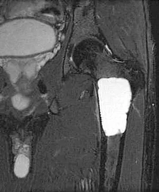
A 21-year-old man has mild but persistent aching pain in his left proximal thigh during impact loading activities. He denies pain at rest and has no other symptoms. Figures 34a through 34e show the radiographs and T1-weighted, T2-weighted, and gadolinium MRI scans of the left hip. What is the most likely diagnosis?


Explanation

The radiographs show a centrally located radiolucent lesion with cortical thinning and mild osseous expansion; these findings are the hallmarks of a simple bone cyst. Whereas this particular lesion does not demonstrate sclerosis, the distinct margin of this lesion with sharp transition to normal bone is common. The MRI scans reveal a purely cystic lesion with bright T2 signal, and the gadolinium image shows the classic rim enhancement of cystic lesions. Fibrous dysplasia with cystic degeneration might have a very similar appearance and should be considered in the differential diagnosis. Parsons TW: Benign bone tumors, in Fitzgerald RH, Kaufer H, Malkani AL (eds): Orthopaedics. St Louis, MO, Mosby, 2002, pp 1027-1035. May DA, Good RB, Smith DK, et al: MR imaging of musculoskeletal tumors and tumor mimickers with intravenous gadolinium: Experience with 242 patients. Skeletal Radiol 1997;26:2-15.


Question 11

What nerve is at greatest risk when developing the superficial plane between the tensor fascia lata and sartorious during the anterior (Smith-Peterson) approach to the hip?


Explanation

The lateral femoral cutaneous nerve pierces the fascia between the tensor fascia lata and the sartorius approximately 2.5 cm distal to the anterosuperior iliac spine and is at risk when the interval is defined. The superior gluteal and femoral nerves define the internervous plane between the tensor fascia lata and the sartorius and are not at risk for injury. Hoppenfeld S, deBoer P: Surgical Exposures in Orthopaedics: The Anatomic Approach. Philadelphia, PA, JB Lippincott, 1984, pp 302-316.

Question 12

An axial T1-weighted MRI scan of the pelvis is shown in Figure 35. Which of the following structures is enclosed by the circle?


Explanation

The obturator vessels and nerve pass along the lateral pelvic wall along the true pelvic brim (nerve lies anterior to the vessels and lies on the obturator internus muscle) and descend into the obturator groove at the upper portion of the obturator foramen. Higuchi T: Normal anatomy and magnetic resonance appearance of the pelvis, in Takahashi HE, Morita T, Hotta T, et al (eds): Operative Treatment of Pelvic Tumors. Tokyo, Japan, Springer-Verlag, 2003, pp 4-21.


Question 13

At the level of the midcalf, the plantaris tendon is found at which of the following locations?


Explanation

The plantaris tendon is often harvested to augment a tendon reconstruction. The origin of the plantaris muscle is on the posterolateral aspect of the distal femur, and the muscle lies lateral to the tibial nerve and the posterior tibial artery. The tendon then courses posteriorly between the soleus and the medial head of the gastrocnemius. Clement CD: Anatomy: A Regional Atlas of Human Anatomy, ed 3. Baltimore, MD, Munich, Germany, Urban and Schwarzberg, 1987, Figure 475.

Question 14

In the posterior approach to the proximal radius (proximal Thompson approach), the supinator is exposed through the interval between what two muscles?


Explanation

The proximal exposure of the radius is most often used for internal fixation of fractures, resection of tumors, or decompression of the posterior interosseous nerve beneath the supinator muscle. The supinator muscle is exposed through the interval between the extensor carpi radialis brevis and the extensor digitorum comminus muscles. This interval can be more easily palpated further distal in the forearm. Hoppenfeld S, deBoer P: Surgical Exposures in Orthopaedics, ed 2. Philadelphia, PA, Lippincott-Raven, 1994, pp 136-146.

Question 15

Figure 36 shows the hip arthrogram of a newborn. Which of the following structures is enclosed by the circle?


Explanation

The structure enclosed by the circle is the acetabular labrum. It is visible as the white point of tissue outlined by the darkly radiopaque contrast. The appearance of the contrast surrounding the sharp white point of a normal labrum is called the "rose thorn sign." The limbus is the term reserved for a rounded, infolded labrum seen with arthrography. The pulvinar is the fatty tissue seen in the empty acetabulum when the hip is dislocated. The ligamentum teres is seen as a white stripe outlined by contrast coursing from the central acetabulum to the dislocated femoral head. The transverse acetabular ligament courses across the inferior portion of the acetabulum and is not clearly seen with arthrography. Herring JA: Tachdjian's Pediatric Orthopaedics, ed 3. Philadelphia, PA, WB Saunders, 2002, vol 1, pp 532-533.


Question 16

Figures 37a and 37b show radiographs of a 24-year-old man who has a humeral bone lesion that was found during a screening chest radiograph. He denies any symptoms despite leading a very active lifestyle. What is the most likely diagnosis?


Explanation

The radiographs reveal a geographic, diaphyseal lesion with very subtle cortical expansion, cortical thinning, relatively sharp demarcation, and angular rather than rounded borders, suggesting a fibrous bone lesion. This lesion demonstrates the classic ground glass appearance of fibrous dysplasia. Ewing's sarcoma, metastases, and aneurysmal bone cyst all typically have a more aggressive appearance. Parsons TW: Benign bone tumors, in Fitzgerald RH, Kaufer H, Malkani AL (eds): Orthopaedics. St Louis, MO, Mosby, 2002, pp 1027-1035.


Question 17

Figures 38a and 38b show the CT scans of a 64-year-old woman. What is the most likely diagnosis?


Explanation

The CT scans show large cystic lesions in the talus and calcaneus with complete subluxation of the subtalar joint, allowing the calcaneus to slide laterally until it becomes blocked by the fibula. The cause of this subluxation is severe posterior tibial tendon dysfunction. Although no fibular fracture has yet appeared, it can occur with continued stress from the calcaneus. There is, however, a pathologic fracture in the medial calcaneus through a medial degenerative cyst. The joint space is irregular and not symmetrical as would be seen in an inflammatory arthropathy. Cystic lesions are not present in the tibia. No stress fracture is seen in the talus. Coughlin MJ: Sesamoids and accessory bones of the foot, in Coughlin MJ, Mann RA (eds): Surgery of the Foot and Ankle, ed 7. St Louis, MO, Mosby, 1999, pp 437-499.


Question 18

The arrow in Figure 39 is pointing to which of the following ligaments?


Explanation

The lunotriquetral interosseous ligament stabilizes the lunotriquetral joint. The scapholunate interosseous ligament stabilizes the scapholunate joint. The ulnolunate ligament originates from the base of the ulnar styloid and inserts in the lunate. The ulnotriquetral ligament originates from the base of the ulnar styloid and inserts on the triquetrum. The ulnolunate and the ulnotriquetral ligaments are important stabilizers to the ulnar side of the wrist. The short radiolunate ligament originates on the volar ulnar margin of the distal radius and inserts in the ulnar margin of the lunate. Berger RA: Ligament anatomy, in Cooney WP, Linscheid RL, Dobyns JH (eds): The Wrist, Diagnosis and Operative Management. St Louis, MO, Mosby, 1998, pp 73-105.


Question 19

The medial collateral ligament complex of the elbow originates on what portion of the medial epicondyle?


Explanation

The medial collateral ligament complex of the elbow consists of three portions: the anterior bundle, the posterior bundle, and a transverse component that has little biomechanic significance. The origin of the ligament is from the central two thirds of the anteroinferior undersurface of the medial epicondyle.

Question 20

Figures 40a and 40b show the pre- and postoperative radiographs of an 82-year-old woman with bilateral hip pain who has had staged total hip arthroplasties. To minimize potential injury to the sciatic nerve at the time of surgery, the surgeon should


Explanation

To improve hip biomechanics and secure more suitable bone for acetabular fixation, the true acetabulum is often resurfaced in patients who have developmental dysplasia of the hip, thus lowering the hip center and lengthening the leg. Acute lengthening of more than 3 cm will place excessive tension on the sciatic nerve and require a femoral shortening to avoid sciatic nerve injury. The other maneuvers will not relieve sciatic nerve tension because of limb lengthening. Koval KJ (ed): Orthopaedic Knowledge Update 7. Rosemont, IL, American Academy of Orthopaedic Surgeons, 2002, pp 430-431.


Question 21

Based on the radiographic findings shown in Figure 41, which of the following wrist ligaments is most likely disrupted?


Explanation

The radiograph shows a diastasis of the scapholunate interval, caused by certain failure of the scapholunate interosseous ligament. The lunotriquetral interosseous ligament stabilizes the lunotriquetral joint. The long radiolunate ligament originates in the volar radius and inserts in the lunate. The short radiolunate ligament originates on the ulnar margin of the radius and inserts on the ulnar margin of the lunate. The ulnolunate ligament originates at the ulnar styloid base and inserts on the volar aspect of the lunate. Linscheid RL, Dobyns JH, Beabout JW, et al: Traumatic instability of the wrist: Diagnosis, classification, and pathomechanics. J Bone Joint Surg Am 1972;54:1612-1632. Mayfield JK, Johnson RP, Kilcoyne RK: Carpal dislocations: Pathomechanics and progressive perilunar instability. J Hand Surg Am 1980;5:226-241.


Question 22

Which of the following extensor tendons commonly have multiple slips?


Explanation

The extensor digiti mini quinti is most typically a tendon with two slips. The abductor pollicis longus has multiple slips that insert in order of frequency on the base of the first metacarpal, trapezium, and thenar muscles. The extensor pollicis longus, extensor carpi radialis brevis, and extensor indicis proprius consistantly have only one slip. von Schroeder HP, Botte MJ: Anatomy of the extensor tendons of the fingers: Variations and multiplicity. J Hand Surg Am 1995;20:27-34.

Question 23

The nerve to the abductor digiti quinti, implicated in some patients who have chronic heel pain, is most commonly a branch of what larger nerve?


Explanation

The nerve to the abductor digiti quinti is the first branch of the lateral plantar nerve. It branches off while the nerve is still on the medial side of the foot and also innervates a portion of the plantar fascia. It can become entrapped beneath the deep fascia of the abductor hallucis muscle and has been associated with some forms of chronic heel pain. Baxter DE, Pfeffer GB: Treatment of chronic heel pain by surgical release of the first branch of the lateral plantar nerve. Clin Orthop 1992;279:229-236.

Question 24

Figure 42 shows the sagittal T2-weighted MRI scan of a patient's right knee. These findings are most commonly seen with a complete tear of the


Explanation

The MRI scan reveals disruption of the lateral capsule and ligaments with fluid in the soft tissues laterally. Additionally, there is a large bone bruise on the medial femoral condyle. This combination indicates injury to the posterolateral complex. These injuries often have coexisting anterior and/or posterior cruciate ligament injuries. Failure to recognize the posterolateral corner injury can lead to failure of anterior or posterior cruciate ligament reconstructions. LaPrade RF, Gilbert TJ, Bollom TS, et al: The magnetic resonance imaging appearance of individual structures of the posterolateral knee: A prospective study of normal knees and knees with surgically verified grade III injuries. Am J Sports Med 2000;28:191-199.


Question 25

Thermal capsulorrhaphy of the inferior glenohumeral ligament can cause iatrogenic injury to which of the following nerves?


Explanation

The axillary nerve courses from anterior to posterior just below the inferior shoulder capsule. Thermal energy applied to the inferior aspect of the shoulder capsule can result in injury to this nerve. Wong KL, Williams GR: Complications of thermal capsulorrhaphy of the shoulder. J Bone Joint Surg Am 2001;83:151-155.

Question 26

During a deltopectoral approach to the shoulder, the coracoid process may be osteotomized for better exposure. Which nerve is most at risk if the conjoint tendon is forcefully retracted medially?


Explanation

The musculocutaneous nerve enters the coracobrachialis muscle 3 to 8 cm distal to the coracoid tip. Vigorous medial retraction of the conjoint tendon can cause a stretch or compression injury to this nerve.

Question 27

During a flatfoot reconstruction, the spring ligament is identified and reefed. Which of the following accurately describes the anatomic attachments of the superomedial calcaneonavicular (spring) ligament?


Explanation

The superomedial band of the spring ligament originates on the sustentaculum tali and inserts on the superomedial aspect and plantar surface of the navicular.

Question 28

A patient presents with isolated weakness in external rotation of the shoulder. An MRI reveals a paralabral cyst in the spinoglenoid notch. Which of the following muscles is most likely affected?


Explanation

The suprascapular nerve innervates the supraspinatus, then passes through the spinoglenoid notch to innervate the infraspinatus. Compression at the spinoglenoid notch causes isolated infraspinatus weakness.

Question 29

During a posterolateral corner reconstruction of the knee, the surgeon isolates the popliteus tendon. Where is the femoral attachment of the popliteus tendon located relative to the fibular collateral ligament (FCL)?


Explanation

The popliteus tendon inserts on the lateral femoral condyle distal and anterior to the origin of the fibular collateral ligament (LCL).

Question 30

A 25-year-old falls on an outstretched hand and sustains a scaphoid waist fracture. Which of the following best describes the predominant arterial supply to the proximal pole of the scaphoid?


Explanation

The scaphoid receives its primary blood supply from the dorsal carpal branch of the radial artery, which enters the dorsal ridge distally and supplies the bone in a retrograde fashion.

Question 31

During an anterior thoracolumbar spine approach, the artery of Adamkiewicz is at risk. From which side and vertebral level does this artery most commonly arise?


Explanation

The artery of Adamkiewicz provides the major blood supply to the anterior spinal artery of the lower spinal cord. It most commonly arises on the left side between T9 and L2.

Question 32

During a surgical approach to the proximal radius, the surgeon identifies the posterior interosseous nerve (PIN). The PIN passes between the two heads of which muscle?


Explanation

The posterior interosseous nerve (PIN) passes between the superficial and deep heads of the supinator muscle at the arcade of Frohse.

Question 33

During a plantar approach to the foot for a compartment release, the surgeon encounters the "Master Knot of Henry." Which two tendons cross at this anatomical landmark?


Explanation

The Master Knot of Henry is located in the medial plantar midfoot where the flexor hallucis longus tendon crosses dorsal to the flexor digitorum longus tendon.

Question 34

A surgeon is performing a surgical dislocation of the hip. To protect the main blood supply to the femoral head, the medial circumflex femoral artery (MCFA) must be protected. The deep branch of the MCFA passes between which two muscles?


Explanation

The deep branch of the medial circumflex femoral artery (MCFA) passes between the obturator externus and quadratus femoris muscles before traveling anterior to the conjoint tendon to supply the femoral head.

Question 35

Tears of the triangular fibrocartilage complex (TFCC) often occur in conjunction with distal radius fractures. Which zone of the TFCC is considered avascular and therefore has the poorest healing potential?


Explanation

The central articular disk of the TFCC is avascular, relying on synovial fluid for nutrition, and lacks healing potential. The peripheral portions have a blood supply and can be repaired.

Question 36

A patient with a traction injury to the lower trunk of the brachial plexus will demonstrate deficits in muscles supplied by which two cords?


Explanation

The lower trunk (C8-T1) continues as the anterior division to form the medial cord and sends a posterior division to the posterior cord. Thus, lower trunk injuries affect medial and posterior cord distributions.

Question 37

During an ulnar nerve transposition, the surgeon must release Osborne's ligament. This structure spans between the medial epicondyle and which other bony landmark?


Explanation

Osborne's ligament forms the roof of the cubital tunnel, extending from the medial epicondyle to the olecranon process over the ulnar nerve.

Question 38

During an ilioinguinal approach to the acetabulum, massive bleeding is encountered near the superior pubic ramus. This is most likely due to injury to the "corona mortis," which is an anastomosis between which two vascular systems?


Explanation

The corona mortis is a vascular anastomosis between the obturator (internal iliac system) and external iliac (or inferior epigastric) systems, located over the superior pubic ramus.

Question 39

When inserting a pedicle screw in the lumbar spine, the traditional starting point is at the intersection of the pars interarticularis, the midpoint of the transverse process, and which other structure?


Explanation

The traditional starting point for a lumbar pedicle screw is the intersection of a vertical line through the lateral border of the superior articular facet and a horizontal line bisecting the transverse process.

Question 40

A 22-year-old football player sustains a midfoot injury. The Lisfranc ligament is crucial for midfoot stability. What are the true bony attachments of the Lisfranc ligament?


Explanation

The Lisfranc ligament connects the lateral aspect of the medial cuneiform to the medial base of the second metatarsal, providing critical stability to the tarsometatarsal joint complex.

Question 41

A patient with an axillary nerve injury following a proximal humerus fracture will most likely show denervation in the deltoid and which other muscle?


Explanation

The axillary nerve passes through the quadrangular space and innervates both the deltoid and the teres minor muscles. It also supplies sensory innervation to the lateral shoulder.

Question 42

During a distal femoral osteotomy, retractors are placed medially. Which nerve is at greatest risk of injury as it exits the adductor canal?


Explanation

The saphenous nerve travels through the adductor (Hunter's) canal and exits anterior to the adductor magnus tendon, making it susceptible to injury during medial approaches to the distal femur or knee.

Question 43

A patient complains of lateral leg pain and numbness over the dorsum of the foot following a fibular fracture. The superficial peroneal nerve typically pierces the deep fascia to become subcutaneous at what location?


Explanation

The superficial peroneal nerve pierces the deep crural fascia at the junction of the middle and distal thirds of the lateral leg to provide sensory innervation to the dorsum of the foot.

Question 44

During a carpal tunnel release, the recurrent motor branch of the median nerve is inadvertently injured. This will result in weakness of which of the following actions?


Explanation

The recurrent motor branch of the median nerve innervates the thenar muscles (opponens pollicis, abductor pollicis brevis, and superficial head of flexor pollicis brevis), which are primarily responsible for thumb opposition and palmar abduction.

Question 45

The anterior cruciate ligament (ACL) is composed of two main bundles. Which of the following best describes the function of the anteromedial (AM) bundle?


Explanation

The anteromedial (AM) bundle of the ACL is primarily tight in flexion and controls anterior tibial translation, whereas the posterolateral (PL) bundle is tight in extension and controls rotatory stability.

Question 46

In the cervical spine, the vertebral artery typically enters the transverse foramen at which vertebral level?


Explanation

The vertebral artery typically enters the transverse foramen at the C6 vertebral level and travels superiorly through the foramina of C6 to C1 before entering the foramen magnum.

Question 47

When performing an anterolateral approach to the distal humerus, the radial nerve is at risk. Between which two muscles is the radial nerve located as it crosses the lateral intermuscular septum in the distal third of the arm?


Explanation

In the distal third of the arm, the radial nerve pierces the lateral intermuscular septum to enter the anterior compartment. It lies in the interval between the brachialis medially and the brachioradialis laterally.

Question 48

During an anterior thoracolumbar spine approach, segmental arteries are ligated. Unintended ligation of the great radicular artery of Adamkiewicz typically leads to ischemia of which specific spinal cord region?


Explanation

The artery of Adamkiewicz is the major blood supply to the anterior spinal artery in the lower thoracic and upper lumbar region. Its disruption compromises the anterior two-thirds of the spinal cord, potentially causing anterior cord syndrome.

Question 49

During an ilioinguinal approach for an anterior column acetabular fracture, significant hemorrhage occurs superior to the superior pubic ramus. This bleeding is most likely from an anastomosis (corona mortis) between which two vascular systems?


Explanation

The corona mortis is a critical vascular anastomosis between the external iliac (or its inferior epigastric branch) and the obturator (internal iliac system) vessels. It routinely crosses the superior pubic ramus and is highly vulnerable during pelvic surgery.

Question 50

An MRI of the knee demonstrates an avulsion of the popliteus tendon from its femoral insertion. Where is the normal anatomic footprint of the popliteus tendon on the femur relative to the lateral collateral ligament (LCL) origin?


Explanation

The popliteus tendon inserts on the lateral femoral condyle anterior and inferior to the origin of the lateral collateral ligament (LCL). This spatial relationship is critical during posterolateral corner reconstructions.

Question 51

A patient develops weak shoulder abduction and external rotation following a posterior fracture-dislocation of the proximal humerus. The injured nerve exits the axilla through a space bounded superiorly by which structure?


Explanation

The axillary nerve passes through the quadrangular space to innervate the deltoid and teres minor. This space is bounded superiorly by the teres minor (or subscapularis anteriorly), inferiorly by the teres major, medially by the long head of the triceps, and laterally by the humerus.

Question 52

A patient presents with the inability to cross their index and middle fingers. Which muscle group and nerve combination is primarily responsible for this specific action?


Explanation

Crossing the fingers requires adduction, which is performed by the palmar interossei muscles (PAD). These muscles are innervated by the deep branch of the ulnar nerve.

Question 53

During a surgical release for tarsal tunnel syndrome, the flexor retinaculum is divided. What is the normal anatomic order of structures passing behind the medial malleolus, from anterior to posterior?


Explanation

The order of structures from anterior to posterior is Tom, Dick, AND Very Nervous Harry: Tibialis posterior, Flexor Digitorum Longus, Artery (Posterior tibial), Vein, Nerve (Posterior tibial), and Flexor Hallucis Longus.

Question 54

A 45-year-old sustains a displaced subcapital femoral neck fracture. The primary blood supply to the adult femoral head is disrupted. Which vessel is the predominant contributor to this supply?


Explanation

The medial femoral circumflex artery (MFCA) provides the primary blood supply to the adult femoral head via its lateral epiphyseal branches. The artery of the ligamentum teres provides a negligible supply in adults.

Question 55

A patient exhibits an inability to extend their fingers at the metacarpophalangeal joints but has normal wrist extension following a proximal radius fracture. At what anatomical site is the involved nerve most likely compressed or injured?


Explanation

The posterior interosseous nerve (PIN) is vulnerable to injury at the Arcade of Frohse, the proximal fascial edge of the supinator. PIN palsy results in loss of digit extension but spares the ECRL, allowing radial wrist extension.

Question 56

A runner complains of chronic medial heel and arch pain. Examination reveals a positive Tinel's sign posterior to the medial malleolus radiating to the plantar medial foot. Which muscle is innervated by the medial plantar nerve?


Explanation

The medial plantar nerve innervates the abductor hallucis, flexor hallucis brevis, flexor digitorum brevis, and the first lumbrical. The other listed muscles are innervated by the lateral plantar nerve.

Question 57

During the insertion of a right-sided pedicle screw at T12, the screw breaches the medial cortex of the pedicle. Which structure is most directly at risk from this errant trajectory?


Explanation

A medial pedicle breach endangers the neural elements within the spinal canal. At the T12 level, the conus medullaris and the lower spinal nerve roots are immediately medial to the pedicle.

Question 58

In a patient with neurogenic thoracic outlet syndrome caused by a cervical rib, the lower trunk of the brachial plexus is commonly compressed. This lower trunk is formed by the union of which nerve roots?


Explanation

The lower trunk of the brachial plexus is formed by the union of the C8 and T1 ventral rami. Compression of the lower trunk typically yields ulnar-sided hand symptoms and intrinsic muscle wasting.

Question 59

A patient develops avascular necrosis of the proximal pole of the scaphoid following a nonunion. The predominant blood supply to the scaphoid enters at which anatomical location?


Explanation

The primary blood supply to the scaphoid is derived from the radial artery, with dorsal branches entering the dorsal ridge (waist). The blood then flows in a retrograde fashion to supply the proximal pole, making it vulnerable to avascular necrosis in fractures.

Question 60

During an arthroscopic rotator interval release for adhesive capsulitis, the surgeon must identify the structures comprising the interval. Which two tendons form the superior and inferior borders of the rotator interval, respectively?


Explanation

The rotator interval is a triangular anatomical space bordered superiorly by the anterior margin of the supraspinatus tendon and inferiorly by the superior margin of the subscapularis tendon. It contains the long head of the biceps tendon and coracohumeral ligament.

Question 61

When performing an anterior cervical discectomy and fusion (ACDF), lateral dissection over the uncinate process is limited to avoid injuring the vertebral artery. The vertebral artery typically enters the transverse foramen at which cervical level?


Explanation

The vertebral artery typically enters the transverse foramen of the C6 vertebra and ascends through the transverse foramina of the upper cervical vertebrae. Anomalous entry at C7 or C5 occurs in a small percentage of patients.

Question 62

An orthopedic surgeon is performing an anatomical ACL reconstruction. The anteromedial (AM) bundle of the ACL is tightest in which knee position, and where does it insert on the tibia relative to the posterolateral (PL) bundle?


Explanation

The anteromedial (AM) bundle of the ACL is tightest in flexion and controls anterior translation. It inserts anteromedial to the posterolateral (PL) bundle on the tibial footprint.

Question 63

A patient presents with proximal forearm pain and weakness in flexing the thumb interphalangeal joint. Compression of the median nerve at the ligament of Struthers involves an aberrant anatomical band connecting the medial epicondyle to what structure?


Explanation

The ligament of Struthers is an anomalous band present in about 1% of the population, connecting a bony supracondylar process on the anteromedial humerus to the medial epicondyle. It can compress both the median nerve and the brachial artery.

Question 64

A patient has deep gluteal pain radiating down the posterior thigh. The sciatic nerve normally exits the greater sciatic foramen inferior to the piriformis muscle. In a common anatomical variant, a portion of the nerve pierces the piriformis. Which portion typically pierces the muscle?


Explanation

In approximately 10-15% of individuals, the sciatic nerve splits early, and the common peroneal (fibular) division pierces the piriformis muscle. The tibial division typically continues to pass inferior to the muscle.

Question 65

A rock climber experiences a sudden "pop" in their ring finger, followed by bowstringing of the flexor tendons. Which annular pulley is located over the proximal phalanx and is considered the most biomechanically critical to prevent this bowstringing?


Explanation

The A2 pulley is located over the proximal phalanx and, along with the A4 pulley over the middle phalanx, is the most critical for preventing bowstringing. The A2 pulley withstands the highest forces during grip.

Question 66

During a syndesmotic fixation for an ankle fracture, the surgeon visualizes the anterior inferior tibiofibular ligament (AITFL). At its distal insertion on the fibula, what specific tubercle does the AITFL attach to?


Explanation

The AITFL attaches laterally to Wagstaffe's tubercle on the anterior aspect of the distal fibula. It attaches medially to Chaput's tubercle on the anterolateral distal tibia.

Question 67

A 45-year-old woman presents with shoulder pain and an inability to elevate her arm above 90 degrees following a lymph node biopsy in the posterior cervical triangle. Examination reveals lateral winging of the scapula. Which of the following muscles is primarily denervated?


Explanation

The spinal accessory nerve (CN XI) innervates the trapezius and is at risk during procedures in the posterior cervical triangle. Injury leads to lateral winging of the scapula and profound difficulty with overhead elevation.

Question 68

During open reduction and internal fixation of a medial malleolus fracture, a retractor is placed posterior to the medial malleolus. Which of the following structures is most anterior in the retro-malleolar groove and at highest risk of injury?


Explanation

The structures passing posterior to the medial malleolus from anterior to posterior are the Tibialis posterior tendon, Flexor digitorum longus, Posterior tibial Artery, Tibial Nerve, and Flexor hallucis longus. The tibialis posterior is the most anterior structure.

Question 69

When performing a surgical dislocation of the hip for a femoroacetabular impingement procedure, the deep branch of the medial femoral circumflex artery is protected by preserving which of the following muscles?


Explanation

The deep branch of the MFCA runs anterior to the quadratus femoris and posterior to the obturator externus. Preserving the obturator externus during surgical dislocation protects the main blood supply to the femoral head.

Question 70

A patient complains of an inability to extend the fingers at the metacarpophalangeal joints but has preserved wrist extension with radial deviation. The sensory examination is completely normal. Compression of the involved nerve most commonly occurs between the two heads of which muscle?


Explanation

Posterior interosseous nerve (PIN) syndrome causes weakness of finger extensors and extensor carpi ulnaris, leading to radial deviation during wrist extension. The most common site of compression is the Arcade of Frohse, located at the proximal edge of the superficial head of the supinator.

Question 71

During placement of an L4 pedicle screw, a lateral breach occurs. Which of the following nerve roots is at greatest risk for direct injury from the misdirected screw?


Explanation

The exiting nerve root lies immediately inferior and anterior to the pedicle. A lateral breach of the pedicle puts the exiting nerve root of the same level (L4) at risk as it courses laterally and inferiorly out of the neural foramen.

Question 72

The popliteus tendon inserts on the lateral femoral condyle. Relative to the lateral collateral ligament (LCL) insertion on the femur, where is the femoral footprint of the popliteus tendon located?


Explanation

The popliteus tendon originates on the lateral femoral condyle distal and anterior to the femoral origin of the lateral collateral ligament (LCL). This anatomical relationship is critical during posterolateral corner reconstruction.

Question 73

During an open carpal tunnel release, the recurrent motor branch of the median nerve is inadvertently transected. Which of the following thumb movements will be most significantly impaired?


Explanation

The recurrent motor branch of the median nerve innervates the opponens pollicis, abductor pollicis brevis, and the superficial head of the flexor pollicis brevis. Denervation results in loss of palmar abduction and opposition of the thumb.

Question 74

A patient undergoes arthroscopic shoulder surgery for a biceps pulley lesion. The rotator interval is evaluated. Which of the following structures forms the inferior border of the rotator interval?


Explanation

The rotator interval is bounded superiorly by the anterior margin of the supraspinatus, inferiorly by the superior margin of the subscapularis, and medially by the coracoid process. It contains the long head of the biceps tendon, SGHL, and coracohumeral ligament.

Question 75

A patient sustains a laceration to the palmar aspect of the hand resulting in a "lumbrical plus" posture during attempted finger flexion. Which of the following describes the origin and insertion of the first lumbrical?


Explanation

The lumbricals originate from the flexor digitorum profundus tendons and insert into the extensor expansion (radial lateral band) of the corresponding digit. They flex the metacarpophalangeal joints and extend the interphalangeal joints.

Question 76

In evaluating a patient with a midfoot injury, the Lisfranc ligament is identified on MRI. This ligament securely connects which two osseous structures?


Explanation

The Lisfranc ligament is a strong interosseous ligament that connects the lateral aspect of the medial cuneiform to the medial base of the second metatarsal. It is the primary stabilizer of the second tarsometatarsal joint.

Question 77

During an anterior ilioinguinal approach for an acetabular fracture, significant hemorrhage is encountered posterior to the superior pubic ramus. This bleeding is most likely originating from an anastomosis between which two vascular systems?


Explanation

The corona mortis is a vascular anastomosis between the obturator and external iliac (or inferior epigastric) systems located on the posterior aspect of the superior pubic ramus. It is highly susceptible to injury during anterior pelvic approaches.

Question 78

A patient presents with deep gluteal pain and posterior thigh paresthesias. An anatomical variation in the relationship between the sciatic nerve and the piriformis muscle is suspected. In the most common anatomical configuration, how does the sciatic nerve exit the pelvis?


Explanation

In approximately 80-85% of individuals, the entire sciatic nerve exits the greater sciatic foramen passing inferior to the piriformis muscle. Variations, such as the nerve piercing the muscle, may predispose to piriformis syndrome.

Question 79

A 6-year-old boy sustains a posterolateral displaced supracondylar fracture of the humerus. Which neurovascular structures are at the highest risk of being tethered or injured by the proximal fracture fragment?


Explanation

In a posterolateral displaced supracondylar humerus fracture, the proximal fragment displaces anteromedially. This places the brachial artery and the median nerve at greatest risk of direct injury or tethering.

Question 80

A patient requires posterior instrumentation of the cervical spine. To avoid vertebral artery injury during lateral mass screw placement at C5, the surgeon must be aware of the artery's path. Through which transverse foramen does the vertebral artery typically first enter the cervical spine?


Explanation

The vertebral artery typically enters the transverse foramen of C6 in about 90% of cases and ascends through the transverse foramina of the upper cervical vertebrae. It rarely enters the C7 transverse foramen.

Question 81

A patient has posterolateral rotatory instability (PLRI) of the elbow. Reconstruction of the lateral ulnar collateral ligament (LUCL) is planned. The LUCL normally originates on the lateral epicondyle and inserts on which of the following structures?


Explanation

The lateral ulnar collateral ligament (LUCL) originates on the lateral epicondyle and inserts on the supinator crest of the proximal ulna. It acts as the primary restraint against posterolateral rotatory instability of the elbow.

Question 82

During a Smith-Petersen anterior approach to the hip, the superficial internervous plane is utilized to access the joint safely. This plane is defined by muscles innervated by which of the following nerve pairs?


Explanation

The superficial internervous plane for the anterior approach is between the sartorius (femoral nerve) and the tensor fasciae latae (superior gluteal nerve).

Question 83

A patient presents with inability to actively extend the fingers at the metacarpophalangeal joints following a proximal forearm fracture, though wrist extension is preserved but deviates radially. Which of the following muscles is typically spared in this specific nerve compression syndrome?


Explanation

The clinical picture describes posterior interosseous nerve (PIN) palsy. The extensor carpi radialis longus is spared because it is innervated by the radial nerve proper, proximal to its bifurcation into the PIN and superficial sensory branch.

Question 84

A 45-year-old male presents with severe radicular leg pain, weakness in great toe extension, and numbness along the dorsum of the foot. An MRI reveals a posterolateral disc herniation at the L4-L5 level. Which nerve root is most likely compressed?


Explanation

In the lumbar spine, a posterolateral disc herniation typical impinges the traversing nerve root. Therefore, a herniation at L4-L5 compresses the L5 nerve root.

Question 85

During a standard deltopectoral approach to the shoulder, the cephalic vein is identified in the interval. The vein is classically retracted in which direction, and for what anatomic reason?


Explanation

The cephalic vein is typically retracted laterally to protect the major venous tributaries that drain into it from the deltoid muscle, reducing the risk of bleeding and preserving venous drainage.

Question 86

During a posterior approach to the hip (Kocher-Langenbeck), preservation of the main blood supply to the adult femoral head is critical. To protect the ascending branch of the medial femoral circumflex artery (MFCA), the surgeon should strictly avoid transecting the tendon of the:


Explanation

The ascending branch of the MFCA courses intimately along the inferior border of the obturator externus tendon. Protecting this tendon protects the critical blood supply to the femoral head.

Question 87

A 22-year-old male sustains a proximal pole scaphoid fracture. The high risk of avascular necrosis in this fracture pattern is due to the retrograde intraosseous blood supply. The primary vascular inflow to the scaphoid enters at which location?


Explanation

The major blood supply to the scaphoid enters distally along the dorsal ridge from branches of the radial artery, leaving the proximal pole dependent on tenuous retrograde flow.

Question 88

A 28-year-old volleyball player develops an isolated paralytic ganglion cyst at the spinoglenoid notch. Physical examination is most likely to demonstrate which of the following isolated deficits?


Explanation

Compression of the suprascapular nerve at the spinoglenoid notch occurs distal to the motor branches supplying the supraspinatus. This results in isolated weakness of the infraspinatus, causing an external rotation deficit with preserved abduction.

Question 89

During a posteromedial surgical approach to the tibial plateau for internal fixation of a shear fracture, the surgeon dissects between the medial head of the gastrocnemius and the popliteus. Which major neurovascular structures are immediately at risk in this deep interval?


Explanation

The popliteal artery and tibial nerve pass vertically through the posterior knee, lying deep to the medial head of the gastrocnemius and superficial to the popliteus muscle.

Question 90

In a Latarjet procedure for recurrent anterior shoulder instability, the conjoint tendon is retracted medially. Excessive medial retraction places which of the following nerves at highest risk as it enters the deep surface of the coracobrachialis?


Explanation

The musculocutaneous nerve typically enters the deep surface of the coracobrachialis approximately 3 to 8 cm distal to the coracoid process. Vigorous medial retraction of the conjoint tendon can cause a traction neuropraxia.

Question 91

A patient with De Quervain's tenosynovitis demonstrates a positive Finkelstein test. The involved tendons form the radial (anterior) border of the anatomical snuffbox. Which of the following tendons forms the ulnar (posterior) border of this space?


Explanation

The anatomical snuffbox is bordered radially by the first extensor compartment tendons (APL and EPB) and ulnarly by the third extensor compartment tendon (EPL).

Question 92

In a severe talar neck fracture (Hawkins Type III), the body of the talus is at high risk for avascular necrosis. The primary blood supply to the talar body enters via the artery of the tarsal canal, which is a direct branch of the:


Explanation

The artery of the tarsal canal is a branch of the posterior tibial artery and provides the dominant blood supply to the body of the talus.

Question 93

When utilizing the volar (Henry) approach to the proximal radius, the surgeon enters the internervous plane between the brachioradialis and the pronator teres. Which nerves supply these two muscles, respectively?


Explanation

The brachioradialis is innervated by the radial nerve, while the pronator teres is innervated by the median nerve. This provides a safe, true internervous plane for exposing the proximal radius.

Question 94

During posterior spinal fusion, a surgeon evaluates the accuracy of lumbar pedicle screw tracts using a ball-tipped probe. If the medial cortical wall of the L4 pedicle is breached, which anatomic structure is immediately at risk?


Explanation

The medial wall of the lumbar pedicle lies in direct contact with the thecal sac. The exiting nerve root passes directly inferior to the pedicle, and the traversing root lies medial to the pedicle within the canal.

Question 95

During dissection for a midfoot reconstruction, the surgeon identifies the Master Knot of Henry. Which of the following best describes the anatomic relationship of the tendons at this location?


Explanation

At the Master Knot of Henry in the plantar midfoot, the flexor digitorum longus (FDL) crosses superficial (plantar) to the flexor hallucis longus (FHL) tendon, meaning the FHL is dorsal (deep).

Question 96

A patient suffers a severe compartment syndrome of the anterior leg. Following fasciotomy, the patient has an irreversible loss of function of the deep peroneal nerve. Which of the following sensory deficits will definitively be present?


Explanation

The deep peroneal nerve provides motor function to the anterior leg compartment and isolated sensory innervation to the first dorsal web space of the foot.

Question 97

A weightlifter presents with vague posterior shoulder pain and selective atrophy of the teres minor muscle on MRI. Compression of the axillary nerve in the quadrilateral space is diagnosed. What are the superior and inferior muscular borders of this space?


Explanation

The quadrilateral space is bounded superiorly by the teres minor, inferiorly by the teres major, medially by the long head of the triceps, and laterally by the surgical neck of the humerus.

None

Clinic OS
Medically Verified Content by
Prof. Clinic OS
Consultant Orthopedic & Spine Surgeon
Chapter Index