العربية
Part of the Master Guide

Orthopedic Board Review Mock Exam #603: 100 High-Yield MCQs

Orthopedic Sports Medicine 2026 MCQs: Board Review Questions & Answers (Part 4)

23 Apr 2026 58 min read 98 Views
Figure for Sports Medicine 2001 MCQs - Part 4 - Question 80

Key Takeaway

Looking for accurate information on Orthopedic Sports Medicine 2026 MCQs: Board Review Questions & Answers (Part 4)? Top-rated Orthopedic Sports Medicine 2026 MCQs bank. Practice with clinical case questions, orthopedic surgery board review, and evidence-based answers updated for 2026.

Orthopedic Sports Medicine 2026 MCQs: Board Review Questions & Answers (Part 4)

Comprehensive 100-Question Exam


00:00

Start Quiz

Question 1

A 15-year-old diver has had persistent, activity-related low back pain for the past 2 months. He denies any history of trauma. Examination reveals that the pain is localized to the lumbosacral junction, and there are no radicular symptoms. The pain is worse with back extension. Neurologic examination is normal, as are AP, lateral, and oblique radiographs of the lumbosacral spine. Further evaluation should include


Explanation

Spondylolysis may develop as a stress fracture resulting from repetitive hyperextension during athletic activities. In young people, the pars interarticularis is thin, the neural arch has not yet reached maximum strength, and the intravertebral disk is less resistant to shear. While clinical symptoms may lead to the suspicion of spondylolysis, radiographic confirmation may be difficult in early cases. Plain radiographs may be negative initially, and the plain MRI scan may not offer good visualization of the pars. A bone scan with SPECT is very sensitive initially. CT scans with regular axial and reverse-gantry angled cuts may help determine the type of fracture and the course of treatment. Congeni J, McCulloch J, Swanson K: Lumbar spondylolysis: A study of natural progression in athletes. Am J Sports Med 1997;25:248-253.

Question 2

A 23-year-old college basketball player reports persistent lateral ankle pain after sustaining an inversion injury 6 months ago. Examination reveals pain over the anterolateral ankle, absence of swelling, and no clinical instability. Management consisting of vigorous physical therapy fails to provide relief, and a intra-articular corticosteroid injection provides only temporary relief. Radiographs obtained at the time of injury and subsequent AP and varus stress views are normal. A recent MRI scan fails to show any abnormalities. Management should now include


Explanation

Because the patient has failed to respond to appropriate nonsurgical management and imaging studies are normal, the use of arthroscopy not only aids in the diagnosis of chronic ankle pain, but is also helpful in its treatment. In patients with this condition, typical findings include synovitis in the lateral gutter and fibrosis along the talofibular articulation; syndesmosis chondromalacia of the talus and ankle also may be found. In patients with anterior soft-tissue impingement, approximately 84% who have a poor response to nonsurgical management will have a good to excellent response after arthroscopic synovectomy and debridement. Ferkel RD, Fasulo GJ: Arthroscopic treatment of ankle injuries. Orthop Clin North Am 1994;25:17-32.

Question 3

Which of the following tissues used for anterior cruciate ligament (ACL) reconstruction has the highest maximum load to failure?


Explanation

While the patellar tendon ligament is considered by many to be the tissue of choice for ACL reconstruction, more recent studies have shown that the quadruple semitendinosus and gracilis tendon graft has the greatest stiffness and offers the highest maximum load to failure. Hamner DL, Brown CH Jr, Steiner ME, Hecker AT, Hayes WC: Hamstring tendon grafts for reconstruction of the anterior cruciate ligament: Biomechanical evaluation of the use of multiple strands and tensioning techniques. J Bone Joint Surg Am 1999;81:549-557. Cooper DE, Deng XH, Burstein AL, Warren RF: The strength of the central third patellar tendon graft: A biomechanical study. Am J Sports Med 1993;21:8l8-823. Brown CH Jr, Steiner ME, Carson EW: The use of hamstring tendons for anterior cruciate ligament reconstruction: Technique and results. Clin Sports Med 1993;12:723-756.

Question 4

Creatine is currently being used by athletes as a dietary supplement in an attempt to enhance performance. What is the physiologic basis for its use?


Explanation

Creatine is currently used as a nutritional supplement in an attempt to enhance athletic performance. The physiologic basis for its use is based on its conversion by CK to PCr, which acts as an energy reservoir in muscle cells for the production of ATP. A number of studies that examined the effect of creatine supplementation on performance concluded that while creatine does not increase peak force production, it can increase the amount of work done in the first few anaerobic short duration, maximal effort trials. The mechanism for this enhancement of work is unknown, but it is most likely secondary to the increase in the available PCr pool. Greenhaff PL: Creatine and its application as an ergogenic aid. Int J Sport Nutr 1995;5:S100-S110. Greenhaff PL, Casey A, Short AH, Harris R, Soderlund K, Hultman E: Influence of oral creatine supplementation on muscle torque during repeated bouts of maximal voluntary exercise in man. Clin Sci 1993;84:565-571. Trump ME, Heigenhauser GJ, Putman CT, Spriet LL: Importance of muscle phosphocreatine during intermittent maximal cycling. J Appl Physiol 1996;80:1574-1580.

Question 5

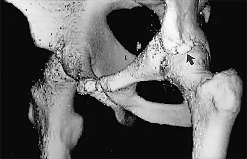
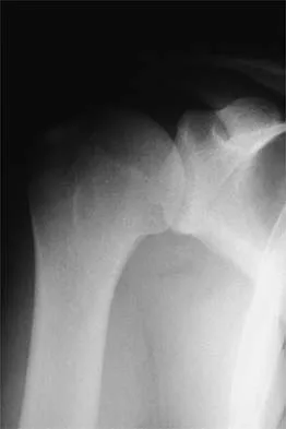
A 16-year-old high school football player sustains an injury to the left hip. The avulsed fragment identified by the arrow in Figure 34 represents the origin of which of the following structures?


Explanation

The avulsed fragment represents the origin of the rectus femoris from the anterior inferior iliac spine and the brim of the acetabulum. Avulsion of the anterior inferior iliac spine is much less common than avulsion of the anterior superior iliac spine with its origin of the sartorius. The origin of the gluteus minimus is from the outer cortex of the iliac wing and has not been reported as a source of bony avulsion. The hip capsule is composed of the ischiofemoral and pubofemoral ligaments, in addition to the iliofemoral ligament. The pelvic attachment of the ischiofemoral ligament is from the ischial part of the acetabulum posteriorly, while the pubofemoral ligament attaches to the pubic portion inferiorly. Technically, ligaments do not have origins and insertions as muscle tendon groups do, but have attachment sites. Metzmaker JN, Pappas AM: Avulsion fractures of the pelvis. Am J Sports Med 1985;13:349-358.

Question 6

Which of the following methods of meniscal repair has the highest load to failure strength?


Explanation

Numerous experimental studies have shown that vertical suture techniques are superior to all of the other noted methods. In fact, vertical sutures have been shown to be twice as strong as several of these techniques. DeHaven KE: Meniscus repair. Am J Sports Med 1999;27:242-250. Dervin GF, Downing KJ, Keene GC, McBride DG: Failure strengths of suture versus biodegradable arrow for meniscal repair: An in vitro study. Arthroscopy 1997;13:296-300.

Question 7

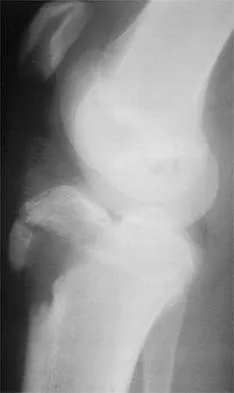
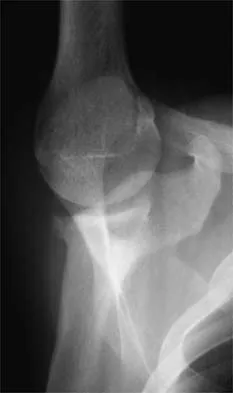
Figure 35 shows the lateral radiograph of a 15-year-old basketball player who felt a dramatic pop in his knee when landing after a lay-up. The patient reports that he cannot bear weight on the injured extremity. Management should consist of


Explanation

Tibial tubercle avulsion is an injury of the adolescent knee that most often occurs just before the end of growth. The fracture usually occurs with jumping, either at push-off or landing. This patient has a type III injury. In type III injuries, the articular surface is disrupted, and meniscal injury and compartment syndrome can occur. Open reduction is the treatment of choice, and anterior fasciotomy should be considered prophylactically at the time of surgery. Although the fracture heals with an anterior epiphysiodesis of the proximal tibia, little growth remains in this patient and no special handling of the physis is warranted. Ogden JA, Tross RB, Murphy MJ: Fractures of the tibial tuberosity in adolescents. J Bone Joint Surg Am 1980;62:205-215.

Question 8

A 52-year-old man has pain in the sternal area after landing on his right shoulder in a fall from his bicycle. In addition, he reports that he had difficulty swallowing and breathing immediately after the fall, but the symptoms resolved. A CT scan reveals a posterior sternoclavicular dislocation. Initial management should include


Explanation

Posterior sternoclavicular dislocations require rapid treatment because of the proximity of major neurovascular structures and the airway. Initial management should consist of closed reduction under general anesthesia in the operating room with a chest surgeon available. A successful closed reduction is usually stable. Internal fixation of sternoclavicular injuries should be avoided because of the likelihood of hardware migration and possible injury to the mediastinal structures. If closed reduction is unsuccessful, open reduction is indicated. Treatment following reduction of the sternoclavicular joint includes the application of a figure-of-8 splint and a sling for 6 weeks, followed by stretching and strengthening exercises. Beaty JH (ed): Orthopaedic Knowledge Update 6. Rosemont IL, American Academy of Orthopaedic Surgeons, 1999, pp 287-297.

Question 9

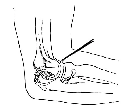
What nerve is at greatest risk of harm from the portal shown in Figure 36?


Explanation

The figure shows the anterolateral portal for elbow arthroscopy, and injury to the radial nerve has been reported in conjunction with this portal site. Studies have shown that closer proximity to the radial nerve is associated with more distal portal sites. The lateral and posterior antebrachial cutaneous nerves are both at less risk of injury. The ulnar and median nerves are both fairly remote to this location. Field LD, Altchek DW, Warren RF, O'Brien SJ, Skyhar MJ, Wickiewicz TL: Arthroscopic anatomy of the lateral elbow: A comparison of three portals. Arthroscopy 1994;10:602-607. Papilion JD, Neff RS, Shall LM: Compression neuropathy of the radial nerve as a complication of elbow arthroscopy: A case report and review of the literature. Arthroscopy 1988;4:284-286.

Question 10

In the majority of patients with chronic anterior cruciate ligament (ACL)-deficient knees, analysis of the gait pattern during level walking will most likely reveal which of the following changes?


Explanation

Patients with chronic ACL-deficient knees typically have lower than normal net quadriceps activity during the middle portion of the stance phase; the net moment about the knee reverses from one that demands quadriceps activity to one that demands increased hamstring activity. This type of gait is termed "quadriceps avoidance." This avoidance is believed to be a functional adaptation to reduce anterior tibial translation, and it is most prevalent as the knee moves from 45 degrees of flexion toward full extension, the arc of motion through which the ACL is most responsible for stability. Hurwitz DE, Andriacchi TP, Bush-Joseph CA, Bach BR Jr: Functional adaptations in patients with ACL-deficient knees. Exerc Sport Sci Rev 1997;25:1-20. Andriacchi TP, Birac D: Functional testing in the anterior cruciate ligament-deficient knee. Clin Orthop 1993;288:40-47.

Question 11

Glenohumeral inferior stability in the adducted shoulder position is primarily a function of the


Explanation

When the arm is adducted, the superior structures, including the superior glenohumeral ligament, are responsible in limiting the inferior translation. With the arm abducted, the inferior glenohumeral ligament complex is responsible for limiting inferior subluxation. Rotator cuff activity can actually depress the humeral head and does not play a role in preventing inferior subluxation. The long head of the biceps and the posterior glenohumeral ligament do not play a role in protecting the shoulder from inferior instability. Warner JJ, Deng XH, Warren RF, Torzilli PA: Static capsuloligamentous restraints to superior-inferior translation of the glenohumeral joint. Am J Sports Med 1992;20:675-685.

Question 12

A 20-year-old football player has repeated episodes of heat cramps during summer training sessions. A deficiency of what electrolyte is most responsible for heat cramps?


Explanation

Sodium deficiency is the cause of heat cramps. It is the principle electrolyte of sweat and is readily lost during training, especially in warmer temperatures. The condition can be avoided by adding extra table salt to food and maintaining good hydration before and after sports activities. Salt tablets are to be avoided when a patient has heat cramps because the high soluble load will cause gastric irritation. Bergeron MF, Armstrong LE, Maresh CM: Fluid and electrolyte losses during tennis in the heat. Clin Sports Med 1995;14:23-32.

Question 13

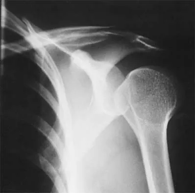
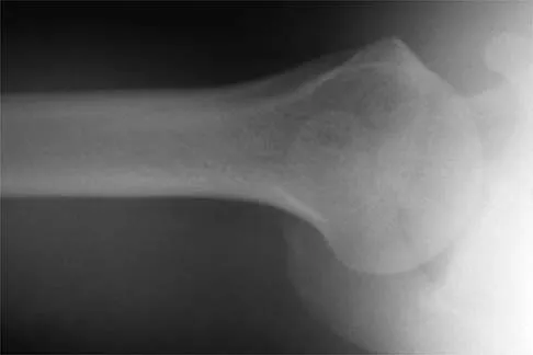
Figure 37 shows the radiograph of a 23-year-old football player who sustained a blow to the anterior aspect of his shoulder. Examination reveals pain and limited rotation. He is unable to flex the arm above the shoulder. Management should include which of the following studies?


Explanation

The patient has a posterior dislocation. The radiograph reveals marked internal rotation, but fails to show whether the humeral head is posteriorly displaced. Therefore, an axillary radiograph should be obtained to help confirm the diagnosis. Transverse view CT or MRI scans also may be useful. The other studies will not help confirm the diagnosis. In addition to a direct posterior blow, a shoulder dislocation may be caused by a seizure disorder or electrocution. Bloom MH, Obata WG: Diagnosis of posterior dislocation of the shoulder with the use of Velpeau axillary and angle-up roentgenographic views. J Bone Joint Surg Am 1967;49:943-949.

Question 14

A 21-year-old football player had severe pain and immediate swelling in the left anteromedial chest wall while bench pressing near maximal weights several days ago. Examination at the time of injury revealed a mass on the anteromedial chest wall. Follow-up examination now reveals decreased swelling, and axillary webbing is observed. The patient has weakness to adduction and forward flexion. The injured muscle originates from the


Explanation

The patient has a pectoralis major rupture, an injury that occurs most commonly during weight lifting. Grade III injuries represent complete tears of either the musculotendinous junction or an avulsion of the tendon from the humerus, the most common injury site. Examination will most likely reveal ecchymoses and swelling in the proximal arm and axilla, and strength testing will show weakness with internal rotation and in adduction and forward flexion. Axillary webbing, caused by a more defined inferior margin of the anterior deltoid as the result of rupture of the pectoralis, can be seen as the swelling diminishes. Surgical repair is the treatment of choice for complete ruptures. Nonsurgical treatment is associated with significant losses in adduction, flexion, internal rotation, strength, and peak torque. The pectoralis major originates from the proximal clavicle and the border of the sternum, including ribs two through six. The pectoralis major inserts (rather than originates) on the humerus. The coracoid process is the insertion site for the pectoralis minor, as well as the origin for the conjoined tendon. The pectoralis major has no attachment or origin from the scapula. The anterior deltoid originates from the lateral one third of the clavicle and the anterior acromion. Miller MD, Johnson DL, Fu FH, Thaete FL, Blanc RO: Rupture of the pectoralis major muscle in a collegiate football player: Use of magnetic resonance imaging in early diagnosis. Am J Sports Med 1993;21:475-477.

Question 15

A 24-year-old baseball pitcher reports pain over the posterior aspect of his shoulder that occurs only during throwing. He notes that the discomfort is greatest during the late cocking and early acceleration phases. Examination reveals localized tenderness with palpation over the external rotators and posterior glenoid. Radiographs are shown in Figures 38a through 38c. What is the most likely diagnosis?


Explanation

38b 38c The radiographs show a posterior glenoid osteophyte, often termed a "thrower's exostosis." These exostoses are best visualized on the Stryker notch view and may be missed on other more standard radiographic views of the shoulder. CT and MRI scans may be used, but usually add little information to the radiographic findings. Arthroscopic examination of patients with this condition commonly reveals undersurface tearing of the rotator cuff and posterior labrum. Treatment of this condition remains somewhat controversial, with avocation of both nonsurgical and surgical techniques. Meister K, Andrews JR, Batts J, Wilk K, Baumgarten T, Baumgartner T: Symptomatic thrower's exostosis: Arthroscopic evaluation and treatment. Am J Sports Med 1999;27:133-136. Ferrari JD, Ferrari DA, Coumas J, Pappas AM: Posterior ossification of the shoulder: The Bennett lesion. Etiology, diagnosis, and treatment. Am J Sports Med 1994;22:171-176.

Question 16

What percent of the adult human meniscus is vascularized?


Explanation

The adult menisci are considered to be relatively avascular structures, with the peripheral blood supply originating predominately from the lateral and medial genicular arteries. Branches of these vessels form the perimeniscal capillary plexus, which supplies the peripheral border throughout its attachment to the joint capsule. Vascular penetration studies have shown that 10% to 30% of the peripheral portion of the medial meniscus and 10% to 25% of the lateral meniscus are vascularized. Arnoczky SP, Warren RF: Microvasculature of the human meniscus. Am J Sports Med 1982;10:90-95.

Question 17

A 30-year-old man who participates in recreational sports reports the spontaneous onset of intermittent pain and swelling about the right knee. Examination reveals a 3+ effusion, with a range of motion of 10 degrees to 60 degrees. He has mild diffuse tenderness but no instability. MRI scans and an arthroscopic view are shown in Figures 39a through 39c. Management should consist of


Explanation

39b 39c The patient has synovial chondromatosis. The MRI scans show multiple small proscribed areas of signal intensity in the gutters and suprapatellar pouch, suggesting very small loose bodies. The arthroscopic view shows the classic appearance of multiple small chondral loose bodies. Synovial chondromatosis is a condition in which the synovium undergoes metaplasia, producing multiple chondral loose bodies that can subsequently ossify. The treatment of choice, removal of the loose bodies and arthroscopic synovectomy, results in a lower incidence of recurrence than other treatment methods. Coolican MR, Dandy DJ: Arthroscopic management of synovial chondromatosis of the knee: findings and results in 18 cases. J Bone Joint Surg Br 1989;71:498-500.

Question 18

Figure 40 shows the plain radiograph of a 30-year-old woman who has had a long history of standing bilateral anterior knee pain and a sense of patellar instability without frank dislocation. Nonsurgical management consisting of anti-inflammatory drugs and physical therapy has failed to provide relief. Examination reveals full range of motion of both knees, with moderate patellofemoral crepitance. Patellar apprehension and patellar grind tests are positive. The Q-angle measures 20 degrees. Management should now consist of


Explanation

The history, physical examination, and radiographs indicate that the patellofemoral pain is most likely caused by excessive lateral patellar pressure and patellar maltracking. Because the radiographs reveal the lateral tilt of the patella and lateral subluxation, the treatment of choice is bilateral lateral releases with anteromedialization of the tibial tubercles. This procedure corrects not only the excessive lateral patellar pressure, but also the lateral subluxation. The use of patella-stabilizing braces or taping may provide temporary relief, but these implements are not well-tolerated and they will not change the underlying biomechanics of the knee. Simple lateral release is indicated for isolated lateral tilt, but it does not correct the lateral subluxation. The use of thermal capsular shrinkage for the medial retinaculum has not been proven to provide long-term correction of the deformity. Boden BP, Pearsall AW, Garrett We Jr, et al: Patellofemoral instability: Evaluation and management. J Am Acad Orthop Surg 1997;5:47-57.

Question 19

A 24-year-old runner who underwent an allograft reconstruction of the anterior cruciate ligament (ACL) 3 years ago now reports anterior knee pain. Examination reveals no swelling or effusion, and the patient has full motion. A Lachman test and a pivot-shift test are negative. Palpation reveals tenderness on the patellar tendon and at the inferior pole of the patella. AP and lateral radiographs are shown in Figures 41a and 41b. Management should consist of


Explanation

41b The radiographs show tunnel enlargement, which is seen after ACL reconstruction, particularly with allografts. Occasionally, there will be formation of an associated subcutaneous pretibial cyst. It has been proposed that the tunnel enlargement and cyst are the result of incomplete incorporation of allograft tissues within the bone tunnels. There may be residual graft necrosis, allowing synovial fluid to be transmitted through the tunnel to collect in the pretibial area, manifesting as a synovial cyst. In the absence of cyst formation, the presence of tunnel enlargement does not appear to adversely affect the clinical outcome. Based on studies by Fahey and associates, continued tunnel expansion does not occur. Victoroff and associates report good results with curettage and bone grafting of the tibial tunnel if a pretibial cyst is present. Because this patient does not have a pretibial cyst, observation with activity modification is the preferred treatment. Fahey M, Indelicato PA: Bone tunnel enlargement after anterior cruciate ligament replacement. Am J Sports Med 1994;22:410-414.

Question 20

What is the most common mechanism of injury that produces turf toe?


Explanation

The most common mechanism of injury for turf toe is a hyperextension injury to the MTP joint. The foot is typically in a dorsiflexed position with the heel raised when an external force drives the MTP joint into further dorsiflexion. The joint capsule usually tears at the metatarsal neck because its attachment is weaker there than at the proximal phalanx. Some compression injuries to the dorsal articular surface of the metatarsal head can result from extension or hyperextension. Clanton TO, Ford JJ: Turf toe injury. Clin Sports Med 1994;13:731-741.

Question 21

A 68-year-old man embarks on a 24-week strength training program. He trains at 80% of his single repetition maximum for both the upper and lower extremities. Which of the following changes can be anticipated?


Explanation

Consistent, long-term exercise training in older athletes has proven very beneficial in reversing both endurance and strength losses that traditionally have been seen with aging. This patient's program will lead to a significant increase in the strength, cross-sectional area, and capillary density of the trained muscles. No major changes in aerobic capacity are anticipated. Strength improvements of up to 5% per day, similar to those for younger athletes, have been identified in this population in one study. Kirkendall DT, Garrett WE Jr: The effects of aging and training on skeletal muscle. Am J Sports Med 1998;26:598-602.

Question 22

A 26-year-old ballet dancer reports posterolateral ankle pain, especially with maximal plantar flexion. Examination reveals maximal tenderness just posterior to the lateral malleolus, and symptoms are heightened with forced passive plantar flexion. Radiographs are shown in Figures 42a and 42b. What is the most likely cause of the patient's symptoms?


Explanation

42b The patient has a symptomatic os trigonum caused by impingement that occurs with maximal plantar flexion of the ankle in the demi-pointe or full-pointe position. Patients frequently report posterolateral pain localized behind the lateral malleolus that may be misinterpreted as a disorder of the peroneal tendon. Pain with passive plantar flexion (the plantar flexion sign) indicates posterior impingement, not a problem with the peroneal tendon. The symptoms are not characteristic of a stress fracture, nor do the radiographs show a stress fracture or an osteochondritis dissecans lesion. The os trigonum is modest in its dimensions. The incidence or magnitude of symptoms does not correlate with the size of the fragment. Large fragments may be asymptomatic, while small lesions may create significant symptoms. Marotta JJ, Micheli LJ: Os trigonum impingement in dancers. Am J Sports Med 1992;20:533-536.

Question 23

A 23-year-old soccer player sustains a grade III complete posterior cruciate ligament (PCL) tear after colliding with another player. In reconstructing the PCL, it is optimal to reconstruct the


Explanation

The PCL is a nonisometric structure with nonuniform tension during knee motion, with maximum tension at 90 degrees of flexion. While the posteromedial PCL fibers have been found to be the most isometric, the anterolateral fibers represent the bulk of the ligament. Studies have suggested that anterior placement of the femoral tunnel is superior to placement in an isometric position. The anterolateral bundle tightens as the knee flexes; therefore, it is optimal to tension the graft at 90 degrees of flexion. Harner CD, Xerogeanes JW, Livesay GA, et al: The human posterior cruciate ligament complex: An interdisciplinary study. Ligament morphology and biomechanical evaluation. Am J Sports Med 1995;23:736-745.

Question 24

Figure 43 shows the lateral radiograph of a patient who underwent anterior cruciate ligament reconstruction. Based on the tunnel placement shown in the radiograph, evaluation of postoperative knee range of motion will most likely show


Explanation

The radiograph shows the correct tibial tunnel and anterior femoral tunnel; therefore, range of motion will most likely show loss of flexion. Brown CH Jr, Carson EW: Revision anterior cruciate ligament surgery. Clin Sports Med 1999;18:109-171.

Question 25

A 10-year-old soccer player has bilateral heel pain and reports that the pain is worse during and immediately after sports. Examination reveals that the calcaneal tuberosities are painful to palpation bilaterally. What is the most likely diagnosis?


Explanation

Calcaneal apophysitis (Sever's disease) is a common cause of heel pain in children who are active in sports. The symptoms are most commonly bilateral and will often respond to a gastrocnemius-soleus complex stretching program. In addition, rest, anti-inflammatory drugs, and heel pads for the shoe may be prescribed. There is no effect on the long-term growth of the calcaneus. Micheli LJ, Ireland ML: Prevention and management of calcaneal apophysitis in children: An overuse syndrome. J Pediatr Orthop 1987;7:34-38.

Question 26

A 22-year-old collegiate baseball pitcher presents with posterior shoulder pain during the late cocking phase of throwing. Physical examination demonstrates a 20-degree glenohumeral internal rotation deficit (GIRD) compared to the contralateral shoulder.

What is the primary pathomechanical cause of this patient's internal impingement?


Explanation

Internal impingement in overhead athletes is primarily driven by a contracture of the posterior band of the inferior glenohumeral ligament (IGHL). This contracture leads to a posterosuperior shift of the humeral head during maximum abduction and external rotation, trapping the posterosuperior rotator cuff against the labrum.

Question 27

A 26-year-old male sustains an isolated posterior cruciate ligament (PCL) rupture in a motorcycle accident and fails nonoperative management. A single-bundle PCL reconstruction is planned.

To correctly recreate the anterolateral (AL) bundle, the femoral tunnel should be placed at which of the following locations?


Explanation

In single-bundle PCL reconstruction, the stronger anterolateral (AL) bundle is typically reconstructed. The native femoral footprint of the AL bundle is located on the anterolateral aspect of the medial femoral condyle within the notch.

Question 28

During surgical reconstruction of a multiligament knee injury, the surgeon isolates the fibular collateral ligament (FCL) to recreate its native anatomy.

Where is the correct anatomical location of the native FCL femoral attachment?


Explanation

The femoral attachment of the fibular collateral ligament (FCL) is situated approximately 1.4 mm proximal and 3.1 mm posterior to the lateral epicondyle. Accurate tunnel placement here is critical to restore native posterolateral corner kinematics.

Question 29

A 45-year-old female experiences a sudden pop in the back of her knee while squatting. MRI reveals a posterior root tear of the medial meniscus with 4 mm of meniscal extrusion.

Biomechanical studies demonstrate that this specific injury pattern alters tibiofemoral contact pressures equivalent to which of the following?


Explanation

A posterior root tear of the medial meniscus completely disrupts the circumferential hoop stresses of the meniscus. This biomechanical failure leads to increased peak contact pressures that are equivalent to those seen after a total meniscectomy.

Question 30

A 30-year-old cyclist sustains a Type V acromioclavicular (AC) joint injury. During open reduction and reconstruction, the surgeon focuses on restoring the primary restraint to superior clavicular translation.

Which ligament acts as this primary restraint?


Explanation

The conoid ligament, which is the more medial of the two coracoclavicular (CC) ligaments, is the primary restraint to superior translation of the clavicle. The trapezoid ligament provides primary resistance to axial compression.

Question 31

A 22-year-old hockey player undergoes hip arthroscopy for symptomatic femoroacetabular impingement (FAI). Radiographs show an alpha angle of 65 degrees.

To address the cam deformity, osteochondroplasty is most commonly performed in which region of the femoral head-neck junction?


Explanation

Cam deformities are characterized by extra bone at the femoral head-neck junction, leading to asphericity. They most commonly occur in the anterosuperior quadrant of the femoral head-neck junction.

Question 32

A 17-year-old female presents with her first episode of lateral patellar dislocation. An MRI confirms rupture of the medial patellofemoral ligament (MPFL).

Where is the native femoral footprint of the MPFL located in relation to the medial epicondyle and adductor tubercle?


Explanation

The native femoral footprint of the MPFL (Schottle's point) is anatomically located in the groove or saddle between the medial epicondyle distally and the adductor tubercle proximally.

Question 33

A 25-year-old professional tennis player undergoes arthroscopic repair of a Type II superior labrum anterior-posterior (SLAP) lesion.

What is the most commonly reported complication following this specific procedure in overhead athletes?


Explanation

Repairing Type II SLAP lesions in overhead athletes is frequently associated with postoperative stiffness, particularly a clinically significant loss of external rotation. Due to poor return-to-play rates, biceps tenodesis is increasingly favored in this demographic.

Question 34

A 13-year-old gymnast complains of vague, activity-related anterior knee pain. Imaging reveals an osteochondritis dissecans (OCD) lesion with intact overlying cartilage.

What is the most common anatomical location for this pathology in the knee?


Explanation

The classic and most common location for an osteochondritis dissecans (OCD) lesion in the knee is the lateral aspect of the medial femoral condyle (LAME). This accounts for approximately 70% of all knee OCD lesions.

Question 35

A 20-year-old collegiate javelin thrower is evaluated for medial elbow pain. The moving valgus stress test is highly positive at 90 degrees of flexion.

Which specific structure is the primary restraint to valgus stress at this angle of elbow flexion?


Explanation

The anterior bundle of the ulnar collateral ligament (UCL) is the primary restraint to valgus stress at the elbow between 30 and 120 degrees of flexion. The posterior bundle is a secondary restraint.

Question 36

A 32-year-old powerlifter feels a tearing sensation in his chest while attempting a maximal bench press. Examination reveals an asymmetric chest wall and weakness in internal rotation.

Which anatomical segment of the involved muscle is most frequently ruptured, and where does it insert on the humerus?


Explanation

Pectoralis major ruptures most commonly occur at the sternocostal (sternal) head near its humeral insertion. Because the tendon twists 180 degrees, the sternal head forms the deep lamina and inserts proximal and posterior to the clavicular head on the humerus.

Question 37

A 19-year-old soccer player is scheduled for ACL reconstruction. The surgeon discusses autograft choices, including bone-patellar tendon-bone (BTB) and hamstring.

In comparative studies, what complication is significantly more common with BTB autograft than with hamstring autograft?


Explanation

BTB autograft is associated with a significantly higher incidence of donor-site morbidity, particularly anterior knee pain and pain with kneeling, when compared to hamstring autograft.

Question 38

Following an arthroscopic repair of a full-thickness supraspinatus tear, the tendon undergoes a complex biological healing process.

How does the healed tendon-to-bone interface typically present histologically?


Explanation

Unlike native tendon insertions which feature a complex four-zone fibrocartilaginous transition, healing following rotator cuff repair typically results in a mechanically inferior fibrovascular scar tissue interface. It does not regenerate the native enthesis.

Question 39

A 24-year-old rugby player sustains an external rotation injury to the ankle. Examination reveals tenderness over the anterior syndesmosis, and radiographs demonstrate widening of the tibiofibular clear space.

Which ligament provides the greatest absolute resistance to diastasis of the distal tibiofibular syndesmosis?


Explanation

Biomechanical studies show that the posterior inferior tibiofibular ligament (PITFL) contributes approximately 42% of the resistance to syndesmotic diastasis, making it the strongest and most important individual stabilizer of the syndesmosis.

Question 40

A 40-year-old male is brought to the trauma bay after a high-speed skiing collision. His knee is grossly deformed with a prominent "dimple sign" over the medial joint line.

What does this specific clinical finding indicate regarding the injury?


Explanation

A "dimple sign" or transverse groove on the medial aspect of a dislocated knee is pathognomonic for an irreducible posterolateral knee dislocation. It is caused by the medial femoral condyle buttonholing through the medial capsule.

Question 41

A 22-year-old soccer player undergoes an anatomic single-bundle anterior cruciate ligament (ACL) reconstruction. Which of the following is the most likely consequence of placing the femoral tunnel too far anteriorly (shallow) in the intercondylar notch?


Explanation

An anteriorly placed (shallow) femoral tunnel in ACL reconstruction results in a graft that is loose in extension and tight in flexion. This non-anatomic placement often leads to a loss of terminal knee flexion and potential graft stretching or failure.

Question 42

A 17-year-old female presents with recurrent lateral patellar dislocations. Imaging shows a TT-TG distance of 14 mm, normal patellar height, and a torn medial patellofemoral ligament (MPFL). She undergoes isolated MPFL reconstruction. Where is the anatomic femoral attachment of the MPFL located?


Explanation

The anatomic femoral footprint of the MPFL is located in the saddle region between the adductor tubercle proximally and the medial epicondyle distally. Radiographically, this correlates closely with Schöttle's point.

Question 43

A 28-year-old bodybuilder feels a pop in his anterior axilla while performing a heavy bench press. Examination reveals loss of the anterior axillary fold and weakness in internal rotation. Which segment of the pectoralis major tendon is under the most tension and most likely to rupture first during the eccentric phase of a bench press?


Explanation

The pectoralis major tendon undergoes a 180-degree twist before inserting on the humerus. The inferior sternocostal fibers insert most proximally and superiorly, making them stretch the most and rupture first during eccentric loading in extension and abduction.

Question 44

A 55-year-old physically active man reports a popping sensation in his posterior knee while squatting, followed by acute posteromedial joint line pain. MRI reveals a medial meniscus posterior root tear.

Biomechanical studies demonstrate that an un-repaired complete posterior root tear alters knee joint contact pressures most similarly to which of the following?


Explanation

A complete tear or avulsion of the medial meniscus posterior root completely compromises the hoop stresses of the meniscus. This results in peak joint contact pressures equivalent to those seen after a total meniscectomy, rapidly accelerating cartilage wear.

Question 45

A 21-year-old collegiate baseball pitcher presents with vague posterior shoulder pain and a decrease in pitching velocity. Examination reveals 15 degrees of internal rotation and 120 degrees of external rotation in the dominant arm, compared to 60 degrees of internal rotation and 90 degrees of external rotation in the non-dominant arm. Which of the following is the most appropriate initial management?


Explanation

Glenohumeral internal rotation deficit (GIRD) is common in overhead athletes due to repetitive microtrauma leading to posterior capsular contracture. The initial treatment of choice is a targeted posterior capsular stretching program, specifically utilizing sleeper stretches.

Question 46

A 45-year-old recreational tennis player has persistent deep shoulder pain. MRI shows an isolated Type II Superior Labrum Anterior Posterior (SLAP) tear. He undergoes arthroscopic evaluation. What is the most reliable predictor of failure if a primary SLAP repair is performed in this patient instead of a biceps tenodesis?


Explanation

In patients over the age of 40, primary repair of a Type II SLAP tear has a significantly high failure rate and frequently results in postoperative stiffness. Biceps tenodesis is generally the preferred surgical treatment in this demographic.

Question 47

A 20-year-old rugby player presents with his third anterior shoulder dislocation. A CT scan with 3D reconstruction is obtained.

Measurements indicate a 25% anterior glenoid bone loss. What is the most appropriate surgical intervention to minimize the risk of recurrent instability?


Explanation

Critical glenoid bone loss exceeding 20-25% in a contact athlete is a strong indication for a bony augmentation procedure, such as the Latarjet procedure. Soft tissue Bankart repairs have unacceptably high failure rates in the setting of significant bone loss.

Question 48

A 19-year-old collegiate swimmer presents with insidious onset bilateral shoulder pain. Physical examination demonstrates a 2+ sulcus sign bilaterally, apprehension at end-range abduction/external rotation without a distinct subluxation event, and generalized ligamentous laxity. What is the most appropriate initial management?


Explanation

Multidirectional instability in an overhead athlete is initially managed non-operatively with a targeted physical therapy program. Surgery (e.g., capsular shift) is reserved for patients who fail an extensive (e.g., 6-month) trial of physical therapy.

Question 49

When comparing bone-patellar tendon-bone (BTB) autograft to hamstring autograft for anterior cruciate ligament (ACL) reconstruction, BTB autograft is associated with a statistically higher incidence of which of the following postoperative complications?


Explanation

BTB autografts are historically associated with a higher incidence of anterior knee pain and donor site morbidity compared to hamstring autografts. Rates of graft rupture, DVT, and infection are generally comparable between the two graft types.

Question 50

A 22-year-old collegiate baseball pitcher is diagnosed with a Type II superior labrum anterior and posterior (SLAP) tear. During which phase of throwing does the 'peel-back' mechanism place maximal strain on the biceps anchor?


Explanation

The 'peel-back' mechanism occurs during the late cocking phase of throwing when the shoulder is in maximum abduction and external rotation. This position causes a posterior and inferior shift of the biceps vector, peeling the labrum off the glenoid.

Question 51

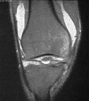
A 50-year-old moderately active female feels a 'pop' in her posterior knee while squatting. An MRI is obtained as seen in the figure.

It demonstrates a medial meniscus posterior root tear with 4 mm of extrusion and minimal osteoarthritis. What is the most appropriate surgical management?


Explanation

Posterior root tears of the medial meniscus lead to loss of hoop stresses and rapid progression of osteoarthritis. Transtibial pull-out repair is indicated in active patients with minimal osteoarthritis to restore joint biomechanics and prevent cartilage degeneration.

Question 52

A 24-year-old competitive rugby player sustains his third anterior shoulder dislocation. A pre-operative CT scan demonstrates 28% anterior glenoid bone loss. Which of the following surgical interventions is most appropriate?


Explanation

The Latarjet procedure (coracoid transfer) is the gold standard for anterior shoulder instability in the presence of critical anterior glenoid bone loss (>20-25%). Arthroscopic soft tissue repairs alone have an unacceptably high failure rate in this setting.

Question 53

A 12-year-old boy presents with vague anterior knee pain. Radiographs demonstrate a 1.5 cm osteochondritis dissecans (OCD) lesion of the medial femoral condyle.

MRI reveals no fluid behind the lesion, and his physes are wide open. What is the recommended initial management?


Explanation

Stable OCD lesions (no fluid behind the fragment on MRI) in patients with open physes have a high rate of spontaneous healing. A trial of non-operative management with activity restriction is the initial standard of care.

Question 54

A 10-year-old skeletally immature female soccer player sustains an anterior cruciate ligament (ACL) tear. She is Tanner stage 1 with significant growth remaining. Which of the following surgical techniques has the lowest risk of causing a growth arrest?


Explanation

In skeletally immature patients with significant growth remaining, physeal-sparing techniques such as all-epiphyseal or over-the-top extra-articular reconstructions are recommended. These avoid drilling across the physes, thereby minimizing the risk of premature physeal closure and angular deformity.

Question 55

A 45-year-old woman sustains a posterior root tear of the medial meniscus. Biomechanically, this injury alters the joint contact pressures to be most equivalent to which of the following scenarios?


Explanation

A complete posterior root tear of the medial meniscus disrupts the hoop stresses, leading to functional extrusion of the meniscus. Biomechanically, this results in increased articular contact pressures equivalent to a total meniscectomy, accelerating joint degeneration.

Question 56

A 22-year-old rugby player undergoes a Latarjet procedure for recurrent anterior shoulder instability with 25% glenoid bone loss. Postoperatively, he presents with an inability to actively flex his elbow or supinate his forearm. Which nerve is most likely injured?


Explanation

The musculocutaneous nerve is at high risk during the Latarjet procedure due to its proximity to the coracoid process and conjoint tendon, which are retracted during exposure. Injury results in weakness of elbow flexion and forearm supination.

Question 57

During medial patellofemoral ligament (MPFL) reconstruction for recurrent patellar instability, the femoral attachment must be accurately positioned. Which of the following describes the correct radiographic landmark (Schöttle's point) for the femoral footprint on a true lateral view?


Explanation

Schöttle's point identifies the anatomic femoral footprint of the MPFL. It is located approximately 1 mm anterior to the posterior femoral cortex line, 2.5 mm distal to the posterior origin of the medial femoral condyle, and proximal to the Blumensaat line.

Question 58

A 28-year-old male is brought to the emergency department after a high-velocity knee dislocation. His knee is reduced, but the ankle-brachial index (ABI) is 0.8. Which of the following is the most appropriate next step in management?


Explanation

An ABI less than 0.9 in the setting of a knee dislocation is highly suggestive of an arterial injury. CT angiography is the gold standard diagnostic tool to confirm a popliteal artery injury and should be performed urgently to guide surgical intervention.

Question 59

A 26-year-old athlete presents with a symptomatic 4.0 cm² osteochondral defect of the medial femoral condyle with 5 mm of subchondral bone loss. Nonoperative management has failed. Which of the following is the most appropriate surgical intervention?


Explanation

Osteochondral allograft transplantation is indicated for large (>2 cm²) defects, especially when there is significant subchondral bone loss. Procedures like MACI and ACI are typically reserved for pure chondral defects without substantial bony involvement.

Question 60

A 25-year-old hockey player presents with anterior hip pain exacerbated by hip flexion, adduction, and internal rotation. Radiographs reveal a crossover sign and an alpha angle of 65 degrees. What is the most likely combination of pathomorphologies?


Explanation

An alpha angle greater than 55 degrees indicates a Cam deformity (femoral head-neck junction abnormality). The crossover sign on an AP pelvis radiograph indicates cranial retroversion of the acetabulum, typical of Pincer impingement. Together, they represent mixed femoroacetabular impingement (FAI).

Question 61

A 32-year-old weightlifter feels a "pop" in his anterior chest while performing a heavy bench press. Examination reveals ecchymosis and loss of the anterior axillary fold. If surgical repair is planned, which portion of the pectoralis major tendon is most commonly ruptured and requires mobilization?


Explanation

Pectoralis major ruptures most commonly occur during maximum eccentric contraction. The sternocostal head is typically avulsed from its insertion on the proximal humerus, while the clavicular head often remains intact.

Question 62

A 21-year-old runner complains of bilateral anterolateral leg pain that reliably begins 15 minutes into a run and resolves 30 minutes after rest. Nonoperative measures have failed. What is the gold standard diagnostic test for this condition?


Explanation

Chronic exertional compartment syndrome is definitively diagnosed using dynamic intracompartmental pressure testing. The Pedowitz criteria include specific pressure thresholds resting, 1 minute after exercise, or 5 minutes after exercise.

Question 63

A 40-year-old male sustains an acute Achilles tendon rupture. When discussing surgical versus nonoperative management with a functional rehabilitation protocol, the patient should be informed that surgical repair provides which of the following advantages?


Explanation

Historically, operative repair of Achilles tendon ruptures is associated with a lower rate of re-rupture compared to nonoperative management. However, operative management carries higher risks of wound complications and iatrogenic sural nerve injury.

Question 64

A 20-year-old collegiate baseball pitcher presents with medial elbow pain and decreased pitching velocity. The moving valgus stress test is positive. MRI confirms a full-thickness tear of the anterior bundle of the ulnar collateral ligament (UCL). During reconstruction, where must the graft be secured on the ulna to recreate the native anterior bundle insertion?


Explanation

The anterior bundle of the UCL is the primary restraint to valgus stress at the elbow. It originates on the anteroinferior surface of the medial epicondyle and inserts on the sublime tubercle at the base of the coronoid process.

Question 65

A 60-year-old male presents with a massive, irreparable posterosuperior rotator cuff tear. He has an intact subscapularis, no glenohumeral arthritis (Hamada grade 1), but demonstrates a positive hornblower's sign. Which of the following joint-preserving procedures is most indicated to restore active external rotation?


Explanation

In an active patient with an irreparable posterosuperior rotator cuff tear and severe active external rotation weakness, a latissimus dorsi tendon transfer is indicated. Superior capsular reconstruction improves superior stability but does not actively restore external rotation.

Question 66

A 45-year-old water skier sustains a hyperflexion injury of the hip with the knee extended. He has severe posterior thigh pain and a palpable defect at the ischial tuberosity. MRI shows a complete 3-tendon proximal hamstring avulsion with 4 cm of retraction. What is the most appropriate management?


Explanation

Complete proximal hamstring avulsions involving all three tendons with significant retraction (>2 cm) in an active patient are best treated with acute surgical repair. This prevents chronic weakness, debilitating pain, and secondary sciatic nerve tethering.

Question 67

A 48-year-old recreational tennis player presents with vague anterior shoulder pain. An MRI arthrogram reveals a Type II SLAP tear. He has failed 6 months of nonoperative management. What is the currently recommended surgical intervention for this specific demographic?


Explanation

In older patients (typically >40 years) with a symptomatic Type II SLAP tear, primary biceps tenodesis is strongly preferred. SLAP repair in this older demographic is associated with higher rates of postoperative stiffness, persistent pain, and revision surgery.

Question 68

A 30-year-old male sustains a severe varus and hyperextension injury to his knee. Examination reveals a positive dial test (increased external rotation) at 30 degrees of knee flexion, but the external rotation normalizes to match the contralateral knee at 90 degrees of flexion. What is the primary injured structure?


Explanation

A positive dial test at 30 degrees of flexion that normalizes at 90 degrees indicates an isolated injury to the posterolateral corner (PLC). If the test remains positive at both 30 and 90 degrees, it suggests a combined PLC and PCL injury.

Question 69

A 22-year-old rugby player undergoes an open Latarjet procedure for recurrent anterior shoulder instability. Postoperatively, he presents with numbness over the lateral aspect of his forearm and weakness in elbow flexion. Which nerve is most likely injured?


Explanation

The musculocutaneous nerve is at high risk during the Latarjet procedure due to its proximity to the conjoint tendon and coracoid process. Vigorous medial retraction of the conjoint tendon places traction on this nerve, causing lateral forearm numbness and biceps weakness.

Question 70

During clinical examination of a multiligamentous knee injury, the dial test is performed. At 30 degrees of knee flexion, the primary restraint to external rotation of the tibia is the:


Explanation

The popliteofibular ligament (PFL) and popliteus complex are the primary restraints to external tibial rotation at 30 degrees of flexion. The LCL is the primary restraint to varus stress.

Question 71

A 30-year-old weightlifter suffers a complete rupture of the pectoralis major tendon. Regarding the normal anatomy of the pectoralis major footprint on the humerus, the sternal head inserts:


Explanation

The pectoralis major tendon undergoes a 180-degree twist before insertion. Consequently, the sternal head inserts proximal and deep, while the clavicular head inserts distal and superficial on the lateral lip of the bicipital groove.

Question 72

If a femoral tunnel is placed too anteriorly (high in the notch in extension) during an anterior cruciate ligament (ACL) reconstruction, what is the resulting biomechanical effect on the graft?


Explanation

An anteriorly placed femoral tunnel (anterior to the anatomic footprint) creates a graft that is excessively tight in flexion and loose in extension, often leading to restricted knee flexion and eventual graft failure.

Question 73

The peel-back mechanism responsible for Type II SLAP lesions in overhead-throwing athletes occurs maximally in which shoulder position?


Explanation

The peel-back mechanism occurs during the late cocking phase of throwing, characterized by abduction and maximal external rotation. In this position, the biceps vector shifts posteriorly, transmitting torsional forces that peel the posterosuperior labrum off the glenoid.

Question 74

Following hip arthroscopy for femoroacetabular impingement utilizing a perineal traction post, the patient reports severe numbness in the groin and scrotum. Neuropraxia of which nerve is the most likely cause?


Explanation

Pudendal nerve neuropraxia is a well-documented complication of hip arthroscopy due to direct compression from the perineal post against the groin during sustained traction.

Question 75

Biomechanical studies show that a complete, unrepaired tear of the posterior root of the medial meniscus alters knee contact mechanics equivalent to:


Explanation

A medial meniscus posterior root tear destroys the meniscal hoop stresses, allowing radial extrusion of the meniscus. Biomechanically, this results in peak tibiofemoral contact pressures identical to a total medial meniscectomy.

Question 76

During ulnar collateral ligament (UCL) reconstruction of the elbow, the graft is typically anchored to the anatomic footprints. The anterior bundle of the UCL originates from the anteroinferior medial epicondyle and inserts onto the:


Explanation

The anterior bundle of the UCL is the primary restraint to valgus stress at the elbow and inserts onto the sublime tubercle, located on the medial aspect of the coronoid process.

Question 77

During anatomical reconstruction of the coracoclavicular (CC) ligaments for a high-grade acromioclavicular joint separation, proper graft placement requires knowledge of the native footprint. The conoid ligament footprint on the clavicle is located:


Explanation

The conoid ligament attaches to the conoid tubercle of the clavicle, which is located posteromedial to the attachment of the trapezoid ligament. The conoid is the primary restraint to superior clavicular translation.

Question 78

A 22-year-old soccer player sustains a non-contact knee injury. Radiographs show an elliptic bone fragment adjacent to the lateral tibial plateau. What structure is typically attached to this avulsed fragment?


Explanation

This describes a Segond fracture, which is an avulsion of the anterolateral ligament (ALL) and lateral capsular structures. It is highly pathognomonic for an anterior cruciate ligament (ACL) tear.

Question 79

A 21-year-old collegiate rugby player presents with recurrent anterior shoulder instability. CT scan demonstrates 28% anterior glenoid bone loss. What is the most appropriate definitive surgical management?


Explanation

In contact athletes with critical glenoid bone loss (typically >20-25%), a Latarjet procedure (coracoid transfer) is indicated. Arthroscopic soft tissue repairs alone have an unacceptably high failure rate in this setting.

Question 80

A 48-year-old female experiences a pop in her posterior knee while deep squatting. MRI reveals a medial meniscus posterior root tear with 3 mm of extrusion. What is the primary biomechanical consequence if this injury is left untreated?


Explanation

A posterior root tear disrupts the structural meniscal ring, leading to a loss of hoop stresses and resultant meniscal extrusion. This biomechanically mimics a total meniscectomy and accelerates medial compartment osteoarthritis.

Question 81

When performing a single-bundle posterior cruciate ligament (PCL) reconstruction, which native bundle is recreated to restore primary restraint against posterior tibial translation in flexion?


Explanation

The native PCL consists of two distinct bundles. The anterolateral bundle is the larger of the two, is tight in flexion, and is the primary target for single-bundle PCL reconstructions.

Question 82

A 22-year-old baseball pitcher presents with medial elbow pain and decreased velocity. Pain is most severe during the late cocking and early acceleration phases of throwing. Which bundle of the ulnar collateral ligament (UCL) is most likely injured and serves as the primary restraint to valgus stress at 90 degrees of flexion?


Explanation

The anterior bundle of the UCL is the primary restraint to valgus stress at the elbow, particularly between 30 and 120 degrees of flexion. It is most vulnerable during the late cocking and early acceleration phases of throwing.

Question 83

A 32-year-old weightlifter felt a tearing sensation in his anterior axilla while performing a heavy bench press. Exam reveals loss of the anterior axillary fold. He undergoes surgical repair. During anatomic repair, what is the correct orientation of the sternocostal head insertion on the humerus relative to the clavicular head?


Explanation

The pectoralis major tendon undergoes a 180-degree twist before inserting on the humerus. This orientation causes the sternocostal head to insert deep (posterior) and proximal to the clavicular head.

Question 84

A 28-year-old female undergoes hip arthroscopy for femoroacetabular impingement. Postoperatively, she reports profound numbness in the perineal region and labia. Which nerve was most likely injured due to intraoperative traction on the perineal post?


Explanation

The pudendal nerve can be compressed against the perineal post during traction in hip arthroscopy. This typically presents as transient numbness or pain in the perineum and genitalia that resolves with conservative management.

Question 85

A 17-year-old female experiences recurrent lateral patellar instability and is scheduled for a medial patellofemoral ligament (MPFL) reconstruction. Where is the exact anatomical location of the femoral origin of the MPFL (Schottle's point)?


Explanation

The anatomic femoral origin of the MPFL lies in a saddle-shaped depression between the medial epicondyle and the adductor tubercle. Proper isometric graft placement at this location is critical to prevent abnormal patellofemoral tracking.

Question 86

A 24-year-old football player sustained a direct blow to the anteromedial aspect of his knee. Physical examination reveals increased external tibial rotation of 15 degrees compared to the contralateral side at 30 degrees of knee flexion, but symmetrical external rotation at 90 degrees. What is the most likely injury pattern?


Explanation

The dial test evaluates for excessive external rotation. Increased rotation at 30 degrees only indicates an isolated posterolateral corner (PLC) injury, whereas increased rotation at both 30 and 90 degrees indicates a combined PLC and PCL injury.

Question 87

A 25-year-old athlete undergoes an arthroscopic microfracture for a 1.5 cm^2 full-thickness chondral defect on the medial femoral condyle. What is the predominant type of collagen in the reparative tissue generated by this procedure?


Explanation

Marrow stimulation techniques like microfracture produce fibrocartilage, which is predominantly composed of Type I collagen. This differs from native hyaline articular cartilage, which is predominantly composed of Type II collagen.

Question 88

A 30-year-old overhead athlete undergoes an arthroscopic Type II SLAP repair. Six months postoperatively, he complains of persistent pain and a significant loss of external rotation. What is the most common iatrogenic cause of this complication?


Explanation

Capturing the anterosuperior capsule or the middle glenohumeral ligament (MGHL) during a SLAP repair can severely restrict external rotation. Suture anchors must be placed meticulously to avoid tethering these essential anterior stabilizing structures.

Question 89

A 19-year-old female collegiate runner presents with an anterior tibial stress fracture and reports irregular menstrual cycles for the past year. Dual-energy x-ray absorptiometry reveals low bone mineral density. What is the primary underlying physiological mechanism for her condition (Relative Energy Deficiency in Sport)?


Explanation

Relative Energy Deficiency in Sport (RED-S), formerly known as the female athlete triad, is driven by low energy availability. This energetic deficit suppresses the hypothalamic-pituitary-ovarian axis, leading to hypoestrogenism and subsequent decreased bone mineral density.

None

Clinic OS
Medically Verified Content by
Prof. Clinic OS
Consultant Orthopedic & Spine Surgeon
Chapter Index