العربية
Part of the Master Guide

AAOS Pediatric Orthopedic MCQs (Set 2): DDH, SCFE & Spinal Deformities | Board Review

Orthopedic Pediatrics 2026 MCQs: Board Review Questions & Answers (Part 3)

27 Apr 2026 71 min read 61 Views
Figure for Pediatrics 2001 MCQs - Part 3 - Question 51.

Key Takeaway

Discover the latest medical recommendations for Orthopedic Pediatrics 2026 MCQs: Board Review Questions & Answers (Part 3). Top-rated Orthopedic Pediatrics 2026 MCQs bank. Practice with clinical case questions, orthopedic surgery board review, and evidence-based answers updated for 2026.

Orthopedic Pediatrics 2026 MCQs: Board Review Questions & Answers (Part 3)

Comprehensive 100-Question Exam


00:00

Start Quiz

Question 1

Figures 29a and 29b show the radiographs of a 13-year-old competitive gymnast who has had elbow pain for the past 2 weeks. The pain is worse with tumbling activities. Examination reveals a mild effusion and slight limitation of extension and forearm rotation with no locking. Initial management should consist of


Explanation

29b The radiographs show a lesion in the capitellum that is consistent with osteochondritis dissecans. There is no evidence of a loose body at this time. Initial management should consist of cessation of gymnastic activities. Nonsteroidal anti-inflammatory drugs and ice may help to alleviate acute symptoms; most symptoms usually resolve in 6 to 12 weeks. The patient may then begin range-of-motion and strengthening exercises, with a slow return to activities once full range of motion and good strength have been achieved. However, the prognosis for a return to high-level competitive gymnastics is guarded. Surgery is indicated for intra-articular loose bodies, a locked elbow, or failure of nonsurgical management. Surgery may be done either open or arthroscopically. Loose bodies should be removed, and cartilage flaps should be debrided. The results of bone grafting and internal fixation generally have been poor. Drilling the base of the defect may stimulate replacement with fibrocartilage, but the benefits of this procedure are not well documented. Maffulli N, Chan D, Aldridge MJ: Derangement of the articular surfaces of the elbow in young gymnasts. J Pediatr Orthop 1992;12:344-350. Bauer M, Jonsson K, Josefsson PO, Linden B: Osteochondritis dissecans of the elbow: A long-term follow-up study. Clin Orthop 1992;284:156-160.

Question 2

A 12-year-old boy who has had a 1-month history of right thigh pain and a limp reports worsening of the pain after a fall, and he can no longer walk or bear weight on the involved extremity. Radiographs of the pelvis reveal a slipped capital femoral epiphysis with moderate to severe displacement. While positioning the patient on the fracture table for screw fixation, partial reduction of the slip is achieved. No further reduction maneuvers are attempted, and the epiphysis is stabilized with a single cannulated screw. What complication is most likely to develop following this procedure?


Explanation

Traditional classification of slipped capital femoral epiphyses is based on the following temporal criteria: acute (symptoms that persist for less than 3 weeks); chronic (symptoms that persist for more than 3 weeks); or acute on chronic (acute exacerbation of long-standing symptoms). A newer classification differentiates between a stable slip where weight bearing is possible, and an unstable slip if it is not. Reduction of an unstable slip often occurs unintentionally with induction of anesthesia and positioning of the patient for surgery. The rate of satisfactory results is lower primarily because of a much higher incidence of osteonecrosis following internal fixation of an unstable slip. Loder RT, Richards BS, Shapiro PS, Reznick LR, Aronson DD: Acute slipped capital femoral epiphysis: The importance of physeal stability. J Bone Joint Surg Am 1993;75:1134-1140.

Question 3

Figure 30 shows the AP radiograph of a 9-month-old girl who has been referred for evaluation of unequal leg lengths. Examination reveals symmetrical abduction of the hips. When the hips are flexed 90 degrees, the right knee height is greater than the left knee. The girth of the right thigh and calf is larger than the contralateral side. There are no cutaneous lesions, and examination of the spine is normal. The infant is moving all extremities equally and spontaneously. Management should consist of


Explanation

Hemihypertrophy or hemihypotrophy is usually idiopathic, and either the leg or the entire side of the body may be involved. In the infant or young child, it is often difficult to determine which side is abnormal if the condition is mild. Because of the association of Wilms' tumor with hemihypertrophy, these patients should undergo a yearly renal ultrasound until at least age 5 years. Other conditions that may exhibit hemihypertrophy include Klippel-Trenaunay-Weber syndrome, Proteus syndrome, and neurofibromatosis. In this patient, the mild hemihypertrophy is idiopathic. Because of the normal spinal examination and absence of neurologic findings, an MRI scan is unnecessary. The absence of clinical and radiographic evidence of hip dysplasia makes both an ultrasound of the hips and application of a Pavlik harness unnecessary. Richards BS (ed): Orthopaedic Knowledge Update: Pediatrics. Rosemont Ill, American Academy of Orthopaedic Surgeons, 1996, pp 185-193. Sponseller PD: Localized disorders of bone and soft tissue, in Morrissy RT, Weinstein SL (eds): Lovell and Winter's Pediatric Orthopaedics, ed 4. Philadelphia, Pa, Lippincott-Raven, 1996, pp 305-344.

Question 4

What is the mechanism of action of an intramuscular injection of botulinum type A toxin in reducing spasticitiy?


Explanation

The use of intramuscular botulinum type A toxin has been shown to be a useful adjuvant in the management of dynamic deformity in patients with cerebral palsy. Botulinum type A toxin is a neurotoxin produced by Clostridium botulinum that works by interfering with presynaptic acetylcholine release at cholinergic nerve terminals. At the cellular level, the mechanism involves endocytosis of the intact botulinum toxin molecule by cells in the end plate, followed by disulfide cleavage and translocation of the light chain into the cytosol where it disrupts the normal binding of the synaptosomal vesicles to the axon terminal membrane. Neither the nerve terminal nor the neuromuscular junction is damaged. The muscle paralysis is reversible and dose-dependent. Baclofen is a neuropharmacologic agent that functions as a GABA agonist. Dorsal rhizotomy is a neurosurgical procedure that reduces spasticity by dividing afferent (excitatory) fibers in the posterior rootlet of the spinal nerves. Koman LA, Mooney JF III, Smith B, Goodman A, Mulvaney T: Management of cerebral palsy with botulinum-A toxin: Preliminary investigation. J Pediatr Orthop 1993;13:489-495.

Question 5

A 5-year-old boy has had right hip pain and a limp for the past 3 months. Examination of the right hip reveals irritability and restricted abduction and internal rotation. AP and lateral radiographs of the hips are shown in Figures 31a and 31b. Initial management should consist of


Explanation

31b A favorable prognosis can be expected in up to 70% of children with Legg-Calve-Perthes disease who are younger than age 6 years. Containment treatment has not been shown to alter the outcome in this age group. The goals of treatment in this patient are to reduce pain (synovitis), restore motion, and improve function. Symptomatic treatment modalities include bed rest, traction, crutches, activity modification, and nonsteroidal anti-inflammatory drugs. Herring JA: The treatment of Legg-Calve-Perthes disease: A critical review of the literature. J Bone Joint Surg Am 1994;76:448-458.

Question 6

Hamstring lengthening and posterior transfer of the rectus femoris will be most successful in a patient with cerebral palsy who has which of the following gait abnormalities?


Explanation

Children with cerebral palsy typically ambulate with a crouched gait characterized by excessive flexion of the hips and knees during stance. Many patients exhibit co-contracture of the quadriceps and hamstrings, causing a stiff-knee gait. Normally, the rectus femoris fires at the initiation of swing and in terminal swing through initial contact. Prolonged activity of the rectus femoris throughout the swing phase interferes with normal knee flexion. This contributes to a stiff knee during swing phase and prevents clearance of the foot. Lengthening of the hamstrings alone will not improve foot clearance. Hamstring lengthening is contraindicated when there is hyperextension during stance. Transfer of the rectus femoris to one of the knee flexors has been shown to improve knee flexion during swing by an average of 15 degrees. This allows improved foot clearance. Gage JR, Perry J, Hicks RR, Koop S, Werntz JR: Rectus femoris transfer to improve knee function of children with cerebral palsy. Dev Med Child Neurol 1987;29:159-166.

Question 7

Figures 32a and 32b show the radiographs of a 13-year-old boy who sustained a fracture while playing football 1 week ago. Management at the time of injury included application of a cast and the use of crutches. A follow-up office visit reveals a normal neurologic examination, and the patient reports no discomfort with the cast and crutches. Management should now include


Explanation

32b Stable fractures and minimally displaced fractures in children can and should be treated by closed methods. Because loss of reduction is common, alignment of tibia fractures must be monitored closely for the first 3 weeks after cast application. This is most easily handled in a cooperative patient by cast wedging. Some children require application of a second cast under general anesthesia 2 to 3 weeks after injury, particularly if the subsidence of swelling has caused the cast to loosen. Surgical indications include the presence of soft-tissue injuries, unstable fracture patterns, fractures associated with compartment syndrome, and the child with multiple injuries. Surgical options in children include percutaneous pins, external fixation, plates and screws, and intramedullary nails. Heinrich SD: Fractures of the shaft of the tibia and fibula, in Rockwood CA, Wilkins KE, Beaty JH (eds): Fractures in Children, ed 4. Philadelphia, Pa, Lippincott-Raven, 1996, pp 1340-1346.

Question 8

A 14-year-old patient with an L3 myelomeningocele underwent anterior and posterior spinal fusion for a curve of 50 degrees. Follow-up examination 1 week after the procedure now reveals persistent drainage from the posterior wound. Results of laboratory cultures show Streptococcus viridans, Staphylococcus aureus, and Enterococcus. In addition to IV antibiotics, surgical irrigation, and debridement, management should include


Explanation

The rate of wound infections has dramatically decreased with the routine use of prophylactic antibiotics. Factors known to increase the risk of infection include instrumentation, prolonged surgical time, excessive blood loss, poor perioperative nutritional status, a history of surgery, and a history of infection. The use of allograft does not result in an increased rate of infection. Adequate treatment requires early diagnosis and intervention. Temperature elevation and persistent wound drainage are highly suspicious for infection. An erythrocyte sedimentation rate and a WBC are not useful in diagnosis unless serial examinations show rising levels. Patients should be taken to the operating room where the entire wound can be reopened, irrigated, and debrided. Bone graft can be washed and replaced. Hardware should not be removed. The wound should be closed over suction drains. IV antibiotics should be given for a period of at least 10 days, followed by 6 weeks orally. Leaving the wound open to granulate with dressing changes results in prolonged hospitalization, inadequate treatment of the infection, and a poor cosmetic result. Lonstein JE: Complications of treatment, in Bradford DS, Lonstein JE, Moe JH, et al (eds): Moe's Textbook of Scoliosis and Other Spinal Deformities, ed 2. Philadelphia, Pa, WB Saunders, 1987, p 476.

Question 9

What is the primary mechanism of injury for the fracture shown in Figures 33a and 33b?


Explanation

33b The radiographs show a triplane fracture of the ankle. In adolescence, closure of the distal tibial physis starts peripherally at the anteromedial aspect of the medial malleolus and extends posteriorly and laterally. The anterolateral quadrant of the physis is the last to close, making this region the most susceptible to separation. When the foot is twisted into external rotation, the anterolateral portion of the epiphysis is avulsed by the pull of the anterior tibiofibular ligament. When this fragment alone is avulsed, the result is a juvenile Tillaux fracture. When the fracture extends to involve the remainder of the physis and posterior metaphysis, as in this patient, the result is a triplane fracture. Richards BS (ed): Orthopaedic Knowledge Update: Pediatrics. Rosemont, Ill, American Academy of Orthopaedic Surgeons, 1996, pp 267-272. Dias LS, Giegerich CR: Fractures of the distal tibial epiphysis in adolescence. J Bone Joint Surg Am 1983;65:438-444.

Question 10

Figure 34 shows the standing AP radiograph of a 2-year-old girl who has a left bowleg deformity. Her mother states that she first noticed the problem when the child began walking at age 10 months, and the deformity has worsened over the past 6 months. Examination reveals a definite lateral thrust of the knee during the stance phase of gait. Management should consist of


Explanation

Infantile tibia vara is a developmental condition characterized by a varus angulation of the proximal end of the tibia that is caused by a growth disturbance of the proximal medial physis. In a study of 42 affected extremities in 24 children younger than age 3 years, it was found that daytime ambulatory brace treatment favorably altered the natural history of tibia vara. Another study of 27 patients with stage II Langenskiöld disease found a success rate of 70% (improved alignment without the need for osteotomy) using brace treatment. These authors also noted that children with unilateral disease were more likely to obtain correction of the deformity compared with those with bilateral disease. In this patient, observation is not warranted because untreated tibia vara has a significant risk for progressive worsening. Osteotomy is best reserved for those patients who, despite bracing, do not show satisfactory clinical and radiographic improvement by age 4 years. Elevation of the medial tibial plateau is a treatment option for older patients who have more advanced disease. An MRI scan would not provide any useful clinical information at this time. Zionts LE, Shean CJ: Brace treatment of early infantile tibia vara. J Pediatr Orthop 1998;18:102-109. Richards BS, Katz DE, Sims JB: Effectiveness of brace treatment in early infantile Blount's disease. J Pediatr Orthop 1998;18:374-380.

Question 11

Figures 35a and 35b show the radiographs of a 7-year-old patient who has progressive deformity of the right thigh accompanied by a dull persistent pain radiating to the knee. Examination reveals an obvious bulge in the right thigh, with flexion of the hip beyond 50 degrees only if the hip is allowed to externally rotate. Management should consist of


Explanation

35b The patient has radiographic signs of osteogenesis imperfecta, including osteopenia, mild acetabular protrusio, cortical thinning, and bowing associated with anterior stress fracturing. The treatment of choice is correction of the bow with osteotomies, followed by intramedullary fixation to prevent further deformity. Biphosphonates, such as pamidronate, may be useful in increasing bone density and preventing fractures. Large multicenter studies on biphosphonate efficacy are currently in progress. Zionts LE, Ebramzadeh E, Stott NS: Complications in the use of the Bailey-Dubow extensible nail. Clin Orthop 1998;348:186-195. Luhmann SJ, Sheridan JJ, Capelli AM, Schoenecker PL: Management of lower-extremity deformities in osteogenesis imperfecta with extensible intramedullary rod technique: A 20-year experience. J Pediatr Orthop 1998;18:88-94.

Question 12

Figures 36a and 36b show the MRI scans of a 15-year-old girl who has had pain and recurrent hemarthrosis in the knee for the past year. Plain radiographs are normal. What is the most likely diagnosis?


Explanation

36b In pediatric patients who have pain and recurrent hemarthrosis in the knee, hemangioma is often seen as an internal derangement of the knee, and long delays in diagnosis are common. An MRI scan is noninvasive and will best aid in diagnosis. In this patient, the MRI scan shows a hemangioma with no evidence of meniscal injury or discoid meniscus. Hemophilia is unlikely because the patient is female. The presence of hemarthrosis makes JRA an unlikely diagnosis.

Question 13

A 2-year-old child has marked hypotonia and depressed reflexes. History reveals that the child was normal at birth and developed normally for the first year. The child also began to ambulate, but lost this ability during the next 6 months. Laboratory studies show a creatine phosphokinase level that is within the normal range. DNA testing confirms a deletion in the survival motor neuron (SMN) gene. What is the most likely diagnosis?


Explanation

The patient has spinal muscular atrophy, type 2. This type is intermediate in severity between the Werdnig-Hoffmann type (type 1) and the Kugelberg-Welander type (type 3). It normally manifests itself between the ages of 3 and 15 months. Survival until adolescence is common. All three types of spinal muscular atrophy have been linked to the SMN gene at the 5q12.2-13.3 locus. DNA testing is available and is preferred to muscle biopsy because it is less invasive and more definitive. Biros I, Forrest S: Spinal muscular atrophy: Untangling the knot? J Med Genet 1999;36:1-8.

Question 14

A 13-year-old boy sustains a valgus stress injury to the knee while playing football, and he is unable to bear weight after the injury. Examination reveals tenderness medially superior to the joint line. The knee is held in flexion, and he has a large effusion and localized medial swelling. Plain radiographs show no obvious fracture. What is the next diagnostic step?


Explanation

In the skeletally mature individual, this mechanism of injury will often result in a sprain of the medial collateral ligament. In skeletally immature patients, the same mechanism can cause a fracture of the distal femoral physis. If the fracture is nondisplaced, the plain radiographs may show only soft-tissue swelling or effusion. While the MRI scan may show edema in the soft tissues on the medial side of the knee and even an abnormality of the physis, stress radiographs provide a quicker and less expensive means of making the diagnosis. Arthroscopy and arthrography would not be helpful in making the diagnosis. Arthroscopy may result in further displacement of the injury. Smith L: Concealed injury to the knee. J Bone Joint Surg Am 1962;44:1659-1660.

Question 15

Figure 37 shows the clinical photograph of a 1-day-old infant who weighed 10.25 lb at birth. Examination reveals an absent right Moro reflex and limited active motion of the right shoulder, elbow, and wrist, but flexion of the fingers. Passive range of motion of the shoulder and elbow is normal. What is the most likely diagnosis?


Explanation

The patient's right upper extremity is held in the "head waiter's" posture with the shoulder internally rotated, the elbow extended, and the wrist in flexion. The Erb type of obstetrical brachial plexus palsy involves the C5 and C6 nerve root, and occasionally, as in this child, the C7 nerve root. Obstetrical palsy is a traction injury, and is associated with a high birth weight, shoulder dystocia, cephalopelvic disproportion, or the use of forceps. Erb palsy is four times more common than injury to the entire plexus or injury to the C8 and T1 nerve roots. It results from the shoulder being depressed while the head and neck are laterally rotated, extended, and tilted in the opposite direction. Most patients recover wrist extension and elbow flexion. Patients with residual weakness of shoulder external rotation and abduction will benefit from release of the pectoralis major, latissimus dorsi, and teres major, with transfer of the latissimus dorsi and the teres major to the posterosuperior aspect of the rotator cuff. Recent studies using arthrograms and CT scans have shown a higher incidence of posterior glenoid deficiency and posterior subluxation than that observed with plain radiographs. The posterior subluxation or dislocation can be effectively reduced by tendon release and transfer procedures. Hoffer MM, Phipps GJ: Closed reduction and tendon transfer for treatment of dislocation of the glenohumeral joint secondary to brachial plexus birth palsy. J Bone Joint Surg Am 1998;80:997-1001. Pearl ML, Edgerton BW: Glenoid deformity secondary to brachial plexus birth palsy. J Bone Joint Surg Am 1998;80:659-667.

Question 16

Figure 38 shows the radiograph of a 5-year-old child who sustained a type III supracondylar fracture. Examination reveals the absence of a radial pulse, but an otherwise well-perfused hand. Following closed reduction and percutaneous pinning, the radial pulse remains absent; however, the hand is pink and well perfused. Management should now include


Explanation

In a study of over 400 patients with displaced supracondylar fractures, 3.2% of the fractures were associated with the absence of the radial pulse with an otherwise well-perfused hand. Based on this study, a period of close observation with frequent neurovascular checks should be completed before attempting invasive correction of the problem. Because of the satisfactory results with expectant management, angiography, exploration, removal of fixation and exploration, and thrombectomy are contraindicated.

Question 17

Figures 39a and 39b show the radiographs of an otherwise healthy 10-year-old boy who has had thigh pain and a limp for the past 9 months. Examination reveals that the left lower extremity is 1 cm shorter, with reduced flexion, abduction, and internal rotation on the left side. The patient is at the 50th percentile for height and the 90th percentile for weight. Serum studies will most likely show


Explanation

39b The patient has a slipped capital femoral epiphysis (SCFE) at a younger than average age (average age 13.5 years for boys and 12.0 years for girls); therefore, an etiology that is not idiopathic must be considered. Hypothyroidism can result in a SCFE, but these children typically fall into the category of less than the 10th percentile for height. SCFE may develop in children with a growth hormone deficiency who have undergone hormonal replacement. Osteodystrophy caused by chronic renal failure may result in a SCFE, but the bone quality is markedly osteopenic on radiographs and the children are chronically ill with both low height and weight percentiles. An elevated estrogen level results in physeal closure and is protective to physeal slippage. Therefore, this child will most likely have normal laboratory values. Loder RT, Hensinger RN: Slipped capital femoral epiphysis associated with renal failure osteodystrophy. J Pediatr Orthop 1997;17:205-211.

Question 18

A 7-year-old patient has had a painless limp for several months. Examination reveals pain and spasm with internal rotation, and abduction is limited to 10 degrees on the involved side. Management consists of 1 week of bed rest and traction, followed by an arthrogram. A maximum abduction/internal rotation view is shown in Figure 40a, and abduction and adduction views are shown in Figures 40b and 40c. The studies are most consistent with


Explanation

40b 40c The radiographs show classic hinge abduction. The diagnostic feature is the failure of the lateral epiphysis to slide under the acetabular edge with abduction, and the abduction view shows medial dye pooling because of distraction of the hip joint. Persistent hinge abduction has been shown to prevent femoral head remodeling by the acetabulum. Radiographic changes are characteristic of severe involvement with Legg-Calve-Perthes disease.The Catterall classification cannot be well applied without a lateral radiograph, but this degree of involvement would likely be considered a grade III or IV. Because the lateral pillar is involved, this condition would be classified as type C using the Herring lateral pillar classification scheme.

Question 19

A 14-year-old football player has had thigh pain and weakness following a full-contact scrimmage 24 hours ago. He recalls that he felt a sharp pain in his back after colliding with a much heavier player. Examination reveals that the spine is minimally tender to palpation in the upper lumbar region. Motor testing reveals quadriceps weakness bilaterally, and a reverse straight leg raising test is positive. Plain radiographs of the thoracolumbar spine are normal. A myelogram, a CT scan with contrast, and an MRI scan are shown in Figures 41a through 41c. What is the most likely diagnosis?


Explanation

41b 41c Fracture of the vertebral end plate is a relatively uncommon injury that is most often seen in adolescent boys. The injury is characterized by traumatic displacement of the vertebral ring-apophysis into the spinal canal and associated disk herniation. Over one third of these injuries are seen in children with lumbar Scheuermann disease. The injury most frequently involves the midlumbar vertebra, and symptoms are often indistinguishable from those associated with a herniated disk. The injury is usually not visible on plain radiographs. The diagnosis is typically made after obtaining MRI or contrast CT scans. Treatment consists of laminotomy and excision of the osteochondral fragments. Beaty JH (ed): Orthopaedic Knowledge Update 6. Rosemont, Ill, American Academy of Orthopaedic Surgeons, 1999, pp 635-651.

Question 20

Figure 42 shows the radiograph of a 12-year-old boy who has a limp and pain in the left hip with athletic activity. Examination reveals decreased abduction and internal rotation of the left hip, with pain at the extremes of motion and a 1-cm limb-length discrepancy. Management should consist of


Explanation

The radiograph shows changes that are most consistent with Legg-Calve Perthes disease. Valgus extension osteotomy is the salvage procedure of choice in patients with late symptomatic Perthes disease with severe joint incongruity. Prerequisites for valgus extension osteotomy include an adequate range of hip adduction and proof of improved congruity in the new position. Total hip arthroplasty is not a good alternative in the young patient. Varus osteotomy would further shorten the extremity and place a flattened portion of the femoral head in the acetabulum. A prerequisite of the innominate osteotomy is a congruent reduction. Skaggs DL, Tolo VT: Legg-Calve-Perthes disease. J Am Acad Orthop Surg 1996;4:9-16.

Question 21

The mother of a 5-year-old child reports that he has had a fever of 103 degrees F (39.4 degrees C), leg swelling, and has been unwilling to bear weight on his right lower leg for the past 7 days. Examination reveals point tenderness at the distal femur. Aspiration at the metaphysis yields 10 mL of purulent fluid, and a Gram stain reveals gram-positive cocci. In addition to hospital admission, management should include


Explanation

The patient has a subperiosteal abscess. Because aspiration revealed 10 mL of purulent fluid, the treatment of choice is surgical incision and drainage of the abscess, followed by immobilization to reduce the risk of pathologic fracture. With an adequate response to IV antibiotics and a susceptible bacteria, the patient may then be switched to oral antibiotics.

Question 22

Figure 43 shows the lateral radiograph of a 12-year-old boy with mild osteogenesis imperfecta who injured his left elbow after pushing his brother. Treatment should consist of


Explanation

The patient has a displaced fracture of the apophysis of the olecranon for which most authorities recommend surgical treatment. In older children, stability of the reduction may be achieved by the use of two parallel medullary Kirschner wires and a figure-of-8 tension band loop of either stainless steel wire or absorbable suture. The use of an absorbable suture does not require removal of the implant. Absorbable suture alone is best used in very young patients who have this type of injury. An intramedullary screw would pose an unnecessary risk of future growth disturbance. A displaced, isolated fracture of the apophysis of the olecranon is an unusual injury in a child. It has been suggested by several authors that children who have osteogenesis imperfecta may be especially prone to this injury. One study reported seven of these fractures occurring in five children who had the mild form of osteogenesis imperfecta (Sillence type IA). The authors of this study suggest that the diagnosis of osteogenesis imperfecta be considered in any child who has a displaced fracture of the apophysis of the olecranon, especially when the injury is associated with relatively minor trauma. Stott NS, Zionts LE: Displaced fractures of the apophysis of the olecranon in children who have osteogenesis imperfecta. J Bone Joint Surg Am 1993;75:1026-1033. Gaddy BC, Strecker WB, Schoenecker PL: Surgical treatment of displaced olecranon fractures in children. J Pediatr Orthop 1997;17:321-324.

Question 23

Figure 44 shows the radiograph of an 11-year-old girl who has hip pain. Further diagnostic workup should include


Explanation

The patient has severe acetabular protrusio, a condition that is frequently associated with Marfan syndrome. An echocardiogram is necessary to rule out the most serious consequence of this syndrome, aortic root widening, which can lead to aortic valve dysfunction or fatal aortic rupture. An electromyogram may be indicated for Charcot-Marie-Tooth disease, which is associated with acetabular dysplasia, but not protrusio. The renal ultrasound, the MRI scan, and the biopsy would be of no value in this patient. Protrusio can also be seen in patients with osteogenesis imperfecta and juvenile rheumatoid arthritis. Steel HH: Protrusio acetabuli: Its occurrence in the completely expressed Marfan syndrome and its musculoskeletal component and a procedure to arrest the course of protrusion in the growing pelvis. J Pediatr Orthop 1996;16:704-718.

Question 24

Figure 45 shows the radiograph of a 2-year-old patient who has progressive lumbar scoliosis as the result of hemivertebra. Examination reveals no associated cutaneous lesions, and an MRI scan shows no associated intraspinal anomalies. Treatment should consist of


Explanation

In a retrospective review of 10 patients treated with hemivertebra excision for hemivertebra in the levels of T12 to L3, the procedure was found to be safe and effective. The procedure provided an average curve correction of 67 degrees and was greatest in patients who were younger than age 4 years at the time of surgery. Long anterior and posterior fusion with instrumentation is not the treatment of choice at this age. Either anterior hemiepiphyseodesis or posterior hemiarthrodesis in this isolated hemivertebra setting would be inadequate. Brace treatment is ineffective in management of the primary curvature.

Question 25

A 10-year-old girl with a history of an obstetrical brachial plexus palsy has been referred for evaluation. Examination reveals a severe adduction internal rotation contracture of the shoulder and a mild flexion contracture of the elbow. Hand function is normal. Radiographs show mild glenohumeral joint incongruity. To achieve the best functional outcome, management should consist of


Explanation

The patient has an upper plexus palsy (Erb palsy) with severe shoulder contracture. While physical therapy for stretching is the treatment of choice to prevent contracture in the newborn, it is unlikely to be of benefit in the older child with an established contracture. Contracture release alone or in combination with muscle transfers can improve the cosmetic appearance, and in the case of a mild deformity, may also improve function. These procedures are less likely to help when there is deformity of the shoulder joint or when arthritic changes are present. The procedure of choice for an older child with joint deformity is rotational osteotomy of the proximal humerus because it can improve cosmesis and function, even in the face of joint deformity. Jahnke AH Jr, Bovill DF, McCarroll HR Jr, James P, Ashley RK: Persistent brachial plexus birth palsies. J Pediatr Orthop 1991;11:533-537. Strecker WB, McAllister JW, Manske PR, Schoenecker PL, Dailey LA: Sever-L'Episcopo transfers in obstetrical palsy: A retrospective review of 20 cases. J Pediatr Orthop 1990;10:442-444.

Question 26

A 4-year-old boy presents with a relapsed right clubfoot. He was initially treated with the Ponseti method as an infant. Examination reveals dynamic supination during the swing phase of gait. Passive range of motion is normal. What is the most appropriate next step in management?


Explanation

Dynamic supination in a relapsed clubfoot previously treated with the Ponseti method is best managed with an anterior tibial tendon transfer (ATTT) to the lateral cuneiform, provided passive correction is achievable. This rebalances the foot to prevent further relapse.

Question 27

A 13-year-old obese boy presents with acute on chronic left hip pain. He is unable to bear weight. Radiographs confirm an unstable slipped capital femoral epiphysis. During surgical fixation, an anterior capsulotomy is performed. What is the primary theoretical purpose of the capsulotomy in this setting?


Explanation

Unstable SCFE is associated with a high rate of avascular necrosis. Capsulotomy is often performed to decompress the hematoma, lower intracapsular pressure, and potentially decrease the risk of AVN, although its ultimate efficacy remains debated.

Question 28

A 6-year-old girl falls from monkey bars and sustains a significantly displaced extension-type supracondylar humerus fracture. On presentation, her hand is well-perfused and pink, but the radial pulse is absent. After closed reduction and percutaneous pinning, the hand remains pink, but the radial pulse is still nonpalpable. What is the most appropriate next step?


Explanation

In a pulseless but well-perfused (pink) hand after closed reduction and pinning of a supracondylar humerus fracture, careful clinical observation is recommended. Collateral circulation is typically adequate, and immediate vascular exploration is not indicated.

Question 29

In a 7-year-old boy diagnosed with Legg-Calvé-Perthes disease, radiographs demonstrate that 60% of the lateral pillar height is maintained. According to the Herring lateral pillar classification, what group does this patient fall into, and what is the general prognosis?


Explanation

According to the Herring lateral pillar classification, Group B indicates that greater than 50% of the lateral pillar height is maintained. It carries a moderate prognosis, and these patients often benefit from containment surgery if they are over 8 years old.

Question 30

A 14-year-old gymnast presents with elbow pain and a locked joint after a fall.

Radiographs demonstrate an elbow dislocation with a missing medial epicondyle on the AP view. What is an absolute indication for open reduction and internal fixation of the medial epicondyle?


Explanation

Incarceration of the medial epicondyle within the elbow joint, often occurring after elbow dislocation, is an absolute indication for operative extraction and fixation. Leaving it incarcerated leads to joint destruction and severe loss of motion.

Question 31

A 5-month-old female with developmental dysplasia of the hip has failed a 4-week trial of Pavlik harness treatment. Ultrasound shows the hip remains persistently dislocated. What is the most appropriate next step in management?


Explanation

If Pavlik harness treatment fails (persistent dislocation after 3-4 weeks), it should be discontinued to prevent "Pavlik harness disease" (posterior lip erosion). Closed reduction with spica casting or transition to a rigid orthosis is the next recommended step.

Question 32

In a 6-year-old non-ambulatory child with spastic quadriplegic cerebral palsy, routine radiographic hip surveillance reveals a Reimers migration percentage of 45%. The child is asymptomatic. What is the most appropriate management?


Explanation

A Reimers migration percentage >40% in a child with spastic CP represents significant hip subluxation with a high risk of progression to dislocation. Bony reconstructive surgery (VDRO and often a pelvic osteotomy) is indicated to restore hip stability.

Question 33

A 3-year-old obese boy presents with progressive bilateral genu varum. Radiographs demonstrate a Langenskiöld stage III deformity of the medial proximal tibia. Bracing has been attempted for 1 year without success. What is the most appropriate surgical treatment?


Explanation

Langenskiöld stage III infantile Blount's disease with failed bracing in a 3-year-old requires surgical correction. A proximal tibial osteotomy (often with fibular osteotomy) is indicated to realign the mechanical axis and relieve stress on the medial physis.

Question 34

A 13-year-old boy presents with frequent ankle sprains and a rigid, painful flatfoot. Radiographs reveal an elongated anterior process of the calcaneus approaching the navicular. What is the most appropriate initial management?


Explanation

The "anteater" sign is pathognomonic for a calcaneonavicular coalition. Initial management of a symptomatic tarsal coalition is nonoperative, consisting of immobilization (cast or boot) to reduce inflammation before considering surgical resection.

Question 35

A 4-year-old boy sustains an isolated midshaft femur fracture after a low-energy fall. He weighs 18 kg (40 lbs). What is the most appropriate definitive management?


Explanation

For children aged 6 months to 5 years weighing less than 20 kg (44 lbs), early spica casting is the gold standard for isolated femoral shaft fractures. Flexible intramedullary nailing is typically reserved for older children.

Question 36

A 15-year-old male athlete presents with sudden anterior knee pain and inability to extend the knee after jumping. Radiographs show a displaced Ogden type III tibial tubercle avulsion fracture extending into the joint. What is the most devastating acute complication associated with this injury that must be closely monitored?


Explanation

Displaced tibial tubercle fractures, particularly Ogden types II and III, carry a significant risk of acute anterior compartment syndrome. This is due to disruption of the anterior tibial recurrent artery and subsequent bleeding into the anterior compartment.

Question 37

A 5-year-old girl presents with a 2-day history of right hip pain, a limp, and a fever of 38.6°C (101.5°F). She is refusing to bear weight. Her serum WBC count is 13,000/mm³, and ESR is 45 mm/hr. According to the Kocher criteria, what is the probability of septic arthritis?


Explanation

The patient meets 4 out of 4 Kocher criteria (fever >38.5°C, non-weight-bearing, WBC >12,000, ESR >40). The probability of septic arthritis with 4 criteria is approximately 93-99%, mandating immediate hip aspiration.

Question 38

A 12-year-old premenarchal girl presents with a right thoracic curve measuring 32 degrees. Her Risser stage is 0. What is the most appropriate management?


Explanation

In a skeletally immature patient (premenarchal, Risser 0-2) with an AIS curve between 25 and 45 degrees, TLSO bracing for a minimum of 16-18 hours per day is indicated to prevent curve progression.

Question 39

A 6-year-old boy with Osteogenesis Imperfecta (Sillence Type III) has sustained multiple femoral bowing deformities and recurrent fractures. He is scheduled for bilateral femoral rodding. Which of the following devices is most appropriate to accommodate longitudinal bone growth?


Explanation

In growing children with severe osteogenesis imperfecta, telescoping intramedullary nails (e.g., Fassier-Duval) are the implants of choice. They provide internal splinting while elongating with the growing bone.

Question 40

A 10-year-old boy with wide-open physes sustains an ACL tear. After a 6-month trial of conservative management, he experiences recurrent giving-way episodes. Surgical reconstruction is planned. Which technique minimizes the risk of growth arrest?


Explanation

In prepubescent children with significant remaining growth (Tanner stage 1 or 2), a physeal-sparing technique, such as an IT band extra-articular tenodesis with an over-the-top intra-articular graft, minimizes the risk of physeal injury.

Question 41

A 3-year-old boy treated with the Ponseti method for idiopathic clubfoot presents with dynamic supination of the foot during the swing phase of gait. Passive range of motion is full, and dorsiflexion is 15 degrees. What is the most appropriate management?


Explanation

Dynamic supination in a previously treated clubfoot with adequate passive dorsiflexion is best treated with an anterior tibial tendon transfer to the lateral cuneiform. This corrects the muscle imbalance causing the deformity.

Question 42

A 6-year-old boy sustains a completely displaced extension-type supracondylar humerus fracture. Examination shows an inability to flex the interphalangeal joint of the thumb and the distal interphalangeal joint of the index finger. Which nerve is most likely injured?


Explanation

The anterior interosseous nerve (AIN) is the most commonly injured nerve in extension-type supracondylar humerus fractures. It innervates the flexor pollicis longus and flexor digitorum profundus to the index and middle fingers.

Question 43

A 4-year-old boy weighing 18 kg sustains an isolated, closed midshaft femur fracture. He has no other injuries and is neurologically intact. What is the most appropriate definitive management?


Explanation

Early spica casting is the gold standard for isolated femur fractures in children aged 6 months to 5 years weighing less than 20 kg. Flexible nailing is generally reserved for children older than 5 years or weighing more than 20 kg.

Question 44

A 5-week-old female infant is treated with a Pavlik harness for a dislocated left hip. After 4 weeks of strict harness wear, ultrasound reveals the hip remains dislocated. What is the next best step in management?


Explanation

If a hip remains dislocated after 3-4 weeks of Pavlik harness treatment, the harness must be discontinued to avoid Pavlik harness disease (acetabular damage). The next appropriate step is a closed reduction and spica casting or transitioning to a rigid abduction orthosis.

Question 45

A 12-year-old boy presents with an acute-on-chronic, unstable slipped capital femoral epiphysis of the left hip. He undergoes uncomplicated in-situ screw fixation. Which of the following is the strongest indication for prophylactic pinning of the contralateral asymptomatic hip?


Explanation

Prophylactic pinning of the contralateral hip is highly recommended in patients with endocrine or metabolic disorders due to the high risk of bilateral involvement. Age less than 10 years or an open triradiate cartilage are also strong indications.

Question 46

A 3-year-old girl is diagnosed with infantile Blount disease (Tibia vara) with a metaphyseal-diaphyseal angle of 18 degrees on standing AP radiographs. What is the most appropriate initial management?


Explanation

For infantile Blount disease in a child under 3-4 years old with a metaphyseal-diaphyseal angle greater than 16 degrees, daytime bracing with KAFOs is the initial treatment of choice. Surgery is indicated if bracing fails or if the child is older than 4 years.

Question 47

A 13-year-old boy presents with rigid flatfeet, frequent ankle sprains, and peroneal spasm. Radiographs show a 'C sign' on the lateral view. Which of the following coalitions is most likely present, and what is the best initial imaging to confirm it?


Explanation

The 'C sign' on a lateral radiograph is indicative of a talocalcaneal coalition, formed by the medial outline of the talar dome and the sustentaculum tali. Computed tomography (CT) is the gold standard for characterizing the size and extent of the coalition.

Question 48

A 9-year-old boy sustains a pathologic fracture through a centrally located, lytic bone lesion in the proximal humerus. The lesion exhibits the 'fallen leaf sign' on radiographs. After the fracture heals, the lesion persists. What is the best initial surgical management?


Explanation

The 'fallen leaf sign' is pathognomonic for a unicameral (simple) bone cyst. After a pathologic fracture heals, persistent active cysts are typically treated with less invasive methods first, such as injections of steroids or bone marrow aspirate.

Question 49

In a 7-year-old child with spastic quadriplegic cerebral palsy, the hip migration percentage is measured at 45% on an AP pelvis radiograph. The child has hip pain and limited abduction. What is the most appropriate surgical intervention?


Explanation

A hip migration percentage greater than 40% in a symptomatic child with cerebral palsy indicates significant subluxation requiring bony reconstruction. Soft tissue releases alone are insufficient, making a proximal femoral VDRO combined with a pelvic osteotomy the standard of care.

Question 50

A 10-year-old boy with spastic diplegic cerebral palsy presents with a crouch gait. Physical examination reveals severe hamstring tightness and knee flexion contractures of 20 degrees. Which of the following interventions can worsen crouch gait if performed in isolation?


Explanation

In patients with crouch gait, isolated Achilles tendon lengthening can drastically worsen the crouch by allowing unregulated tibial progression. This further weakens the plantarflexor-knee extension couple, increasing knee flexion during stance.

Question 51

A 6-year-old sustains the fracture pattern shown in the

radiograph. Upon evaluation in the emergency department, the child's hand is pink and well-perfused, but the radial pulse is absent. What is the most appropriate next step in management?


Explanation

A pink, pulseless hand after a severely displaced supracondylar humerus fracture is a clinical indication for urgent closed reduction and percutaneous pinning. Vascular exploration is generally only indicated if the hand remains white and ischemic after adequate reduction.

Question 52

A 6-week-old infant treated with a Pavlik harness for developmental dysplasia of the hip presents to the clinic. The mother reports that the infant has stopped kicking her left leg. On examination, there is decreased active extension of the left knee, though hip flexion and ankle movements are intact. What is the most likely cause of this finding?


Explanation

Femoral nerve palsy is a known complication of Pavlik harness treatment caused by excessive hip flexion. The harness should be temporarily discontinued or adjusted; recovery of the nerve is typically spontaneous within a few days to weeks.

Question 53

A 3-year-old boy, who was treated successfully for idiopathic clubfoot as an infant using the Ponseti method, now presents with recurrent dynamic supination of the foot during the swing phase of gait. Passive range of motion is fully correctable. What is the most appropriate surgical management?


Explanation

Dynamic supination during gait in a relapsed Ponseti-treated clubfoot, when passively correctable, is best treated with a full tibialis anterior tendon transfer to the lateral cuneiform. This functionally balances the foot during dorsiflexion and prevents further relapse.

Question 54

An 11-year-old girl with obesity presents with a unilateral slipped capital femoral epiphysis (SCFE). Which of the following factors most strongly indicates the need for prophylactic in situ fixation of her contralateral, asymptomatic hip?


Explanation

Prophylactic pinning of the contralateral hip is strongly indicated in patients with endocrine disorders (e.g., hypothyroidism), previous pelvic radiation, or an open triradiate cartilage (modified Oxford bone age score < 16). Endocrine disorders dramatically increase the risk of bilateral involvement.

Question 55

An 8-year-old boy is diagnosed with Legg-Calvé-Perthes disease. Radiographs reveal fragmentation with exactly 50% maintenance of the lateral pillar height. According to the Herring lateral pillar classification, into which group does this patient fall, and what is the current treatment recommendation?


Explanation

Maintaining exactly 50% lateral pillar height defines the Herring B/C border group. In children over 8 years of age at disease onset, patients in the B or B/C border groups have significantly better radiographic outcomes with surgical containment (e.g., proximal femoral varus osteotomy).

Question 56

A 13-year-old boy presents with recurrent ankle sprains and a painful, rigid flatfoot. A lateral foot radiograph

reveals the classic "anteater nose" sign. What is the most appropriate initial surgical management if conservative treatment with immobilization has failed?


Explanation

The "anteater nose" sign (elongated anterior process of the calcaneus) is pathognomonic for a calcaneonavicular coalition. After failed conservative treatment in an adolescent without severe arthritic changes, the gold standard is resection of the cartilaginous/bony bar with interposition of the EDB muscle or fat.

Question 57

A 6-year-old girl with spastic quadriplegic cerebral palsy (GMFCS Level V) is found to have a Reimer's migration index of 50% on screening pelvic radiographs. She is completely asymptomatic. What is the most appropriate management?


Explanation

In a child with cerebral palsy, a migration percentage >40% typically requires bony reconstructive surgery (VDRO and frequently a pelvic osteotomy). Soft tissue releases alone are inadequate for this degree of subluxation and will not prevent progression to painful dislocation.

Question 58

A newborn is noted to have a congenital hemivertebra at T8 causing early scoliotic deformity. Because of the high association of this condition with other anomalies, which of the following additional screening tests are mandatory in the routine workup?


Explanation

Congenital scoliosis is frequently associated with VACTERL anomalies. Renal abnormalities occur in up to 30% of patients, and cardiac anomalies in 10-15%, making a renal ultrasound and echocardiogram mandatory in the diagnostic workup.

Question 59

A 9-year-old boy sustains mild trauma and presents with shoulder pain. Radiographs reveal a minimally displaced pathologic fracture through a centrally located, completely radiolucent lesion in the proximal humerus metaphysis demonstrating a "fallen leaf" sign. What is the most appropriate initial management?


Explanation

The "fallen leaf" or "fallen fragment" sign is pathognomonic for a unicameral (simple) bone cyst. Initial management of a stable pathologic fracture through a UBC is conservative (immobilization) to allow the fracture to heal; healing of the fracture often stimulates partial or complete resolution of the cyst.

Question 60

A 10-year-old girl falls while skiing and sustains a completely displaced (Meyers and McKeever Type III) fracture of the anterior tibial spine. Attempted closed reduction is unsuccessful. During arthroscopic management, what structure is most commonly found entrapped, blocking anatomic reduction?


Explanation

The anterior horn of the medial meniscus is the most common soft-tissue structure to become entrapped beneath a displaced Type III tibial eminence fracture. This interposition prevents anatomic closed reduction and necessitates arthroscopic or open reduction.

Question 61

An infant is evaluated for a congenitally short lower extremity. Radiographs reveal a short femur with severe coxa vara and a radiolucent defect in the subtrochanteric region. The femoral head is seated well within the acetabulum (Aitken Class A Proximal Focal Femoral Deficiency). What is the expected long-term natural history of the subtrochanteric defect?


Explanation

In Aitken Class A PFFD, there is initially a cartilaginous (radiolucent) connection between the femoral neck and the shaft. With time, this defect characteristically ossifies spontaneously, establishing a continuous bony connection but typically resulting in a severe residual coxa vara deformity.

Question 62

A 13-year-old boy with a 2-day history of inability to bear weight on his right leg is diagnosed with an unstable slipped capital femoral epiphysis (SCFE). He is taken to the operating room for urgent in situ pinning. Which of the following intraoperative maneuvers or findings most significantly increases the risk of developing avascular necrosis (AVN)?


Explanation

Inadvertent or forceful reduction of an unstable SCFE dramatically increases the risk of avascular necrosis by disrupting the already compromised retinacular blood supply. Pinning in situ is the gold standard to minimize this risk.

Question 63

A 5-year-old girl presents with a Gartland type III supracondylar humerus fracture. On initial examination, her hand is pink but lacks a palpable radial pulse. Following closed reduction and percutaneous pinning, the hand remains well-perfused and pink, but the radial pulse is still absent on palpation and Doppler. What is the most appropriate next step in management?


Explanation

A "pink, pulseless" hand following adequate reduction and pinning of a supracondylar fracture typically has sufficient collateral circulation and should be observed. Open vascular exploration is indicated if the hand remains "white and pulseless" after reduction.

Question 64

A 6-week-old infant is undergoing treatment for developmental dysplasia of the hip (DDH) with a Pavlik harness. During a routine follow-up, the parents report that the infant is no longer kicking the affected leg as much. On examination, there is decreased active extension of the knee on the affected side, but hip flexion remains active. What is the most likely cause of this finding?


Explanation

Femoral nerve palsy is a known complication of Pavlik harness treatment, usually caused by excessive hyperflexion of the hip. It presents as decreased active knee extension and usually resolves after temporary harness adjustment or removal.

Question 65

An infant is undergoing serial casting for an idiopathic clubfoot utilizing the Ponseti method. To ensure successful correction and minimize the risk of a rocker-bottom deformity, what is the correct sequence of deformity correction?


Explanation

The Ponseti method corrects the clubfoot deformities in a specific sequence: Cavus, Adductus, Varus, and finally Equinus (the mnemonic CAVE). Attempting to correct equinus early can lead to a rocker-bottom foot.

Question 66

A 13-year-old girl sustains an external rotation injury to her ankle. Radiographs demonstrate a Salter-Harris type III fracture of the anterolateral aspect of the distal tibial epiphysis. What anatomical structure is responsible for the avulsion of this specific fracture fragment?


Explanation

This describes a juvenile Tillaux fracture, which occurs due to avulsion by the anterior inferior tibiofibular ligament (AITFL). It happens in adolescents because the distal tibial physis closes from central to anteromedial to posteromedial, leaving the anterolateral physis open and vulnerable last.

Question 67

A 4-year-old boy presents with severe, progressive bilateral bowing of his legs. Standing radiographs reveal depression of the medial tibial plateaus with metaphyseal beaking. The metaphyseal-diaphyseal angle is measured at 20 degrees bilaterally, and he is classified as Langenskiold stage III. What is the most appropriate management?


Explanation

In infantile Blount disease, children older than 3 years or those with a metaphyseal-diaphyseal angle > 16 degrees and Langenskiold stage III or higher are unlikely to respond to bracing. Proximal tibial corrective osteotomy is the standard of care to prevent permanent joint deformity.

Question 68

When evaluating a patient with Legg-Calve-Perthes disease, which of the following radiographic or clinical findings is considered the most reliable predictor of a poor long-term outcome regarding hip joint congruency?


Explanation

The Herring lateral pillar classification is highly prognostic in Legg-Calve-Perthes disease. Collapse of the lateral pillar greater than 50% (Group C) predicts severe deformity and a poor long-term outcome.

Question 69

A 2-year-old child presents with a history of recurrent fractures after minimal trauma, blue sclerae, and dentinogenesis imperfecta. Genetic testing is ordered to confirm the suspected diagnosis. A defect in the gene coding for which of the following is most likely to be found?


Explanation

The clinical presentation is classic for Osteogenesis Imperfecta (OI), which is primarily caused by autosomal dominant mutations in the COL1A1 or COL1A2 genes. These genes are responsible for the synthesis of Type I collagen.

Question 70

A 9-month-old infant, who does not yet walk, is brought to the emergency department for crying when his diaper is changed. Radiographs reveal a spiral fracture of the left femoral diaphysis. The parents state the child caught his leg in the crib slats. What is the most critical initial step in management?


Explanation

A diaphyseal femur fracture in a non-ambulatory infant is highly suspicious for non-accidental trauma (child abuse). The immediate priority, alongside pain control, is securing the child's safety, consulting child protective services, and obtaining a skeletal survey.

Question 71

A 14-year-old boy presents with recurrent ankle sprains and rigid, painful flatfeet bilaterally. Clinical examination reveals absent subtalar motion. A computed tomography (CT) scan confirms a talocalcaneal coalition. Which specific articular facet is most commonly involved in this condition?


Explanation

Talocalcaneal coalitions most frequently involve the middle facet of the subtalar joint. It often presents in early adolescence as the cartilaginous coalition ossifies and restricts hindfoot motion.

Question 72

A 12-year-old elite baseball pitcher complains of vague right shoulder pain that worsens during pitching. Radiographs demonstrate widening, demineralization, and sclerosis of the proximal humeral physis on the dominant arm. What is the most appropriate initial management?


Explanation

Little League Shoulder (proximal humeral epiphysiolysis) is an overuse injury in skeletally immature throwers. The mainstay of treatment is absolute rest from throwing for typically 3 months, followed by a gradual return-to-throwing program.

Question 73

A 6-month-old boy is diagnosed with infantile idiopathic scoliosis. A radiograph reveals a left-sided thoracic curve of 35 degrees. Which of the following parameters is the most important radiographic predictor of curve progression in this patient?


Explanation

The Rib-vertebral angle difference (RVAD), or Mehta's angle, is the most reliable predictor of progression in infantile idiopathic scoliosis. An RVAD greater than 20 degrees strongly indicates a high likelihood of progressive deformity requiring intervention.

Question 74

An 11-year-old boy falls while skiing and sustains a twisting injury to his knee. Radiographs show a completely displaced, non-comminuted avulsion fracture of the anterior tibial eminence (Meyers and McKeever Type III). What is the preferred definitive treatment?


Explanation

Meyers and McKeever Type III (completely displaced) anterior tibial eminence fractures fail to heal properly with closed management due to interposition of structures like the anterior horn of the medial meniscus. They require arthroscopic or open reduction and internal fixation.

Question 75

A 10-year-old boy who plays competitive soccer presents with bilateral posterior heel pain that is exacerbated by running. Examination shows point tenderness over the calcaneal insertion of the Achilles tendon and tight heel cords. Radiographs show increased sclerosis and fragmentation of the calcaneal apophysis. What is the most appropriate management?


Explanation

Sever's disease (calcaneal apophysitis) is a self-limiting traction apophysitis common in active, growing children. Management is purely conservative, consisting of Achilles stretching, heel cord lifts/cups, and temporary activity modification.

Question 76

A 4-year-old girl with spastic quadriplegic cerebral palsy (GMFCS Level V) is evaluated in a routine hip surveillance clinic. Anteroposterior pelvis radiographs reveal a Reimers migration percentage of 45% bilaterally. What is the most appropriate surgical intervention to prevent painful hip dislocation?


Explanation

In children with cerebral palsy, a hip migration percentage greater than 40% usually indicates established dysplasia that will not respond to soft tissue releases alone. Bony reconstruction with VDRO and often a concurrent pelvic osteotomy is required.

Question 77

A 4-week-old girl is being treated for developmental dysplasia of the hip with a Pavlik harness. During a follow-up visit, the mother notes that the child is no longer kicking her right leg as much. Examination reveals an inability to actively extend the right knee, though sensation appears intact. What is the most appropriate next step in management?


Explanation

The patient has developed a femoral nerve palsy, a known complication of excessive hip flexion in a Pavlik harness. The most appropriate management is to temporarily remove the harness to allow the nerve to recover before resuming treatment.

Question 78

A 6-year-old boy sustains a completely displaced extension-type supracondylar humerus fracture. On presentation, the hand is pink, warm, and has brisk capillary refill, but the radial pulse is absent on palpation and Doppler. What is the most appropriate next step in management?


Explanation

A pulseless but well-perfused (pink) hand associated with a supracondylar humerus fracture should initially be managed with urgent closed reduction and percutaneous pinning. Vascular exploration is only indicated if the hand becomes poorly perfused (white/ischemic) after reduction.

Question 79

During the initial application of the Ponseti method for a rigid idiopathic clubfoot in a 2-week-old infant, which of the following maneuvers is the essential first step in correcting the deformity?


Explanation

The first step in the Ponseti method is correcting the cavus deformity by supinating the forefoot (elevating the first ray) in alignment with the hindfoot. Pronating the forefoot will worsen the cavus and block subsequent correction.

Question 80

A 2-year-old boy presents with anterolateral bowing of the tibia. Radiographs demonstrate a diaphyseal narrowing with a sclerotic medullary canal. Which of the following conditions is most strongly associated with this clinical presentation?


Explanation

Anterolateral bowing of the tibia with diaphyseal narrowing is characteristic of congenital pseudarthrosis of the tibia (CPT). CPT is highly associated with Neurofibromatosis type 1 (NF1), which is present in over 50% of these patients.

Question 81

A 13-year-old boy is diagnosed with a unilateral slipped capital femoral epiphysis (SCFE) and undergoes in-situ screw fixation. Prophylactic pinning of the asymptomatic contralateral hip is most strongly indicated if the patient has a history of which of the following?


Explanation

Prophylactic pinning of the contralateral hip in SCFE is indicated in patients with endocrine disorders, such as hypothyroidism or renal osteodystrophy. These conditions significantly increase the risk of developing a subsequent contralateral slip.

Question 82

According to the Gross Motor Function Classification System (GMFCS), what is the recommended hip surveillance radiographic interval for a 6-year-old child with cerebral palsy classified as GMFCS level V?


Explanation

Children with GMFCS level IV and V cerebral palsy have the highest risk of progressive hip displacement. Standard hip surveillance protocols recommend an AP pelvis radiograph every 6 to 12 months for this high-risk group.

Question 83

A 12-year-old girl with a slipped capital femoral epiphysis undergoes in-situ pinning. Six months later, she presents with severe hip stiffness and a painful limp. Radiographs demonstrate significant concentric joint space narrowing without focal collapse of the femoral head. What is the most likely underlying cause of this complication?


Explanation

Concentric joint space narrowing following SCFE fixation is the hallmark of chondrolysis. The most common iatrogenic cause of chondrolysis is unrecognized penetration of the fixation screw into the hip joint space.

Question 84

A 3-year-old boy presents with bilateral varus bowing of his legs. Radiographs reveal medial metaphyseal beaking at the proximal tibia. Which of the following radiographic measurements indicates the highest risk for progression of infantile Blount disease?


Explanation

The metaphyseal-diaphyseal angle (Drennan angle) is critical in evaluating infantile tibia vara. An angle greater than 16 degrees indicates a high likelihood of progression to true infantile Blount disease rather than physiologic bowing.

Question 85

A 4-year-old girl with blue sclerae and a history of multiple low-energy fractures is treated with intravenous pamidronate. What is the primary mechanism of action of this medication in treating her underlying genetic condition?


Explanation

The patient has osteogenesis imperfecta. Bisphosphonates, such as pamidronate, treat the osteopenia associated with this condition by inhibiting osteoclast-mediated bone resorption, thereby increasing bone mineral density.

Question 86

A 14-year-old non-ambulatory patient with spastic quadriplegic cerebral palsy presents with a progressive neuromuscular scoliosis of 75 degrees and a pelvic obliquity of 25 degrees. Surgical correction is planned. What is the most appropriate distal extent of the spinal fusion construct?
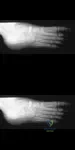
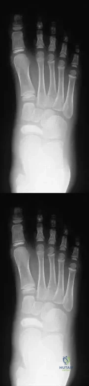
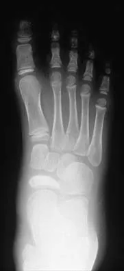


Explanation

In non-ambulatory patients with neuromuscular scoliosis and significant pelvic obliquity (typically >15 degrees), extending the spinal fusion to the pelvis is required to achieve and maintain a level pelvis for optimal sitting balance.

Question 87

Figure 5 shows the oblique radiograph of a 13-year-old boy with a painful, rigid flat foot and recurrent ankle sprains. Conservative management has failed.

What is the most appropriate surgical treatment for this condition?


Explanation

The classic presentation of a calcaneonavicular coalition includes a rigid flatfoot and an 'anteater sign' on the oblique radiograph. For symptomatic cases failing conservative care, the gold standard treatment is resection of the coalition with interposition of the extensor digitorum brevis or fat.

Question 88

An 8-year-old boy weighing 35 kg (77 lb) sustains an isolated, closed transverse midshaft femur fracture. What is the most appropriate definitive management?


Explanation

Flexible intramedullary nailing is the treatment of choice for length-stable midshaft femur fractures in children aged 5 to 11 years weighing under 50 kg (110 lb). It offers excellent outcomes, rapid mobilization, and minimizes the risk of avascular necrosis of the femoral head seen with rigid rigid trochanteric entry nails in this age group.

Question 89

A 14-year-old girl sustains a juvenile Tillaux fracture of the ankle during a soccer match. What is the pathomechanics responsible for this specific fracture pattern?


Explanation

A Tillaux fracture is a Salter-Harris III fracture of the anterolateral distal tibial epiphysis. It is caused by an external rotation force leading to an avulsion by the anterior inferior tibiofibular ligament (AITFL) as the medial physis has already closed.

Question 90

A 6-year-old boy presents with a painless clicking and snapping in his lateral knee. MRI demonstrates a thickened lateral meniscus extending into the intercondylar notch with absent posterior meniscotibial attachments. What is the diagnosis?


Explanation

The Wrisberg variant of a discoid meniscus is characterized by an abnormal or absent posterior meniscotibial attachment (coronary ligament), leaving only the ligament of Wrisberg. This hypermobility causes the classic 'snapping knee' syndrome.

Question 91

Figure 10 shows the AP pelvis radiograph of an 8-year-old boy with a persistent limp. He is diagnosed with Legg-Calvé-Perthes disease.

Which of the following factors represents the most significant prognostic indicator for long-term hip outcome in this patient?


Explanation

Age at the onset of Legg-Calvé-Perthes disease is the most critical clinical prognostic factor. Patients who develop the disease after age 8 have a significantly worse prognosis due to having less remaining growth for femoral head remodeling.

Question 92

A 5-year-old boy presents with an inability to bear weight on his right leg. His oral temperature is 38.6°C (101.5°F), ESR is 45 mm/hr, and peripheral WBC is 13,500/mm³. Radiographs of the hip are normal. According to the Kocher criteria, what is the most appropriate next step in management?


Explanation

This patient meets all four Kocher criteria (fever, inability to bear weight, ESR >40, WBC >12,000), giving him a 99% probability of septic arthritis. The essential next step is an urgent joint aspiration to confirm the diagnosis and obtain cultures before surgical debridement.

Question 93

A 13-year-old boy presents with vague anterior knee pain and intermittent catching. Radiographs reveal an osteochondritis dissecans (OCD) lesion. Where is the most common anatomical location for this lesion in the knee?


Explanation

The classic and most common location for an osteochondritis dissecans (OCD) lesion in the knee is the lateral aspect of the medial femoral condyle. It accounts for approximately 70-80% of all knee OCD lesions.

Question 94

A newborn is noted to have deep circumferential skin creases around the distal right lower extremity with distal limb edema, as well as acrosyndactyly of the toes. What is the recommended surgical management for the deep constriction bands to prevent distal ischemia?


Explanation

The patient has Amniotic Band Syndrome (Streeter dysplasia). Deep constriction bands causing distal edema or vascular compromise are treated with surgical excision utilizing multi-stage Z-plasties to prevent circumferential scarring and contracture.

Question 95

A 15-year-old boy with achondroplasia presents with progressively worsening lower extremity radicular pain and weakness triggered by walking. What is the primary anatomical cause of his spinal symptoms?


Explanation

Patients with achondroplasia have a mutation in the FGFR3 gene resulting in defective endochondral ossification. In the spine, this manifests as shortened pedicles and a narrowed interpedicular distance, leading to severe congenital spinal stenosis.

Question 96

An 18-month-old girl who recently started walking presents with a painless limp and a positive Galeazzi sign on the left. Radiographs confirm a completely dislocated left hip with a dysplastic acetabulum. What is the most appropriate management?


Explanation

In a child over 18 months of age with developmental dysplasia of the hip, conservative treatments and isolated closed reductions have high failure rates. Open reduction, often combined with a pelvic or femoral osteotomy to correct the dysplasia, is the standard of care.

Question 97

A 3-month-old girl with developmental dysplasia of the hip (DDH) is being treated with a Pavlik harness. During her first follow-up visit, the parents report that she has stopped kicking her left leg. On physical examination, the infant exhibits a lack of active knee extension on the left side, but withdrawal to pain is intact. What is the most appropriate next step in management?


Explanation

Femoral nerve palsy is a known complication of Pavlik harness treatment and is caused by hyperflexion of the hip. Management requires immediate reduction of hip flexion by adjusting the anterior straps or temporarily discontinuing the harness until active quadriceps function returns.

Question 98

A 14-year-old boy presents to the emergency department after sustaining a knee injury while jumping for a rebound during a basketball game. He has severe anterior knee pain and cannot actively extend the knee against gravity. A radiograph is shown in Figure 5.

Assuming the imaging confirms an Ogden Type III tibial tubercle avulsion fracture with intra-articular extension, what is the most devastating acute complication associated with this specific injury pattern?


Explanation

Tibial tubercle avulsion fractures can cause tearing of the anterior tibial recurrent artery, which bleeds directly into the tight anterior compartment of the lower leg. Surgeons must maintain a high index of suspicion for acute anterior compartment syndrome in these patients.

Question 99

A 30-month-old boy is evaluated for worsening unilateral bowing of the left leg. Radiographs demonstrate a metaphyseal-diaphyseal angle (Drennan angle) of 18 degrees with focal medial metaphyseal beaking, consistent with infantile Blount disease (Langenskiöld stage II). What is the most appropriate initial management?


Explanation

In a child younger than 3 years with infantile Blount disease (Langenskiöld stage I or II), initial treatment consists of using a knee-ankle-foot orthosis (KAFO) during weight-bearing activities. Surgical intervention is typically reserved for children older than 4 years, those who fail conservative management, or those presenting with advanced stages (III and above).

Question 100

A 13-year-old girl with a history of a slipped capital femoral epiphysis (SCFE) treated with in-situ pinning 6 months ago presents with worsening hip stiffness and pain. Physical examination reveals a significant global loss of hip motion. Radiographs are shown in Figure 10.

Assuming the radiograph demonstrates diffuse joint space narrowing without focal collapse of the femoral head, what is the most likely iatrogenic cause of this patient's current condition?


Explanation

Global loss of motion and diffuse joint space narrowing after SCFE fixation is the hallmark of chondrolysis. The most common iatrogenic cause of chondrolysis in this setting is unrecognized pin or screw penetration into the hip joint, necessitating prompt hardware removal.

None

Clinic OS
Medically Verified Content by
Prof. Clinic OS
Consultant Orthopedic & Spine Surgeon
Chapter Index