Orthopedic Osteoid Osteoma: Epidemiology, Spinal Anatomy & Biomechanics Review
Key Takeaway
Osteoid osteoma is a benign bone tumor often causing severe nocturnal pain, dramatically relieved by NSAIDs. It's common in young adults, frequently affecting long bones and the spine. Spinal lesions, often in posterior elements, can cause painful scoliosis or mimic disc pain. Understanding its epidemiology, precise surgical anatomy, and biomechanics is crucial for effective management and preventing instability.
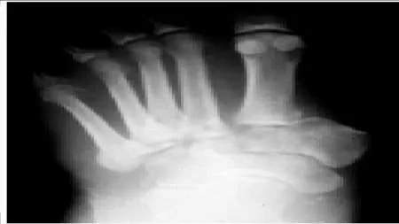
A 16-year-old male presents with a 6-month history of worsening mid-thoracic back pain. He notes that the pain is worse at night and is partially relieved by ibuprofen. On examination, he has a subtle thoracic scoliosis. You order the following imaging.
What is your primary diagnosis, and what is the pathophysiology behind his classic nocturnal pain?
Candidate: The diagnosis is an osteoid osteoma. The pain is caused by prostaglandins, specifically PGE2, which are found in high concentrations within the nidus.
A failing candidate simply states "it's a bone tumor" or focuses only on the diagnosis. They often fail to mention the specific inflammatory mediators or the mechanism of the scoliosis (muscular spasm), which demonstrates a deeper understanding of the clinical presentation.
The diagnosis is an osteoid osteoma, classically presenting with nocturnal pain responsive to NSAIDs. The pathophysiology involves high local concentrations of Prostaglandin E2 (PGE2) and prostacyclin produced by the proliferating osteoblasts and associated inflammatory cells within the nidus. This leads to intense vasodilation and sensitization of nociceptors. Regarding the scoliosis, it is a painful, compensatory response to persistent paravertebral muscle spasm on the side of the lesion.
The patient has failed 3 months of strict NSAID therapy. He now requests definitive treatment. How do you decide between percutaneous radiofrequency ablation (RFA) and open surgical excision in this case?
Candidate: I would choose RFA if it's in an accessible location. If the tumor is too close to the spinal cord or nerve roots, I would consider open surgery to avoid nerve damage.
Candidates often provide a vague answer. A high-scoring candidate must structure the response around "Patient/Lesion/Anatomical" factors and explicitly mention the risk of thermal injury to neural structures versus the risk of iatrogenic instability from open surgery.
The choice is based on a risk-benefit assessment: 1. Anatomical Proximity: If the nidus is <1cm from the spinal cord or traversing nerve roots, RFA carries a high risk of thermal neural injury, favoring open excision. 2. Stability: If the lesion's size or location requires removing >50% of a facet joint or compromises the pedicle, open surgery is preferred to allow for concurrent stabilization/fusion. 3. Diagnostic Uncertainty: If imaging features are atypical for a classic osteoid osteoma, open biopsy and resection are mandatory for histopathological confirmation. 4. Failure of prior treatment: Recurrence after RFA generally mandates open excision.
During the open resection of a thoracic pedicle lesion, you are worried about iatrogenic spinal instability. What defines the "three-column" stability and which structures, if compromised, would trigger your decision to instrument?
Candidate: I would use Denis's three-column theory. The columns are anterior, middle, and posterior. I'd stabilize if I had to remove the facet or the pedicle significantly.
Failing to define the columns clearly (Anterior: ALL + Ant 2/3 body; Middle: PLL + Post 1/3 body; Posterior: Pedicles, facets, laminae, ligamentum flavum, interspinous ligaments). Missing the "Posterior Ligamentous Complex (PLC)" as a key stabilizer.
According to Denis's classification: - Anterior column: ALL and anterior 2/3 of the vertebral body/disc. - Middle column: PLL and posterior 1/3 of the vertebral body/disc. - Posterior column: Pedicles, facets, laminae, and the posterior ligamentous complex (PLC). I would elect for instrumentation if the resection involves a bilateral facetectomy, an extensive unilateral facetectomy combined with pedicle resection, or any injury to the PLC, as these significantly compromise the posterior tension band. Proactive fixation prevents post-operative deformity, especially in a young, skeletally immature patient.
