Distal Femoral Osteosarcoma: A Detailed Clinical Case Study & Diagnostic Imaging
Key Takeaway
Distal femoral osteosarcoma is diagnosed through a comprehensive approach. It typically involves a detailed clinical history of progressive knee pain and swelling, followed by physical examination revealing a firm, tender mass. Imaging, including plain radiographs showing aggressive lytic/blastic lesions with periosteal reaction (e.g., sunburst, Codman's triangle) and MRI for precise local staging and soft tissue extension, is crucial.
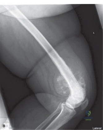
You are presented with a 17-year-old male athlete complaining of worsening left knee pain. He reports 4 months of intermittent pain, now constant with nocturnal awakening. On examination, there is a large, firm, non-tender, warm mass in the distal thigh. Examine the provided radiograph.
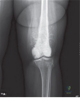
Describe the radiographic findings and provide your top differential diagnosis.
Candidate: The X-ray shows an aggressive lesion in the distal femoral metaphysis. There is cortical destruction, a sunburst periosteal reaction, and a Codman's triangle. There is also a large soft tissue mass with some ossification. The most likely diagnosis is osteosarcoma.
Candidates often miss the opportunity to describe the lesion systematically (location, margins, periosteal reaction, matrix, and soft tissue involvement). Failing to mention "tumor bone" or "matrix production" in the soft tissue, or simply guessing the diagnosis without describing the radiographic features, significantly lowers your score.
A structured response is essential: "The radiograph demonstrates a large, poorly defined, mixed lytic and sclerotic lesion centered in the distal femoral metaphysis. Key features include an aggressive permeative pattern of destruction, a sunburst periosteal reaction, and a Codman’s triangle. Crucially, there is a large extraosseous soft tissue component containing cloud-like opacities representing osteoid matrix production. This pattern is pathognomonic for high-grade osteosarcoma."
The patient has been diagnosed with a stage IIB conventional osteosarcoma. You are planning the local staging and surgical approach. Describe the utility of the MRI in this setting and what specific features you are looking for in the sagittal and coronal views.
Candidate: MRI is used to look at the extent of the tumor. I would look at the intramedullary extent to plan the osteotomy and check if the neurovascular bundle is involved. I also need to rule out skip metastases.
Failing to mention the "fat plane" between the tumor and neurovascular bundle, or ignoring the need for "whole femur" imaging to rule out skip lesions. Candidates often forget to mention that the biopsy tract must be included in the resection block.
Structure your answer by anatomical priorities: 1) Intramedullary Extent: Assessing marrow signal abnormality on T1 to determine proximal osteotomy level. 2) Neurovascular Involvement: Checking for the displacement or encasement of the popliteal artery/sciatic nerve; a visible fat plane suggests resectability. 3) Joint Involvement: Evaluating the epiphyseal plate and articular capsule integrity. 4) Skip Metastases: Imaging the entire femur to rule out synchronous intraosseous lesions, which would change the surgical strategy.
Discuss the importance of the biopsy procedure in the management of this patient. If you were performing the biopsy, what technical principles would you adhere to?
Candidate: I would perform a core needle biopsy. It's important to make a small incision and make sure it can be removed later when the tumor is taken out. I would avoid the neurovascular bundle.
Candidates often fail to state that the biopsy *must* be discussed with the oncological surgeon *before* it occurs. They also frequently fail to mention the risk of hematoma, which can track through tissue planes and contaminate the surgical field.
The biopsy is the most critical step in oncological planning. 1) Planning: The biopsy must be performed by the surgeon who will do the definitive resection. 2) Approach: Use a longitudinal incision that avoids vital structures (the "safe zone") and is placed so it can be completely excised during definitive surgery. 3) Technique: Core needle is preferred over open biopsy to minimize contamination. 4) Hemostasis: Meticulous closure to prevent hematoma formation, which acts as a vector for tumor cell dissemination.
