Undifferentiated Pleomorphic Sarcoma (UPS): A Detailed Diagnostic Case Study of the Thigh
Key Takeaway
Diagnosis of Undifferentiated Pleomorphic Sarcoma (UPS) in the thigh typically involves detailed patient history (progressive, painless mass), thorough clinical examination (deep-seated, firm, fixed lesion), and critical imaging. MRI is definitive, revealing a large, heterogeneous, infiltrative mass with avid, heterogeneous enhancement and potential muscular involvement. These findings necessitate urgent biopsy to confirm high-grade soft tissue sarcoma.
A 68-year-old male presents with a large, firm, non-tender mass in the posterior thigh. It has been slowly enlarging over the last 6 months. He has no history of trauma. Based on the clinical presentation, how do you manage the initial investigation of this soft tissue mass, and what is your primary concern?
Candidate: I would examine the mass for location and mobility. I would order an urgent MRI with contrast and plain radiographs to rule out bone involvement. My primary concern is a soft tissue sarcoma, as any deep-seated mass over 5 cm in an adult should be treated as malignant until proven otherwise.
Candidates often forget to state the importance of the "5 cm rule" or fail to mention the specific danger of performing an early, poorly planned excisional biopsy, which can jeopardize future limb-salvage surgery.
A high-scoring answer follows the "S-I-B" protocol: Staging (MRI with contrast, CXR/CT for systemic staging), Imaging-guided biopsy (Core needle, carefully planned tract), and Biopsy precautions (ensuring the tract is marked for later excision). Acknowledge the "5 cm rule" and emphasize that this is a diagnosis of exclusion requiring a multidisciplinary sarcoma board.
You have performed an MRI of the thigh. The lesion is a large, heterogeneous, infiltrative mass in the posterior compartment with T2 hyperintensity. How do you interpret these findings and what is the next step in your biopsy planning?
Candidate: These findings are highly suspicious for a high-grade soft tissue sarcoma. The heterogeneity and infiltrative margins are characteristic. I would plan an ultrasound-guided core needle biopsy. It is crucial to ensure the biopsy path is longitudinal and planned so that it can be completely excised en bloc during the definitive resection.
Suggesting an incisional biopsy without mentioning the track placement, or failing to discuss the necessity of imaging-guided core biopsy over a "quick look" surgical biopsy.
Structure the answer around: (1) **Radiological Interpretation**: High-grade features (heterogeneity/necrosis). (2) **Biopsy Strategy**: Core needle is gold standard to provide tissue architecture for IHC. (3) **Surgical Planning**: The "biopsy track is part of the tumor" concept—must be directly over the mass, longitudinally oriented, and away from neurovascular bundles to allow for subsequent wide resection.
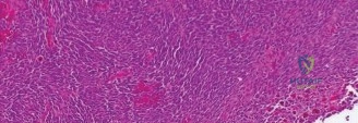
Final pathology confirms an Undifferentiated Pleomorphic Sarcoma (UPS). The multidisciplinary team recommends neoadjuvant radiation. Why do we choose to radiate prior to surgery rather than post-operatively for this specific case?
Candidate: Neoadjuvant radiation is used here because the tumor is large and high-grade. It helps to sterilize the pseudocapsule and microscopic extensions, potentially downstaging the tumor to make surgery easier, and it is generally better tolerated by the patient than post-operative radiation, which has a higher risk of wound complications.
Ignoring the "Wound Complication" trade-off. Failing to mention that while neoadjuvant radiation improves local control, it is associated with higher rates of surgical wound healing complications compared to adjuvant therapy.
Discuss the rationale clearly: (1) Biological: Downsizing, sterilization of the tumor bed, and treating the peripheral microscopic disease. (2) Technical: Allowing for a smaller radiation field and potentially improved functional outcomes. (3) Clinical: Neoadjuvant treatment avoids the fibrotic, irradiated tissue in the post-operative bed, though I would balance this with the known risk of increased surgical site wound complications.
