Orthopedic Hand & Wrist MCQs: Online Exam & Study Questions

Key Takeaway
This interactive board review contains 100 randomly selected orthopedic surgery questions with clinical images, immediate feedback, and detailed references.
Orthopedic Hand & Wrist MCQs: Online Exam & Study Questions
Comprehensive 100-Question Exam
00:00
Start Quiz
Question 1
A 24-year-old male falls on an outstretched hand and sustains a fracture of the scaphoid waist. He is evaluated for surgical fixation due to displacement.
Regarding the vascular anatomy of the scaphoid, which of the following is true?
Explanation
Question 2
A 35-year-old skier injures his thumb after catching his pole in the snow. Physical examination reveals laxity of the thumb metacarpophalangeal (MCP) joint when stressed in radial deviation with the joint in 30 degrees of flexion. Which of the following anatomic structures prevents spontaneous healing of the injured ligament in a Stener lesion?
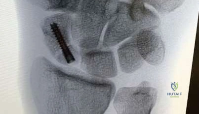
Explanation
Question 3
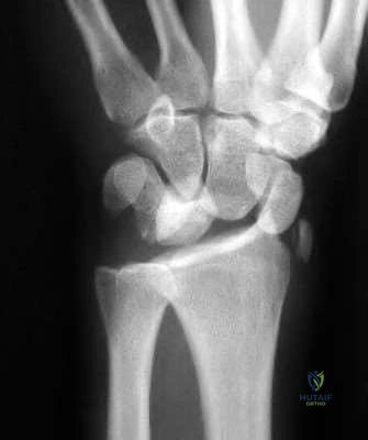
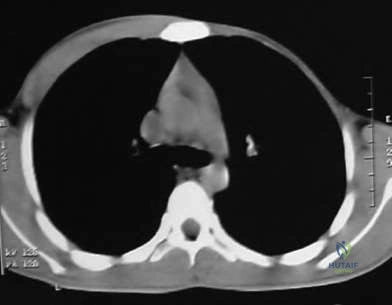
A 55-year-old manual laborer presents with chronic wrist pain. Radiographs reveal advanced scaphoid nonunion advanced collapse (SNAC).
In the expected progression of SNAC wrist arthritis, which of the following articulations is classically spared?
Explanation
Question 4
During a surgical repair of a Zone II flexor tendon laceration in the index finger, the surgeon must carefully repair both the flexor digitorum superficialis (FDS) and flexor digitorum profundus (FDP). At Camper's chiasm, what is the anatomical relationship of the FDS and FDP tendons?
Explanation
Question 5
A 42-year-old diabetic male presents with severe swelling, erythema, and pain in his right middle finger after a minor puncture wound. You suspect pyogenic flexor tenosynovitis.
According to Kanavel's criteria, which of the following signs is typically the earliest and most sensitive finding?
Explanation
Question 6
A 28-year-old male sustained a midshaft humerus fracture 6 months ago resulting in a complete radial nerve palsy with no signs of recovery on recent EMG. For surgical reconstruction, a tendon transfer is planned. The pronator teres (PT) is most commonly transferred to which structure to restore wrist extension?
Explanation
Question 7
A 50-year-old female presents with triggering and pain at the base of her right ring finger. She complains that her finger locks in flexion. The primary site of pathology in typical primary stenosing tenosynovitis (trigger finger) is stenosis at the level of which pulley?
Explanation
Question 8
A 30-year-old male presents with dorsal wrist pain and decreased grip strength. Radiographs show sclerosis and partial collapse of the lunate.
According to the Lichtman classification for Kienböck disease, what distinguishes stage IIIA from stage IIIB?
Explanation
Question 9
A 25-year-old patient suffers a severe crush injury to the hand. Clinical evaluation raises suspicion for compartment syndrome of the hand. How many distinct fascial compartments are recognized in the hand that may require release during fasciotomy?
Explanation
Question 10
During embryonic development, the limb bud grows in three different axes. Which signaling center and its corresponding molecule are primarily responsible for the anteroposterior (radioulnar) patterning of the limb, dictating the development of the thumb to the small finger?
Explanation
Question 11
A 60-year-old female presents with base of thumb pain. Radiographs demonstrate severe joint space narrowing, sclerosis, and osteophytes at the trapeziometacarpal joint, as well as narrowing of the scaphotrapezial (ST) joint. According to the Eaton-Littler classification for thumb carpometacarpal (CMC) arthritis, what stage is this?
Explanation
Question 12
A 65-year-old man of Northern European descent is scheduled for fasciectomy for severe Dupuytren contracture of the ring and small fingers. Which of the following pathological cords is responsible for displacing the neurovascular bundle centrally and placing it at highest risk for iatrogenic injury during surgical excision?
Explanation
Question 13
A 22-year-old male fell onto his extended, ulnarly deviated wrist. A lateral radiograph reveals a perilunate dislocation.
According to Mayfield's stages of perilunate instability, a Stage III injury indicates a complete disruption of which specific ligamentous interval?
Explanation
Question 14
A 45-year-old female sustains a comminuted intra-articular distal radius fracture.
The presence of a separate volar marginal fragment of the lunate facet (volar Barton's variant) is highly important to identify because:
Explanation
Question 15
A 30-year-old male presents with a deformity of his index finger 4 weeks after a jamming injury while playing basketball. Examination reveals the proximal interphalangeal (PIP) joint in fixed flexion and the distal interphalangeal (DIP) joint in hyperextension. This deformity is due to rupture of which structure?
Explanation
Question 16
A 40-year-old cyclist complains of numbness and tingling in the small and ulnar half of the ring finger. He also has weakness in finger abduction and adduction. Examination reveals normal sensation on the dorsum of the ulnar hand.
This clinical presentation is most consistent with compression of the ulnar nerve in Guyon's canal at which specific zone?
Explanation
Question 17
A 27-year-old male presents with an isolated closed rupture of the extensor pollicis longus (EPL) tendon. Which of the following conditions or prior injuries is classically associated with this specific delayed tendon rupture?
Explanation
Question 18
Which of the following intrinsic hand muscles is typically innervated by the median nerve?
Explanation
Question 19
A 14-month-old child presents with an extra digit on the radial side of the thumb (preaxial polydactyly). Radiographs demonstrate that both the proximal and distal phalanges are duplicated, but they articulate with a single, normally formed first metacarpal.
According to the Wassel classification, which type is this?
Explanation
Question 20
A hand surgeon is evaluating a patient with a suspected triangular fibrocartilage complex (TFCC) tear. When considering potential for spontaneous healing or surgical repair of a TFCC tear, the surgeon must account for its vascular supply. Which portion of the TFCC is considered vascularized and capable of healing?
Explanation
Question 21
A 30-year-old manual laborer presents with chronic dorsal wrist pain. Radiographs reveal sclerosis and collapse of the lunate, but the overall carpal height is maintained, and there is no fixed scaphoid rotation. The patient's radiographic evaluation also demonstrates an ulnar negative variance of 3 mm. Which of the following is the most appropriate surgical management?
Explanation
Question 22
A 45-year-old male presents with the inability to actively extend his middle finger at the metacarpophalangeal (MCP) joint after a traumatic 'flicking' injury. On examination, he is able to maintain full MCP extension if the digit is passively extended by the examiner. There is focal swelling dorsally over the MCP joint. What is the primary anatomic structure injured?
Explanation
Question 23
A 60-year-old woman with a long-standing history of rheumatoid arthritis presents with a progressive inability to actively extend her ring and small fingers at the MCP joints. Passive extension is full and intact, but the tenodesis effect is absent. What is the underlying pathophysiology of her condition?
Explanation
Question 24
A 55-year-old female with poorly controlled rheumatoid arthritis presents to the clinic having acutely lost the ability to actively flex the interphalangeal joint of her thumb. Passive flexion remains intact. Which of the following bony prominences is most likely responsible for this spontaneous tendon rupture?
Explanation
Question 25
When evaluating the biomechanical principles of flexor tendon repair to allow for early active mobilization protocols, which of the following factors contributes most to the immediate mechanical strength of the repair site?
Explanation
Question 26
A newborn is diagnosed with a unilateral radial longitudinal deficiency (radial club hand) characterized by an absent radius (Type IV). Before proceeding with any orthopedic interventions, which of the following screening tests is most critical for determining the patient's immediate survival risk?
Explanation
Question 27
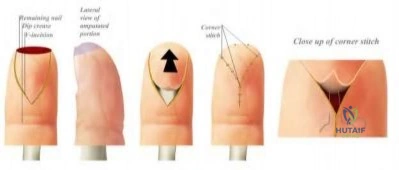
A 9-year-old boy presents to the emergency department after his long finger was crushed in a door. Examination reveals a clinically deformed distal phalanx with the nail plate avulsed proximally, resting dorsal to the eponychial fold. Radiographs show a displaced Salter-Harris I fracture of the distal phalanx. What is the most appropriate management?
Explanation
Question 28
A 35-year-old carpenter presents with a sudden inability to perform a tip-to-tip pinch with his thumb and index finger. When attempting an 'OK' sign, he forms a flat pinch due to an inability to flex the interphalangeal joint of the thumb and the distal interphalangeal joint of the index finger. Sensation over the hand is completely normal. Which muscle group is affected, and what nerve is implicated?
Explanation
Question 29
A 40-year-old male with a chronic low ulnar nerve palsy demonstrates severe clawing of his ring and small fingers. During the physical exam, the examiner stabilizes his MCP joints in flexion, and the patient is subsequently able to actively extend the proximal interphalangeal (PIP) joints of those digits. What is this clinical test, and what does a positive result indicate regarding surgical planning?
Explanation
Question 30
During an electrodiagnostic evaluation for suspected carpal tunnel syndrome, the neurologist notes an anomalous innervation pattern where motor axons cross from the median nerve to the ulnar nerve in the forearm. This anomaly most commonly innervates which of the following muscles?
Explanation
Question 31
A 22-year-old gymnast presents with persistent ulnar-sided wrist pain after a fall. An MRI arthrogram reveals a tear of the triangular fibrocartilage complex (TFCC) directly at its insertion into the fovea at the base of the ulnar styloid. According to the Palmer classification, what type of tear is this, and what is the typical treatment approach?
Explanation
Question 32
A patient presents with an abducted posture of the small finger at rest and is unable to actively adduct it to the ring finger. This condition (Wartenberg's sign) is caused by the unopposed action of which muscle, due to weakness of which other muscle?
Explanation
Question 33
A 34-year-old female presents with severe pain in her thumb pulp, which is highly sensitive to cold, and excruciating point tenderness over the nail bed. There is a faint bluish discoloration under the nail plate. Radiographs show a small, smooth, scalloped radiolucency in the dorsal aspect of the distal phalanx. What is the most likely diagnosis?
Explanation
Question 34
A 28-year-old industrial painter presents to the emergency room 2 hours after accidentally injecting his non-dominant index finger with a high-pressure paint gun. The entry wound is a tiny 2 mm puncture on the volar tip of the digit. The finger is swollen, pale, and mildly painful. What is the most appropriate next step in management?
Explanation
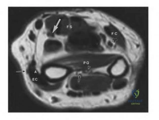
Question 35
A patient develops a purulent infection spreading from the flexor tendon sheath of the small finger into the palm, which subsequently travels up into the flexor tendon sheath of the thumb. This specific pattern, known as a 'horseshoe abscess,' occurs through an anatomical connection between which two spaces?
Explanation
Question 36
A 1-year-old child presents with congenital webbing between two fingers of both hands. Radiographs demonstrate soft tissue connections only, with no bony fusion between the affected digits. Which of the following describes the most common classification and location of this deformity?
Explanation
Question 37
A 5-year-old boy presents with an enlarged index finger. His parents note the finger has grown proportionally faster than the rest of his digits since birth. On exam, the finger is significantly larger in girth and length, with a palpable volar mass and radial deviation. Which of the following pathological findings is most characteristic of this condition?
Explanation
Question 38
A 60-year-old woman presents with severe, chronic base-of-thumb pain. Radiographs demonstrate advanced destruction and sclerosis of the trapeziometacarpal joint. There is also significant narrowing and arthritic change in the scaphotrapezial (STT) joint space. According to the Eaton-Littler classification, what stage of thumb carpometacarpal arthritis does this represent?
Explanation
Question 39
A 45-year-old man presents with chronic wrist pain years after a remote, untreated scaphoid fracture. Radiographs demonstrate a scaphoid nonunion with advanced carpal collapse. There are degenerative changes at the radioscaphoid joint and the scaphocapitate joint, but the radiolunate joint is distinctly preserved. What stage of SNAC (Scaphoid Nonunion Advanced Collapse) wrist is this?
Explanation
Question 40
During digital replantation following an acute traumatic amputation, successful revascularization and functional outcomes depend on a systematic surgical approach. Which anatomical structure should ideally be repaired or stabilized first to provide a foundation for the remainder of the microsurgical reconstruction?
Explanation
Question 41
A 55-year-old female presents with the inability to actively flex her thumb interphalangeal joint 6 months after open reduction and internal fixation of a distal radius fracture with a volar locking plate. Radiographs show the plate is positioned distally, slightly over the watershed line. Which complication is most likely responsible for her current presentation?
Explanation
Question 42
A 32-year-old construction worker presents with central dorsal wrist pain. Radiographs reveal sclerosis and fragmentation of the lunate with a negative ulnar variance of 3 mm, but no carpal collapse.
According to Lichtman's classification of Kienbock's disease, what is the most appropriate surgical intervention?
Explanation
Question 43
A 45-year-old male presents with chronic wrist pain. Radiographs show a scaphoid nonunion with advanced collapse (SNAC). There is significant osteoarthritis at the radioscaphoid and capitolunate joints, but the radiolunate joint space is well preserved. Which of the following is the most appropriate surgical treatment?
Explanation
Question 44
In optimizing a zone II flexor digitorum profundus (FDP) tendon repair to allow for early active motion protocols, which of the following technical modifications provides the greatest increase in the tensile strength of the repair?
Explanation
Question 45
A 28-year-old basketball player jams his finger and presents with a swollen proximal interphalangeal (PIP) joint. Over the next 3 weeks, he develops a characteristic Boutonniere deformity. What is the primary pathoanatomy leading to the progressive hyperextension of the distal interphalangeal (DIP) joint in this deformity?
Explanation
Question 46
During the surgical release of a trigger thumb, the A1 pulley is divided to relieve triggering. Which adjacent structure is at greatest risk of iatrogenic injury if the surgical approach and dissection are placed too far radially?
Explanation
Question 47
A patient with severe carpal tunnel syndrome exhibits profound thenar atrophy but normal sensation over the dorsum of the hand. Electromyography reveals normal motor function of the first dorsal interosseous muscle, but paradoxically normal thenar function on proximal nerve stimulation due to a Martin-Gruber anastomosis. Where does this specific neural connection occur anatomically?
Explanation
Question 48
A cyclist presents with isolated weakness in finger abduction and adduction. Sensation is perfectly intact in the ring and small fingers, and over the hypothenar eminence. Compression of the ulnar nerve in Guyon's canal is diagnosed. In which anatomical zone of Guyon's canal is the compression located?
Explanation
Question 49
A 60-year-old female presents with pain at the base of her thumb. Radiographs demonstrate advanced trapeziometacarpal joint destruction, subchondral sclerosis, and greater than 1/3 radial subluxation of the first metacarpal base. The scaphotrapezial (ST) joint is perfectly preserved. According to the Eaton-Littler classification, what stage is this disease?
Explanation
Question 50
In the progressive sequence of perilunate instability described by Mayfield, what specific anatomical disruption defines Stage II?
Explanation
Question 51
A 25-year-old elite gymnast presents with ulnar-sided wrist pain and instability of the distal radioulnar joint (DRUJ). An MRI reveals a traumatic avulsion of the triangular fibrocartilage complex (TFCC) from its bony insertion at the ulnar fovea. What is the correct Palmer classification for this specific injury?
Explanation
Question 52
In the surgical treatment of Dupuytren's contracture, release of the spiral cord is often necessary to correct proximal interphalangeal (PIP) joint flexion deformities. Which of the following normal fascial structures collectively form the pathological spiral cord?
Explanation
Question 53
A 40-year-old female complains of severe, excruciatingly localized pain under her thumbnail, which is exacerbated by cold weather. On physical examination, inflating a blood pressure cuff on her proximal arm temporarily relieves the pain in the digit. What is the name of this clinical sign?
Explanation
Question 54
A patient presents with a swollen, painful index finger 3 days after a minor puncture wound to the volar crease. Which of the following is NOT one of Kanavel's four cardinal signs of suppurative flexor tenosynovitis?
Explanation
Question 55
A newborn is evaluated for bilateral absence of the radius and hypoplastic thumbs (radial longitudinal deficiency). The pediatrician diagnoses Holt-Oram syndrome. Which associated systemic abnormality must be aggressively ruled out or treated in this child?
Explanation
Question 56
The most common form of congenital syndactyly in the upper extremity typically involves which specific web space, and what is its most common inheritance pattern when familial?
Explanation
Question 57
A 30-year-old mechanic sustains a high-pressure injection injury to his non-dominant index finger while using an industrial sprayer. He presents to the emergency department 2 hours later. Which of the following factors is most strongly associated with an increased ultimate risk of amputation?
Explanation
Question 58
In the setting of traumatic amputations, ischemia time dictates the viability of the replantation. What is the generally accepted maximum cold ischemia time for replantation of an isolated completely amputated digit, compared to a major proximal limb amputation (e.g., at the proximal forearm)?
Explanation
Question 59
A 65-year-old patient with long-standing rheumatoid arthritis presents with a new inability to actively extend the small and ring fingers at the metacarpophalangeal (MCP) joints. Extension at the PIP joints is preserved, and passive MCP extension is full. This clinical picture (Vaughan-Jackson syndrome) is most often caused by attrition and rupture of the extensor tendons over which specific bony prominence?
Explanation
Question 60
A newborn infant presents with the right upper extremity held rigidly in internal rotation, adduction, elbow extension, and wrist flexion (the classic 'waiter's tip' posture). The hand grasp reflex is remarkably preserved. Which brachial plexus nerve roots are primarily injured?
Explanation
Question 61
A 45-year-old male presents with chronic wrist pain and a history of remote trauma. Radiographs reveal advanced scapholunate advanced collapse (SLAC). In the progression to Stage III SLAC wrist, which of the following articulations is newly involved?
Explanation
Question 62
A 30-year-old chef sustains a volar laceration to the index finger. Surgical exploration reveals a 40% partial laceration of the flexor digitorum profundus (FDP) tendon in Zone II, with an intact flexor digitorum superficialis (FDS). What is the most appropriate management?
Explanation
Question 63
A 60-year-old female with long-standing rheumatoid arthritis is unable to actively extend her small and ring fingers at the metacarpophalangeal (MCP) joints. Passive extension is intact. The tenodesis effect does not produce active extension of these digits. What is the primary pathophysiology?
Explanation
Question 64
Lichtman Stage IIIB Kienbock's disease is distinguished from Stage IIIA by the presence of which of the following radiographic findings?
Explanation
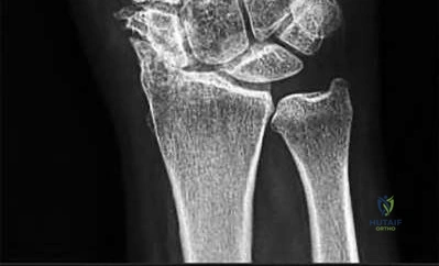
Question 65
A 22-year-old boxer sustains a Bennett fracture. The small volar-ulnar fragment of the thumb metacarpal base is held anatomically in place by which of the following structures?
Explanation
Question 66
A patient presents with atrophy of the dorsal interossei and weakness of finger abduction. Sensation over the volar ulnar aspect of the small finger and the hypothenar eminence is completely preserved. The flexor digitorum profundus to the small finger has normal strength. Where is the most likely site of ulnar nerve compression?
Explanation
Question 67
During surgical release for Dupuytren's contracture, the neurovascular bundle is noted to be displaced centrally and superficially. Which of the following pathological structures is primarily responsible for this displacement?
Explanation
Question 68
In a patient with an irreversible high radial nerve palsy, multiple tendon transfers are planned. What is the standard transfer utilized to restore active wrist extension?
Explanation
Question 69
To minimize the recurrence rate when surgically treating a digital mucous cyst located at the distal interphalangeal (DIP) joint, which step is most essential?
Explanation
Question 70
A rugby player sustains a flexor digitorum profundus (FDP) avulsion from the ring finger (Jersey finger). MRI confirms the tendon has retracted completely into the palm (Leddy-Packer Type 1). What is the optimal timeframe for surgical repair?
Explanation
Question 71
According to the Eaton-Littler classification for thumb carpometacarpal (CMC) joint osteoarthritis, what defines a Stage III radiographic presentation?
Explanation
Question 72
At what age should a complete, simple syndactyly involving the thumb and index finger be surgically released to prevent angular growth deformities?
Explanation
Question 73
A patient cannot fully flex their normal middle, ring, and small fingers after undergoing an index finger flexor digitorum profundus (FDP) tendon repair. What is the primary cause of this phenomenon?
Explanation
Question 74
A 45-year-old female assembly line worker presents with worsening numbness in her thumb, index, and middle fingers. She notes the symptoms are particularly severe when she repetitively makes a prolonged full fist at work. What anatomic phenomenon most likely contributes to her work-related exacerbation of carpal tunnel syndrome?
Explanation
Question 75
During primary repair of a Zone II flexor digitorum profundus (FDP) laceration, preservation of the vincula is emphasized to optimize primary tendon healing. The vincula brevia to the FDP is primarily supplied by branches directly originating from which of the following?
Explanation
Question 76
A 62-year-old man presents with chronic wrist pain and limited range of motion. Radiographs demonstrate scapholunate advanced collapse (SLAC) with arthritic changes involving the radioscaphoid and capitolunate joints, while the radiolunate joint remains widely spaced and pristine.
According to the Watson classification, what is the stage of this patient's SLAC wrist, and what is the primary biomechanical reason the radiolunate joint is spared?
Explanation
Question 77
In a patient with advanced trapeziometacarpal (CMC) joint arthritis of the thumb, progressive attenuation of a specific primary stabilizing ligament leads to the characteristic dorsoradial subluxation of the metacarpal base. Which of the following ligaments is historically referred to as the 'beak' ligament and is directly implicated in this subluxation?
Explanation
Question 78
A 32-year-old carpenter presents with persistent dorsal wrist pain. Imaging confirms avascular necrosis of the lunate (Kienbock disease). Radiographs show complete lunate collapse and a fixed flexed posture of the scaphoid (cortical ring sign), but no degenerative carpal arthritic changes are noted. Which of the following is the most appropriate surgical management?
Explanation
Question 79
A 22-year-old rugby player grabs an opponent's jersey and sustains an isolated hyperextension injury to his ring finger distal interphalangeal (DIP) joint. Imaging demonstrates an avulsed bony fragment located at the level of the proximal interphalangeal (PIP) joint. What Leddy-Packer type is this injury, and what is its vincular status?
Explanation
Question 80
A 45-year-old woman presents with a chronic, untreated mallet finger of the middle digit. She has progressively developed a severe swan neck deformity. What is the primary pathomechanical cause of this secondary deformity?
Explanation
None
