Orthopedics Hand Review | Dr Hutaif Hand & Wrist Review -...

Key Takeaway
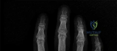
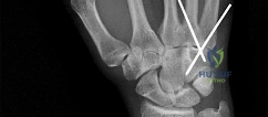
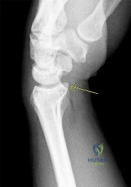
Looking for accurate information on Orthopedics Mcqs Hand0019? Based on the question of figures, a 43-year-old bricklayer's left hand weakness, atrophy, positive Froment sign, and ulnar nerve motor denervation distal to the wrist, without sensory loss, points to ulnar nerve motor branch compression. MRI reveals a ganglion cyst near the hook of the hamate compressing this branch. The best next step is excision of the ganglion cyst.
Orthopedics Hand Review | Dr Hutaif Hand & Wrist Review -...
Comprehensive 100-Question Exam
00:00
Start Quiz
Question 1
A 55-year-old female with long-standing rheumatoid arthritis presents with a finger deformity characterized by PIP joint hyperextension and DIP joint flexion.
What is the primary pathophysiologic event initiating this specific deformity?
Explanation
Question 2
A 25-year-old carpenter sustained a laceration over the volar aspect of the proximal phalanx of his index finger. Physical examination reveals an inability to flex both the PIP and DIP joints. The injury is classified as being in Zone II.
Which of the following is true regarding repairs in this zone?
Explanation
Question 3
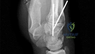
A 30-year-old male presents after a fist fight with thumb base pain. Radiographs reveal a two-part intra-articular fracture of the base of the first metacarpal with subluxation of the metacarpal shaft.
Which deforming forces are primarily responsible for the displacement of the metacarpal shaft in this injury pattern?
Explanation
Question 4
A 40-year-old manual laborer presents with chronic wrist pain. Radiographs reveal a scaphoid nonunion with radioscaphoid arthritis and capitolunate arthritis, but the radiolunate joint is spared.
Which stage of SNAC wrist does this represent, and what is the most appropriate surgical treatment?

Explanation
Question 5
A patient presents with a swollen, erythematous, and painful index finger 3 days after a puncture wound. Which of the following is considered the most sensitive and earliest, albeit least specific, of Kanavel's signs for pyogenic flexor tenosynovitis?
Explanation
Question 6
A 52-year-old male presents with wrist pain. Radiographs show scapholunate dissociation with associated radiocarpal and midcarpal arthritis. The radiolunate joint is notably preserved.
Why is the radiolunate joint typically spared in SLAC wrist?
Explanation
Question 7
A 35-year-old male sustains a midshaft humerus fracture. Examination reveals an inability to extend the wrist and digits. He is diagnosed with a radial nerve palsy. If tendon transfers are required due to lack of recovery, which of the following combinations is the classic Boyes transfer for radial nerve palsy?

Explanation
Question 8
Trigger finger typically involves constriction of the flexor tendon as it passes through which pulley?

Explanation
Question 9
A 28-year-old manual worker presents with dorsal wrist pain. X-rays show sclerosis of the lunate with no collapse. MRI confirms avascular necrosis of the lunate. Radiographs also demonstrate ulnar minus variance.
Which of the following is the most appropriate initial surgical intervention for this patient (Lichtman Stage II)?
Explanation
Question 10
In congenital syndactyly, what is the most common anatomical web space involved?
Explanation
Question 11
A 60-year-old female presents with pain at the base of her thumb, worsened by pinch grasp. A positive 'grind test' is elicited. Eaton-Littler classification on radiograph shows pantrapezial arthritis.
Which ligament attenuation is the primary initiator of thumb carpometacarpal (CMC) joint osteoarthritis?

Explanation
Question 12
A cyclist complains of numbness in his small finger and the ulnar half of his ring finger, along with intrinsic muscle weakness.
Which of the following zones of Guyon's canal is most likely compressed if the patient presents with purely motor symptoms (weakness of hypothenar and interossei muscles) with NO sensory deficits?
Explanation
Question 13
Which of the following is considered an absolute contraindication for finger replantation?
Explanation
Question 14
Which of the following cords is primarily responsible for proximal interphalangeal (PIP) joint contracture in Dupuytren's disease?

Explanation
Question 15
A 40-year-old female presents with severe, lancinating pain in her fingertip, exquisitely sensitive to cold. Examination reveals a bluish discoloration beneath the nail matrix.
Which of the following clinical tests is most specific for diagnosing this condition?

Explanation
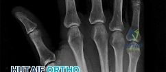
Question 16
A 25-year-old male struck the tip of his finger while playing basketball and now presents with an inability to actively extend the distal interphalangeal (DIP) joint. Radiographs show a small dorsal avulsion fracture of the distal phalanx involving 20% of the articular surface.
What is the recommended primary treatment?

Explanation
Question 17
A patient complains of numbness in the small finger. On examination, there is weakness of the flexor digitorum profundus to the small finger (FDP) and a positive Froment's sign.
This presentation indicates ulnar nerve compression at what level?

Explanation
Question 18
A 45-year-old female presents with a painless, slowly enlarging, firm, lobulated mass on the volar aspect of her index finger. X-rays show soft tissue swelling with no bone involvement.
Histological examination following excision is most likely to show:
Explanation
Question 19
A patient presents with a throbbing, severe pain in the pulp of the thumb. Examination reveals a tense, swollen, and erythematous thumb pulp.
If surgical drainage is required, which of the following incision techniques is generally avoided due to the risk of neuroma formation and loss of sensation on the working surface?

Explanation
Question 20
A 22-year-old rugby player grabbed an opponent's jersey and felt a pop in his ring finger. He cannot actively flex the distal interphalangeal (DIP) joint. Radiographs reveal a small bony fragment retracted to the level of the proximal interphalangeal (PIP) joint.
According to the Leddy and Packer classification, what type of injury is this, and what is its blood supply status?
Explanation
Question 21
A 60-year-old man presents with chronic wrist pain. Radiographs show severe narrowing of the radioscaphoid and capitolunate joints, but the radiolunate joint is preserved. Which of the following surgical procedures is definitively contraindicated in this patient?
Explanation
Question 22
A 62-year-old female undergoes a ligament reconstruction and tendon interposition (LRTI) using the flexor carpi radialis (FCR) tendon for advanced Eaton-Littler Stage III thumb CMC arthritis. During the surgical approach to the CMC joint, which of the following nerves is at greatest risk of iatrogenic injury?
Explanation
Question 23
A 32-year-old male rower presents with pain and swelling approximately 4 to 6 cm proximal to the dorsal wrist joint. Examination reveals localized swelling and crepitus with active wrist flexion and extension. This condition is caused by friction between the muscle bellies and tendons of which two extensor compartments?
Explanation
Question 24
During the repair of a lacerated flexor digitorum profundus (FDP) tendon in Zone II, the surgeon notes damage to the flexor tendon sheath. To prevent biomechanical failure and 'bowstringing' of the flexor tendon, which two annular pulleys are the most critical to preserve or reconstruct?
Explanation
Question 25
A 35-year-old male sustains a high ulnar nerve transection above the elbow. After primary repair, the surgeon decides to perform a distal supercharged nerve transfer to rapidly restore intrinsic hand function before irreversible muscle atrophy occurs. Which of the following nerve transfers is most commonly used for this purpose?
Explanation
Question 26
A 55-year-old woman with long-standing rheumatoid arthritis presents with an acute inability to actively flex the interphalangeal joint of her thumb. Passive range of motion is full and painless. She has a prominent, soft volar wrist mass. What is the most likely mechanism of this deficit?
Explanation
Question 27
A neonate presents with bilateral shortened forearms, radially deviated hands, and absent radii on radiographs. Interestingly, both thumbs are present and appear structurally normal. Which of the following genetic syndromes is most likely in this patient?
Explanation
Question 28
A 32-year-old jackhammer operator presents with chronic dorsal wrist pain. X-rays reveal sclerosis, fragmentation, and early collapse of the lunate. Carpal height is maintained, and there is no fixed rotation of the scaphoid. MRI confirms avascular necrosis of the lunate. According to the Lichtman classification, what is the stage of this Kienbock's disease?
Explanation
Question 29
A 30-year-old male presents with a high radial nerve palsy following a humerus fracture 12 months ago. Nerve exploration was unsuccessful. For the surgical restoration of active wrist extension, which tendon transfer is most commonly utilized and provides the best biomechanical advantage?
Explanation
Question 30
A 45-year-old diabetic patient presents with a pyogenic flexor tenosynovitis of his small finger following a puncture wound. Two days later, he develops massive swelling in the thumb and thenar eminence despite having no direct trauma to the thumb. What anatomical structure facilitates this specific spread of infection?
Explanation
Question 31
A 22-year-old athlete sustains a dorsal fracture-dislocation of the proximal interphalangeal (PIP) joint of the middle finger, involving 45% of the volar articular base of the middle phalanx. The joint is highly unstable to extension block splinting. What is the most appropriate surgical management for this injury?
Explanation
Question 32
A 40-year-old female presents with ulnar-sided wrist pain that worsens with pronation and gripping. Radiographs demonstrate a positive ulnar variance of +4 mm and cystic changes in the lunate and triquetrum. MRI confirms a central perforation of the TFCC but an intact distal radioulnar joint (DRUJ) cartilage. What is the most appropriate surgical intervention?
Explanation
Question 33
In the surgical treatment of Dupuytren's contracture, a specific pathological cord is responsible for flexing the PIP joint and simultaneously displacing the neurovascular bundle centrally and superficially, placing it at high risk of transection. Which of the following normal fascial structures is NOT a precursor to this cord?
Explanation
Question 34
A 25-year-old carpenter suffers a fingertip amputation of his index finger. The amputation is volar-oblique, exposing 4 mm of the distal phalanx. Which of the following is the most appropriate soft tissue coverage option to provide durable, sensate padding?
Explanation
Question 35
A 30-year-old skier presents with severe pain and weakness of pinch in the right thumb following a fall on an outstretched hand with a ski pole. Examination shows 45 degrees of radial deviation of the MCP joint with no firm endpoint. A Stener lesion is suspected. What defines the anatomy of a Stener lesion?
Explanation
Question 36
A 35-year-old male presents with wrist pain 10 years after an untreated fall. Radiographs show a scaphoid nonunion. There is joint space narrowing of the radioscaphoid joint and the capitolunate joint, but the radiolunate joint is perfectly preserved. What stage of Scaphoid Nonunion Advanced Collapse (SNAC) does this represent?
Explanation
Question 37
Six weeks following a non-displaced distal radius fracture treated successfully with a short arm cast, a 58-year-old female presents with a sudden inability to actively lift her thumb into extension. Which tendon is the standard choice for transfer to restore this lost function?
Explanation
Question 38
During a carpal tunnel release, the surgeon must carefully avoid injury to the recurrent motor branch of the median nerve. According to Poisel's classification, what is the most common anatomical relationship of this nerve branch to the transverse carpal ligament?
Explanation
Question 39
A 42-year-old female presents with severe, paroxysmal pain in her left index fingertip that is exquisitely sensitive to cold temperatures. Examination reveals a bluish hue and pinpoint tenderness beneath the nail plate. Which of the following clinical tests is most specific for diagnosing the suspected lesion?
Explanation
Question 40
A 65-year-old female sustains a fracture of the distal radius. Radiographs reveal a volar marginal intra-articular fracture fragment that is displaced proximally and volarly, carrying the carpus with it (Volar Barton's fracture). The continued attachment of which of the following stout ligaments is responsible for pulling the carpus in this direction?
Explanation
Question 41
A 24-year-old male presents with persistent wrist pain 6 months after a fall. Radiographs demonstrate a proximal pole scaphoid nonunion with sclerosis and avascular necrosis.
What is the most appropriate surgical treatment?
Explanation
Question 42
A 22-year-old male presents with a nonunion of the proximal pole of the scaphoid 6 months after a fall. What is the primary arterial supply to the proximal pole of the scaphoid that makes it susceptible to avascular necrosis?
Explanation
Question 43
A patient undergoes early active mobilization following a zone II flexor tendon repair. Which of the following rehabilitation protocols relies on the concept of active extension and passive flexion using rubber band traction?
Explanation
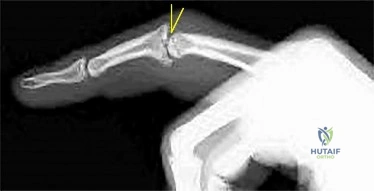
Question 44
A 35-year-old basketball player presents with a drooping distal phalanx of his middle finger after a direct blow. Radiographs show a small dorsal avulsion fracture of the distal phalanx involving 15% of the articular surface without subluxation. What is the most appropriate management?
Explanation
Question 45
A 40-year-old manual laborer presents with dorsal wrist pain. Imaging demonstrates sclerosis and fragmentation of the lunate, but the scaphoid is properly aligned and the carpal height is maintained. What Lichtman stage does this represent?
Explanation
Question 46
A 45-year-old female is unable to make an "OK" sign with her thumb and index finger, demonstrating a flattened pinch. Sensation in her hand is completely normal. Which muscle is most likely affected by this isolated nerve palsy?
Explanation
Question 47
In an acute rupture of the ulnar collateral ligament of the thumb MCP joint (skier's thumb), what anatomical structure typically interposes between the torn ligament ends, preventing healing and necessitating surgical repair?
Explanation
Question 48
A 60-year-old patient with rheumatoid arthritis presents with an inability to actively extend the small and ring fingers at the MCP joints. Passive extension is intact. What is the most common cause of this deformity?
Explanation
Question 49
During evaluation of a patient with suspected carpal tunnel syndrome, the examiner places the wrist in maximum flexion for 60 seconds to reproduce paresthesias in the median nerve distribution. What is the name of this provocative test?
Explanation
Question 50
A 32-year-old male presents with a swollen, erythematous, and exquisitely painful index finger after a puncture wound. Which of the following is NOT one of Kanavel's cardinal signs of flexor tenosynovitis?
Explanation
Question 51
A patient sustained a humerus fracture resulting in a persistent high radial nerve palsy. When planning a standard set of tendon transfers to restore function, which muscle is typically transferred to the extensor carpi radialis brevis (ECRB) to restore wrist extension?
Explanation
Question 52
A 28-year-old male presents with dorsal radial wrist pain following a fall onto an outstretched hand. Radiographs show a widened scapholunate interval (>3 mm) on the AP view and a dorsal tilt of the lunate on the lateral view. What specific deformity is present?
Explanation
Question 53
In the surgical management of Dupuytren's contracture, which fascial structure is typically spared from involvement and helps protect the neurovascular bundle during dissection?
Explanation
Question 54
A 45-year-old female complains of severe, paroxysmal pain in her thumbnail bed, exacerbated by cold temperatures. Exquisite point tenderness is noted on physical exam. What is the most likely diagnosis?
Explanation
Question 55
A 55-year-old female presents with base of thumb pain. Radiographs reveal Eaton-Littler Stage III trapeziometacarpal osteoarthritis. She has failed conservative management. Which surgical procedure is considered the gold standard for long-term pain relief and functional improvement?
Explanation
Question 56
A patient presents with a finger deformity characterized by PIP joint flexion and DIP joint hyperextension. What is the primary anatomic defect responsible for this deformity?
Explanation
Question 57
A full-term infant is born with complete simple syndactyly of the long and ring fingers. When is the optimal time for surgical release to prevent angular deformity and optimize functional outcome?
Explanation
Question 58
A cyclist complains of numbness in his ring and small fingers and weakness in hand grip. Examination reveals clawing of the ring and small fingers. Compression of the ulnar nerve is suspected in Guyon's canal. Which of the following forms the floor of Guyon's canal?
Explanation
Question 59
How many distinct fascial compartments are typically recognized in the human hand when evaluating for compartment syndrome?
Explanation
Question 60
A 60-year-old female presents with a small, clear, fluid-filled nodule on the dorsal aspect of her right index finger DIP joint. Radiographs show underlying osteoarthritis with a prominent osteophyte. What is the most appropriate definitive surgical management to minimize recurrence?
Explanation
Question 61
A patient presents with median nerve distribution paresthesias. Which clinical finding best differentiates Pronator Syndrome from Carpal Tunnel Syndrome?
Explanation
Question 62
A 45-year-old heavy laborer presents with chronic wrist pain. Radiographs demonstrate advanced arthritis of the radioscaphoid and capitolunate joints, while the radiolunate joint is entirely spared. Which of the following is the most appropriate surgical management?
Explanation
Question 63
In a patient undergoing tendon transfers for a high radial nerve palsy, the pronator teres (PT) is typically transferred to restore wrist extension. Why is the extensor carpi radialis brevis (ECRB) preferred as the recipient tendon over the extensor carpi radialis longus (ECRL)?
Explanation
Question 64
A 55-year-old female with severe rheumatoid arthritis presents with an inability to actively extend her small and ring fingers. Passive extension is full, and tenodesis effect demonstrates intact extensor tendons proximal to the wrist. What is the most likely etiology of this presentation?
Explanation
Question 65
During surgical fasciectomy for a complex Dupuytren's contracture of the proximal interphalangeal (PIP) joint, the surgeon must identify and protect the neurovascular bundle, which is often displaced by the spiral cord. Which of the following structures does NOT contribute to the formation of the spiral cord?
Explanation
Question 66
A 32-year-old cyclist presents with weakness in his hand after a long-distance race. Examination reveals weak finger abduction and adduction, and a positive Froment's sign. Sensation over the entire little finger and the ulnar half of the ring finger is completely normal. In which zone of Guyon's canal is the compression most likely located?
Explanation
None
