Orthopedic Basic 2026 MCQs: Board Review Questions & Answers (Part 4)
Key Takeaway
Your ultimate guide to Orthopedic Basic 2026 MCQs: Board Review Questions & Answers (Part 4) starts here. Top-rated Orthopedic Basic 2026 MCQs bank. Practice with clinical case questions, orthopedic surgery board review, and evidence-based answers updated for 2026.
Orthopedic Basic 2026 MCQs: Board Review Questions & Answers (Part 4)
Comprehensive 100-Question Exam
00:00
Start Quiz
Question 1
A 30-year-old woman has pain in her right hand. The radiograph, CT scan, and biopsy specimen are seen in Figures 38a through 38c. What is the most likely diagnosis?
Explanation
Question 2
Which of the following agents have been shown to reduce the incidence of skeletal events in patients with multiple myeloma?
Explanation
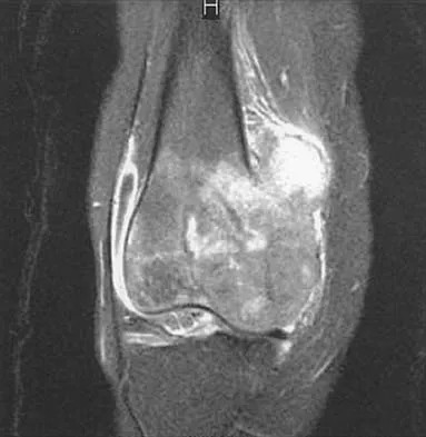
Question 3
A 12-year-old girl has had progressive left knee pain for the past 4 months. She reports that the pain is unrelated to activity, and she has no history of fever or recent infections. Examination reveals full range of motion of the knee but tenderness along the medial joint line. Plain radiographs and MRI scans are shown in Figures 39a through 39d. A biopsy specimen of the lesion is shown in Figure 39e. Treatment should include
Explanation
Question 4
An open biopsy specimen of a radiodense distal clavicle lesion in a 12-year-old girl shows chronic polyclonal inflammatory cells without granuloma formation. Laboratory studies show that bacterial, fungal, and acid-fast bacillus cultures are negative. Subsequently, a similar lesion is noted in the fibula. The next most appropriate step in management should consist of
Explanation
Question 5
Figure 40 shows the radiograph of a 30-year-old woman who has a painful elbow. Examination reveals a deformed skull, multiple cafe-au-lait spots, and bone deformities. What is the most likely diagnosis?
Explanation
Question 6
Figure 41a shows the AP radiograph of a 15-year-old boy who reports lateral knee pain. Figures 41b and 41c show a radiograph of the distal femur that was obtained 5 years ago and a current CT scan. The indication for surgery in this patient would be
Explanation
Question 7
In what decade does the peak incidence of conventional osteosarcoma occur?
Explanation
Question 8
A 10-year-old boy has a painful thigh mass. A radiograph, MRI scan, and biopsy specimen are shown in Figures 42a through 42c. What is the most likely diagnosis?
Explanation
Question 9
A 21-year-old man with neurofibromatosis and multiple cutaneous neurofibromas has a rapidly enlarging painless mass on his buttock. Examination reveals a nontender, well-defined 6- x 6-cm soft-tissue mass that is deep to the fascia. The best course of action should be to order
Explanation
Question 10
A 21-year-old man has had progressive right knee pain for the past 2 months that is exacerbated with weight-bearing activities. A plain radiograph and an MRI scan are shown in Figures 43a and 43b. A biopsy specimen is shown in Figure 43c. According to the Enneking staging system of tumor classification, the lesion should be classified as what stage?
Explanation
Question 11
What is a common clinical finding in patients with severe hypercalcemia secondary to bony metastasis?
Explanation
Question 12
What cell type causes the bone destruction in metastatic lesions?
Explanation
Question 13
What is the most common malignant bone tumor seen in patients with multiple hereditary exostosis?
Explanation
Question 14
An athletic 55-year-old man reports a painless mass in the anterior aspect of the thigh that appeared 3 weeks ago and has not changed in size. The patient denies any history of trauma. Examination reveals a firm, well-defined nontender mass in the anterior thigh and no inguinal adenopathy or cutaneous changes. Plain radiographs are unremarkable. T1- and T2-weighted MRI scans are shown in Figures 44a and 44b. What is the most likely diagnosis?
Explanation
Question 15
Epithelioid sarcoma most commonly occurs in which of the following anatomic locations?
Explanation
Question 16
What common cytologic abnormality is associated with Ewing's sarcoma?
Explanation
Question 17
Figures 45a and 45b show the radiographs of a 46-year-old man who reports the acute onset of right knee pain and is unable to bear weight on the extremity. His medical history is unremarkable. The next most appropriate step in management should consist of
Explanation
Question 18
Figure 46 shows the MRI scan of a patient who has a mass in the calf that has been fluctuating in size. Radiographs are negative. Which of the following procedures will most quickly aid in confirming the diagnosis?
Explanation
Question 19
What are the five most common tumors that metastasize to bone?
Explanation
Question 20
Pain associated with a proximal medial tibial osteochondroma in a 10-year-old patient is most commonly the result of
Explanation
Question 21
Figures 47a through 47f show the AP radiograph, bone scan, CT scan, MRI scan, and biopsy specimens of a 30-year-old woman who has had vague left shoulder pain for 1 year. Management should consist of
Explanation
Question 22
What is the 5-year overall survival rate for adults with high-grade soft-tissue sarcomas?
Explanation
Question 23
Figures 48a through 48c show the lateral radiograph and MRI scans of a 60-year-old man who has had pain in his thigh for 1 month. The next most appropriate step in management should consist of
Explanation
Question 24
A 17-year-old boy has had a mass in his right thigh for the past 6 months. He denies any history of trauma. Examination reveals that the mass is painless and firm. A radiograph and axial MRI scan are shown in Figures 49a and 49b. What is the most likely diagnosis?
Explanation
Question 25
Evaluation of the percent of necrosis in the resected specimen after preoperative chemotherapy is of prognostic value for what type of sarcoma?
Explanation
Question 26
A 65-year-old woman is evaluated for osteoporosis. She is started on denosumab. What is the precise mechanism of action of this medication?
Explanation
Question 27
A 35-year-old man sustains a midshaft humerus fracture resulting in an isolated radial nerve palsy. Electromyography at 4 weeks shows fibrillation potentials in the brachioradialis but no voluntary motor unit action potentials. What is the underlying pathophysiological process occurring at the distal axon?
Explanation
Question 28
Articular cartilage has a complex hierarchical structure that allows it to withstand compressive and shear forces. In the superficial zone of articular cartilage, what is the primary orientation of type II collagen fibers and what is its biomechanical function?
Explanation
Question 29
A 15-year-old boy presents with a painful mass in the distal femur. A biopsy reveals small, round blue cells. Molecular testing demonstrates a t(11;22)(q24;q12) chromosomal translocation. This specific genetic alteration results in the fusion of which two genes, characteristic of the diagnosed lesion?
Explanation
Question 30
A 45-year-old patient undergoes an open reduction and internal fixation of a diaphyseal forearm fracture using absolute stability techniques (lag screw and neutralization plate). Which type of bone healing is expected, and what is the primary biological mediator initiating this process?
Explanation
Question 31
Aseptic loosening is the most common cause of late failure in total joint arthroplasty. The biological cascade leading to periprosthetic osteolysis is primarily initiated by the phagocytosis of wear debris. Which cell type is the principal driver of this initial inflammatory response, and what is the primary cytokine released?
Explanation
Question 32
A 60-year-old man develops a chronic prosthetic joint infection 2 years after a total knee arthroplasty. Intraoperative cultures grow Staphylococcus epidermidis. The organism's ability to persist on the implant surface despite appropriate systemic antibiotic therapy is primarily due to:
Explanation
Question 33
During tensile testing of a human anterior cruciate ligament (ACL) graft, a load-elongation (stress-strain) curve is plotted. What does the non-linear 'toe region' of this curve represent physiologically?
Explanation
Question 34
A patient is diagnosed with familial hypophosphatemic rickets (X-linked dominant). The underlying pathophysiology involves an overproduction of Fibroblast Growth Factor 23 (FGF23). What is the primary renal effect of excessive FGF23 in this condition?
Explanation
Question 35
A 72-year-old woman is scheduled for an elective total hip arthroplasty. Her surgeon prescribes apixaban for postoperative deep vein thrombosis (DVT) prophylaxis. By which precise mechanism does apixaban achieve its primary anticoagulant effect?
Explanation
Question 36
A 65-year-old man with a long-standing history of bone pain, increasing head size, and hearing loss presents with a subtrochanteric femur fracture after a minor fall. What is the characteristic histologic finding of the abnormal bone at the fracture site?
Explanation
Question 37
A 55-year-old woman with metastatic breast cancer to the spine and pelvis is prescribed an agent to reduce the incidence of skeletal-related events (pathologic fractures, spinal cord compression). The prescribed agent is a fully human monoclonal antibody. What is its specific mechanism of action?
Explanation
Question 38
A 14-year-old boy presents with a 2-month history of pain and swelling in his left thigh. Plain radiographs show a permeative, destructive diaphyseal lesion in the femur with an 'onion skin' periosteal reaction. Biopsy reveals uniform, small, round blue cells that stain positive for CD99. Cytogenetic analysis is most likely to show which of the following chromosomal translocations?
Explanation
Question 39
In a 16-year-old patient diagnosed with localized, conventional high-grade osteosarcoma of the distal femur, which of the following clinical or pathologic factors is considered the most significant prognostic indicator for overall long-term survival?
Explanation
Question 40
A 28-year-old man presents with a slow-growing, painful mass around his right knee, deep to the fascia. MRI shows a soft tissue mass, and plain radiographs demonstrate focal calcifications within the lesion. Core needle biopsy demonstrates a biphasic pattern of epithelial and spindle cells. What is the most appropriate initial management for this localized disease?
Explanation
Question 41
A 32-year-old woman presents with worsening knee pain. Radiographs reveal an eccentric, purely lytic lesion in the distal femoral epiphysis extending to the subchondral bone without a sclerotic rim. Biopsy confirms Giant Cell Tumor of bone (GCT). Which of the following accurately describes the molecular pathogenesis and targeted therapy for this tumor?
Explanation
Question 42
A 55-year-old man presents with an enlarging, painful mass in his proximal humerus. Radiographs show a lytic lesion with intralesional 'popcorn' calcifications and deep endosteal scalloping greater than two-thirds of the cortical thickness. Biopsy confirms a Grade II (intermediate) conventional chondrosarcoma. What is the most appropriate definitive treatment?
Explanation
Question 43
A 62-year-old man presents with diffuse back and rib pain. Radiographs show multiple punched-out lytic lesions in the skull and pelvis. Laboratory evaluation reveals hypercalcemia, renal insufficiency, and anemia. Bone marrow aspirate shows >10% clonal plasma cells. Which of the following laboratory abnormalities is most strongly associated with a poor overall survival prognosis in this disease?
Explanation
Question 44
A 20-year-old woman is incidentally found to have a proximal femoral lesion during imaging for a hip strain. The radiograph shows a diaphyseal 'ground-glass' appearance surrounded by a sclerotic rim, with mild varus bowing. She has no endocrine abnormalities or cafe-au-lait spots. The underlying genetic mutation for this bony lesion involves which of the following?
Explanation
Question 45
A 16-year-old boy complains of persistent anterior thigh pain that has worsened over the past 4 months. He notes the pain is significantly worse at night and is dramatically relieved within 30 minutes of taking ibuprofen. Radiographs demonstrate a diaphyseal cortical thickening with a 1-cm central radiolucent nidus. What is the pathophysiologic mechanism responsible for the characteristic pain in this condition?
Explanation
Question 46
A 28-year-old man presents with a slow-growing, painless mass in the popliteal fossa of his left knee. Biopsy reveals a biphasic tumor with both epithelial and spindle cell components. Immunohistochemistry is positive for cytokeratin and epithelial membrane antigen (EMA). Which of the following chromosomal translocations is most characteristic of this lesion?
Explanation
Question 47
A 32-year-old woman presents with a lytic lesion in the distal femur extending to the subchondral bone. Biopsy confirms the diagnosis of a giant cell tumor of bone. She is treated with denosumab preoperatively to downstage the tumor. What is the mechanism of action of this medication?
Explanation
Question 48
A 15-year-old boy presents with knee pain and a destructive, bone-forming lesion in the proximal tibia. Biopsy confirms high-grade intramedullary osteosarcoma. Mutations in which of the following pairs of tumor suppressor genes are most frequently associated with the pathogenesis of this disease?
Explanation
Question 49
During internal fixation of a diaphyseal femur fracture, a surgeon aims to maximize the pullout strength of a cortical screw. Which of the following alterations to screw design or insertion technique most effectively increases pullout strength in dense cortical bone?
Explanation
Question 50
In typical adult hyaline articular cartilage, which zone is characterized by the highest concentration of proteoglycans, the lowest concentration of water, and chondrocytes arranged in vertical columns?
Explanation
Question 51
A 65-year-old man presents with aseptic loosening of his cementless total hip arthroplasty 12 years after the index procedure. Radiographs show extensive periprosthetic osteolysis. The primary biological mechanism driving this osteolysis involves the phagocytosis of wear particles by which of the following cell types, leading to the release of TNF-alpha and IL-1?
Explanation
Question 52
During secondary fracture healing, the soft callus is gradually replaced by hard callus through the process of endochondral ossification. Which of the following collagen types is most highly expressed by hypertrophic chondrocytes as the cartilage matrix calcifies before being replaced by woven bone?
Explanation
Question 53
A 72-year-old woman with severe osteoporosis and a history of a vertebral compression fracture is started on daily subcutaneous injections of teriparatide. Which of the following best describes the physiological effect of this specific dosing regimen?
Explanation
Question 54
A 9-year-old boy presents with a painful, swollen mid-thigh. Radiographs reveal a permeative diaphyseal lesion in the femur with a prominent 'onion-skin' periosteal reaction.
Histology shows sheets of uniform small round blue cells that stain strongly positive for CD99. Which of the following fusion proteins is most likely driving this malignancy?

Explanation
Question 55
A 60-year-old man with metastatic prostate cancer to the spine is treated with intravenous zoledronic acid to reduce the risk of skeletal-related events. Nitrogen-containing bisphosphonates like zoledronic acid inhibit osteoclast function primarily by interfering with which of the following intracellular pathways?
Explanation
Question 56
A 14-year-old boy presents with pain and swelling in his left thigh. Radiographs reveal a permeative, lytic lesion in the diaphysis of the femur with an 'onion-skin' periosteal reaction. A biopsy is performed, showing sheets of uniform, small, round blue cells. Which of the following chromosomal translocations is most strongly associated with this diagnosis?
Explanation
Question 57
Which of the following zones of articular cartilage has the highest concentration of collagen, lowest concentration of proteoglycans, and chondrocytes aligned parallel to the joint surface?
Explanation
Question 58
Bone morphogenetic proteins (BMPs) induce bone formation primarily by signaling through which of the following intracellular pathways?
Explanation
Question 59
A 19-year-old man presents with severe right leg pain that is worse at night and dramatically improves with NSAIDs. Radiographs show a sclerotic lesion in the tibial diaphysis with a small central radiolucent nidus. The intense pain associated with this lesion is primarily mediated by high levels of which of the following within the nidus?
Explanation
Question 60
A 32-year-old woman presents with a slowly enlarging, painless mass around her right knee that has been present for 2 years. MRI reveals a well-circumscribed, heterogeneous soft-tissue mass adjacent to, but not within, the joint space. A biopsy is performed, revealing a biphasic pattern of spindle cells and epithelial cells. What chromosomal abnormality is most characteristic of this tumor?
Explanation
Question 61
Denosumab is utilized in the management of giant cell tumor of bone and metastatic bone disease. It achieves its therapeutic effect by directly binding to and inhibiting which of the following?
Explanation
Question 62
A 55-year-old man presents with dull aching pain in his right shoulder. Radiographs demonstrate a lytic lesion in the proximal humerus with intralesional 'popcorn' calcifications and focal endosteal scalloping greater than two-thirds of the cortical thickness. Which of the following features most strongly suggests a diagnosis of secondary chondrosarcoma rather than a benign enchondroma?
Explanation
Question 63
A 65-year-old man complains of lower back pain and generalized fatigue. Radiographs show multiple 'punched-out' lytic lesions in his skull and vertebral bodies. Laboratory evaluation reveals hypercalcemia, anemia, and an M-spike on serum protein electrophoresis. Which of the following is the most likely finding on bone marrow biopsy?
Explanation
Question 64
A 12-year-old girl is evaluated for a noticeable leg length discrepancy and a limp. Radiographs of her left femur show an expansive, purely lytic lesion with a 'ground-glass' appearance and a characteristic 'shepherd's crook' deformity of the proximal femur. This condition is caused by a somatic activating mutation in which of the following?
Explanation
Question 65
A 24-year-old male sustains an anterior shoulder dislocation during a rugby match. After successful closed reduction, he is noted to have weakness in shoulder abduction and decreased sensation over the lateral aspect of his shoulder. Injury to which of the following nerves is the most likely cause of these findings?
Explanation
Question 66
A 15-year-old boy presents with knee pain and a destructive lesion in the distal femur. Core needle biopsy confirms high-grade intramedullary osteosarcoma. He undergoes neoadjuvant chemotherapy followed by limb-salvage surgery. Which of the following is the most important prognostic factor for long-term survival in this patient?
Explanation
Question 67
A 28-year-old woman is diagnosed with a recurrent giant cell tumor of the proximal tibia after previous curettage and cementation. Her surgeon recommends systemic therapy with denosumab to downstage the tumor prior to a second procedure. What is the mechanism of action of this medication in the context of this lesion?
Explanation
Question 68
A 9-year-old boy presents with a 3-month history of deep, aching pain in his proximal femur that worsens at night. He reports dramatic relief of his symptoms 30 minutes after taking ibuprofen. Radiographs and a CT scan reveal a 7-mm radiolucent nidus surrounded by dense, reactive cortical sclerosis. The intense pain experienced by this patient is most directly mediated by elevated levels of which of the following substances produced by the nidus?
Explanation
Question 69
A 45-year-old man presents with chronic lower back and perineal pain associated with a palpable presacral mass. A biopsy of the lesion reveals cells with copious, vacuolated (bubbly) cytoplasm arranged in cords within a myxoid stroma. Immunohistochemistry is strongly positive for cytokeratin, epithelial membrane antigen (EMA), and brachyury. What is the most likely diagnosis?
Explanation
Question 70
A 22-year-old man presents with a slow-growing, painless soft-tissue mass in the popliteal fossa. MRI demonstrates a deeply seated mass that is intimately associated with the joint capsule but does not involve the intra-articular space. Biopsy reveals a biphasic spindle cell neoplasm. Which of the following chromosomal translocations is most characteristic of this tumor?
Explanation
Question 71
A 65-year-old woman with a history of breast cancer presents with a new metastatic lesion in her femur. You are evaluating her risk for an impending pathologic fracture using the Mirels' criteria. According to this scoring system, which combination of lesion characteristics yields the highest score, indicating the strongest recommendation for prophylactic internal fixation?
Explanation
Question 72
A 14-year-old girl is diagnosed with Ewing sarcoma of the diaphyseal fibula following a biopsy. As part of her initial staging workup, a chest CT, whole-body MRI, and bone marrow aspirate are ordered. What is the most common site of distant metastasis for Ewing sarcoma at the time of initial presentation?
Explanation
Question 73
A 35-year-old man presents with a locally aggressive, osteolytic lesion in the epiphysis of his distal radius. Histopathologic examination reveals abundant, evenly distributed multinucleated osteoclast-like giant cells in a background of round to oval mononuclear cells. Which of the following genetic alterations is highly specific for the neoplastic cells in this tumor?
Explanation
Question 74
A 10-year-old boy presents with swelling and pain in his left thigh. Radiographs reveal a destructive permeative lesion in the femoral diaphysis with an "onion-skin" periosteal reaction. Biopsy demonstrates sheets of uniform, small round blue cells with scant cytoplasm. Immunohistochemical analysis is requested. Staining for which of the following cell surface markers is most characteristically positive in this tumor?
Explanation
Question 75
A 55-year-old man undergoes wide surgical resection of a painless, 8-cm mass located deep within the vastus lateralis of his anterior thigh. Histopathology reveals an undifferentiated pleomorphic sarcoma with high-grade features. The surgical margins are reported as negative (R0). To minimize the risk of local recurrence, what is the most appropriate adjuvant therapy for this patient?
Explanation
Question 76
A 14-year-old boy presents with an aggressive diaphyseal lesion in his femur with a large soft tissue mass. Biopsy reveals small round blue cells. Cytogenetics confirm a t(11;22) translocation. Which of the following is the resulting fusion protein responsible for driving the pathogenesis?
Explanation
Question 77
A 32-year-old woman has a recurrent giant cell tumor of the proximal tibia. She is started on denosumab prior to surgical intervention. What is the precise mechanism of action of this pharmacological agent?
Explanation
Question 78
A 40-year-old man presents with chronic, recurrent hemarthrosis of the knee without a history of trauma or coagulopathy. MRI demonstrates a large, nodular synovial mass with blooming artifact on gradient-echo sequences. Which genetic alteration is most characteristic of this condition?
Explanation
Question 79
A diaphyseal tibia fracture is treated with a well-fitted unlocked intramedullary nail. According to Perren's strain theory, what tissue will predominantly form in the fracture gap if the interfragmentary strain is maintained between 2% and 10%?
Explanation
Question 80
A 65-year-old woman with severe postmenopausal osteoporosis is treated with romosozumab. This medication primarily increases bone mineral density by neutralizing a protein that typically inhibits which of the following signaling pathways?
Explanation
Question 81
A 60-year-old man is diagnosed with multiple myeloma. He has diffuse osteolytic lesions in his axial skeleton. The osteolytic nature of these bone lesions is primarily driven by multiple myeloma cells secreting factors that heavily upregulate which of the following?
Explanation
Question 82
A 55-year-old man presents with increasing thigh pain. Radiographs reveal a large calcified lesion in the medullary canal of the proximal femur with endosteal scalloping greater than two-thirds of the cortical thickness and a focal area of cortical breakthrough. Biopsy demonstrates a Grade II (intermediate grade) conventional chondrosarcoma. What is the most appropriate management?
Explanation
Question 83
A surgeon uses a massive structural cortical allograft for intercalary reconstruction following a tumor resection. Over the subsequent years, the allograft undergoes the process of 'creeping substitution.' In a cortical allograft, which of the following accurately describes this physiological process?
Explanation
Question 84
A 28-year-old woman presents with a slow-growing, painless mass at the posterior aspect of her distal femur. Radiographs show a dense, heavily ossified mass attached to the posterior cortex by a broad base, with a 'string sign' radiolucent cleft separating the tumor from the underlying cortex. A biopsy confirms a low-grade malignant bone-forming tumor. What is the classic genetic amplification associated with this specific tumor?
Explanation
Question 85
A 72-year-old woman on oral alendronate for 10 years presents with a transverse, non-comminuted fracture of the femoral shaft with a medial cortical spike, following a minor trip and fall. She reported having lateral thigh pain for three months prior to the fall. The pathogenesis of her fracture is primarily related to which of the following mechanisms?
Explanation
Question 86
A 15-year-old boy presents with a painful mass in his diaphyseal femur. Radiographs show a permeative lesion with a "moth-eaten" appearance and a "periosteal onion-skin" reaction. Biopsy reveals small round blue cells. Immunohistochemistry is strongly positive for CD99. Which of the following genetic translocations is most characteristic of this tumor?
Explanation
Question 87
A 34-year-old woman presents with knee pain. Radiographs reveal an eccentric, lytic, epiphyseal lesion in the distal femur extending to the subchondral bone. Biopsy confirms multinucleated giant cells interspersed with mononuclear stromal cells. Which of the following targeted therapies acts by binding to RANKL, and what is its specific role in the medical management of this lesion?
Explanation
Question 88
A 14-year-old boy is diagnosed with high-grade conventional osteosarcoma of the proximal tibia. He undergoes neoadjuvant chemotherapy followed by wide surgical resection. Which of the following factors obtained from the surgical specimen is the most important prognostic indicator for his long-term survival?
Explanation
Question 89
A 45-year-old man presents with a deep, painless, slowly growing mass in his thigh. MRI reveals a large intramuscular lesion with high signal intensity on T2-weighted images and a lipomatous component. Biopsy confirms myxoid liposarcoma. Due to the specific biology of this tumor, which of the following imaging modalities should be included in the staging workup that is not typically routine for other high-grade soft tissue sarcomas of the extremity?
Explanation
Question 90
A 9-year-old boy presents with a pathologic fracture of the proximal humerus after throwing a baseball. Radiographs show a centrally located, completely lytic, expansile lesion in the metaphysis of the proximal humerus with a 'fallen leaf' sign. No periosteal reaction is noted outside the fracture site. What is the most appropriate initial surgical management after the fracture has healed?
Explanation
Question 91
A 68-year-old man presents with dull, aching pain in his right thigh and an enlarging hat size. Radiographs of the femur demonstrate cortical thickening, increased bone density, and bowing. Laboratory studies show markedly elevated serum alkaline phosphatase, but normal calcium and phosphorus. The primary defect in this condition is characterized by which of the following?
Explanation
Question 92
A 55-year-old man presents with deep pelvic pain. Radiographs show a large destructive lytic lesion in the right ilium with 'popcorn' intralesional calcifications. Biopsy demonstrates atypical chondrocytes with binucleated cells and myxoid stroma, consistent with grade II chondrosarcoma. What is the standard of care for this localized tumor?
Explanation
Question 93
A 14-year-old girl presents with a rapidly enlarging mass in her proximal fibula. Radiographs reveal an eccentric, expansile, purely lytic metaphyseal lesion with a thin 'eggshell' cortical rim. MRI demonstrates multiple fluid-fluid levels. Genetic analysis of the primary lesion would most likely reveal a translocation involving which of the following genes?
Explanation
Question 94
A 20-year-old man complains of persistent left thigh pain that is worse at night and dramatically relieved by ibuprofen. Radiographs demonstrate cortical thickening in the diaphyseal femur with a small radiolucent nidus. What is the mechanism by which NSAIDs relieve the pain associated with this lesion?
Explanation
Question 95
A 62-year-old man presents with back pain and fatigue. Laboratory investigations reveal anemia, hypercalcemia, and a monoclonal spike on serum protein electrophoresis. Plain radiographs of the spine are negative for obvious lytic lesions. What is the most sensitive imaging modality to detect skeletal involvement in this patient?
Explanation
Question 96
A 35-year-old woman presents with persistent right knee pain. Radiographs reveal an eccentric, purely lytic lesion in the distal femoral epiphysis extending into the metaphysis and reaching the subchondral bone. A core needle biopsy demonstrates numerous multinucleated giant cells uniformly dispersed among round-to-oval mononuclear stromal cells. Due to the high risk of joint collapse with surgical curettage, targeted medical therapy is considered. The most appropriate pharmacological agent for this lesion primarily targets which of the following molecular pathways?
Explanation
Question 97
A 15-year-old boy presents with an osteosarcoma of the proximal tibia. His family history is notable for a mother who developed early-onset breast cancer at age 32 and an uncle diagnosed with a soft-tissue sarcoma at age 25. A germline mutation in which of the following genes is the most likely underlying cause of this patient's familial cancer syndrome?
Explanation
Question 98
A 10-year-old boy is evaluated for worsening pain and swelling in his left thigh. Plain radiographs show a permeative diaphyseal lesion in the femur with an associated 'onion-skin' periosteal reaction. A biopsy is performed, revealing uniform sheets of small, round, blue cells with scant cytoplasm. Immunohistochemistry is positive for CD99. Which of the following chromosomal translocations is most strongly associated with this neoplasm?
Explanation
Question 99
A 65-year-old man presents with severe back pain, fatigue, and generalized weakness. Laboratory tests reveal a normocytic anemia, hypercalcemia, and an elevated serum creatinine. Radiographs of the spine and skull demonstrate multiple, well-circumscribed, 'punched-out' lytic lesions without reactive sclerosis. The extensive bone destruction seen in this condition is primarily mediated by which of the following cellular mechanisms?
Explanation
Question 100
A 22-year-old man has a 6-month history of a dull, aching pain in his posterior neck. He notes that the pain is constant and is not consistently relieved by over-the-counter NSAIDs. A CT scan of the cervical spine reveals a 2.5-cm radiolucent nidus surrounded by a thin rim of reactive sclerosis located in the posterior elements of C5. Histological examination of a biopsy specimen reveals interconnected, disorganized trabeculae of woven bone lined by prominent osteoblasts within a highly vascularized connective tissue stroma. What is the most likely diagnosis?
Explanation
None
