Lambotte’s Principles and the AO-ASIF Philosophy of Surgical Fracture Management
Key Takeaway
Lambotte’s historical principles form the foundation of modern AO-ASIF fracture management guidelines. These dictate anatomical reduction, stable internal fixation, preservation of vascularity, and early active mobilization. Successful surgical intervention requires meticulous preoperative planning, respect for soft tissue biology, and precise execution of provisional and definitive stabilization. Understanding the biomechanical demands and inherent risks of operative osteosynthesis is critical for orthopedic surgeons to optimize patient outcomes and prevent nonunion or infection.
Comprehensive Introduction and Patho-Epidemiology
The surgical management of fractures represents one of the most profound evolutions in modern medicine, governed by a delicate, unforgiving balance between mechanical stabilization and biological preservation. While the conceptual desire to align and stabilize broken bones spans millennia, it was the pioneering work of Albin Lambotte—universally heralded as the "Father of Modern Osteosynthesis"—that formalized the principles of operative fracture treatment in the early 20th century. Lambotte’s foundational tenets established the necessity of surgical exposure, anatomical reduction, and robust internal fixation. Building upon this historical and intellectual bedrock, the Arbeitsgemeinschaft für Osteosynthesefragen (AO-ASIF), founded in 1958 by Maurice Müller, Martin Allgöwer, Robert Schneider, and Hans Willenegger, refined these concepts into a universal, biologically sound philosophy. The AO-ASIF principles dictate contemporary orthopedic trauma surgery, shifting the paradigm from mere mechanical "carpentry" to a sophisticated understanding of mechanobiology.
The ultimate goal of operative fracture management is the rapid, complete, and painless restoration of limb function. This objective is achieved through a profound understanding of bone biology, the preservation of the soft tissue envelope, and the precise calculation of the biomechanical demands placed upon the injured extremity. The epidemiology of fractures requiring surgical intervention demonstrates a classic bimodal distribution. High-energy trauma (motor vehicle collisions, falls from height, ballistic injuries) predominantly affects the younger demographic, resulting in complex, comminuted, and often open fracture patterns. Conversely, low-energy mechanisms (ground-level falls) disproportionately affect the geriatric population, where diminished bone mineral density and altered trabecular microarchitecture present unique challenges for hardware purchase and construct longevity.
Understanding the pathophysiology of fracture healing is paramount to executing the AO-ASIF principles. Bone healing occurs via two distinct pathways, dictated entirely by the mechanical environment created by the surgeon. Primary (direct) bone healing occurs under conditions of absolute stability, where interfragmentary strain is reduced to less than 2%. This environment allows osteoclasts to cross the fracture site via cutting cones, followed by osteoblasts depositing lamellar bone in a process of direct Haversian remodeling, notably without the formation of a periosteal callus. Secondary (indirect) bone healing occurs under conditions of relative stability, where interfragmentary strain is maintained between 2% and 10%. This controlled micro-motion stimulates a robust biological cascade, progressing from hematoma formation to a fibrocartilaginous soft callus, which subsequently undergoes endochondral ossification to form a hard, woven bone callus before final remodeling.
The modern orthopedic trauma surgeon must function as a "biological watchmaker." The decision to pursue absolute versus relative stability is not arbitrary; it is dictated by the fracture personality. Simple articular and diaphyseal fractures demand anatomical reduction and absolute stability to restore joint congruity and allow direct remodeling. Conversely, highly comminuted diaphyseal and metaphyseal fractures require relative stability via bridging constructs to preserve the fragmented blood supply and stimulate robust secondary healing. Failure to respect this mechanobiological relationship—such as attempting to rigidly plate a highly comminuted diaphyseal fracture—inevitably leads to devascularization, stress shielding, and catastrophic nonunion.
Detailed Surgical Anatomy and Biomechanics
The execution of successful osteosynthesis is inextricably linked to a profound understanding of osteovascular anatomy and the biomechanical principles governing implant-bone constructs. The vascular supply to a long bone is highly specialized, relying on a dual system comprising the nutrient artery and the periosteal plexus. The nutrient artery, entering through the diaphyseal cortex, arborizes into the medullary canal and supplies the inner two-thirds of the cortical bone via centrifugal flow. The periosteal vascular network, derived from the surrounding muscular attachments (angiosomes), supplies the outer one-third of the cortex. In the event of a fracture, the medullary supply is frequently disrupted, temporarily reversing the flow to a centripetal direction reliant entirely on the periosteal envelope. Iatrogenic destruction of this periosteal supply through aggressive surgical stripping devitalizes the cortical bone, transforming functional fragments into necrotic sequestra and precipitating atrophic nonunion.
Stephan Perren’s Strain Theory remains the biomechanical cornerstone of the AO-ASIF philosophy. Strain is defined mathematically as the change in gap length divided by the original gap length ($/Delta L/L$). Tissues within the fracture gap can only survive if the strain does not exceed their specific physiological tolerance. Granulation tissue can tolerate 100% strain, fibrocartilage up to 10%, and lamellar bone only 2%. Therefore, if a fracture gap is extremely narrow but inherently unstable, the microscopic motion results in a massive percentage change in gap length (high strain), which destroys forming osteons and leads to a hypertrophic nonunion. The surgeon must manipulate this mechanical environment by either decreasing the motion (increasing stability via compression) or increasing the functional working length of the construct (distributing the motion over a larger area via bridge plating or intramedullary nailing) to bring the strain within the optimal window for bone formation.
Implant biomechanics dictate the selection and application of hardware. Plates can function in five distinct biomechanical modes: tension band, compression, neutralization (protection), buttress, and bridge. The tension band principle converts deleterious tensile forces into beneficial compressive forces at the opposite cortex, provided the bone can withstand compression. Compression plates utilize eccentrically placed screws within oval holes to generate axial interfragmentary compression. Neutralization plates protect a primary lag screw from torsional, bending, and shear forces. Buttress plates resist axial shear forces, typically at metaphyseal-epiphyseal junctions. Bridge plates bypass a comminuted zone entirely, providing relative stability and preserving the fracture hematoma. Intramedullary nails function as load-sharing devices situated at the mechanical axis of the bone. Their resistance to bending is proportional to the fourth power of their radius (polar moment of inertia), making larger-diameter nails exponentially stiffer, though this must be balanced against the biological cost of excessive endosteal reaming.
The concept of "working length" is critical when utilizing bridging constructs. The working length is defined as the distance between the two closest points of fixation spanning the fracture. A short working length concentrates stress over a small segment of the implant, increasing the risk of fatigue failure (hardware breakage) before biological union occurs. Conversely, a longer working length distributes the bending stresses, increasing the flexibility of the construct and promoting robust callus formation via controlled micro-motion. The surgeon must meticulously balance the stiffness of the implant material (e.g., titanium alloy versus stainless steel), the geometry of the construct, and the biological capacity of the host tissue to optimize the mechanobiological environment for fracture consolidation.
Exhaustive Indications and Contraindications
The decision to proceed with operative fracture management requires a rigorous assessment of the fracture pattern, the soft tissue envelope, and the patient’s physiological reserve. While surgical stabilization is the gold standard for many injuries, inappropriate intervention can result in catastrophic outcomes that far exceed the morbidity of the initial trauma. The indications for surgery must be weighed against the inherent "second hit" of surgical trauma.
Absolute indications for immediate or early surgical intervention include open fractures, fractures associated with vascular compromise requiring repair, impending compartment syndrome, and irreducible joint dislocations. Polytrauma patients with multiple long-bone fractures benefit significantly from Early Total Care (ETC) or Damage Control Orthopedics (DCO) to facilitate upright positioning, optimize pulmonary mechanics, and mitigate the risk of Acute Respiratory Distress Syndrome (ARDS). Intra-articular fractures with displacement or step-off exceeding 2 millimeters represent a strict indication for anatomical reduction and absolute stability to prevent early, rapid-onset post-traumatic osteoarthritis. Diaphyseal fractures with unacceptable shortening, angulation, or rotational malalignment that cannot be maintained in a cast or functional brace also necessitate operative stabilization.
Patient-specific factors heavily influence the surgical decision-making process. The physiological state of the patient, categorized by the American Society of Anesthesiologists (ASA) classification, dictates their ability to tolerate anesthesia and surgical blood loss. In the polytraumatized patient, the systemic inflammatory response syndrome (SIRS) triggered by the initial injury can be exacerbated by prolonged surgical intervention, pushing the patient into a lethal triad of coagulopathy, hypothermia, and metabolic acidosis. In these scenarios, the indication shifts from definitive fixation to rapid, provisional stabilization using external fixators (Damage Control Orthopedics). Bone quality is another critical determinant; severe osteoporosis compromises hardware purchase, necessitating the use of fixed-angle locking plates, intramedullary load-sharing devices, or polymethylmethacrylate (PMMA) cement augmentation.
Contraindications to definitive internal fixation are equally critical to recognize. Operating through compromised soft tissues—such as active fracture blisters, severe contusions, burns, or active dermatitis—inevitably leads to wound dehiscence, deep hardware infection, and potential amputation. Active osteomyelitis or gross contamination within the surgical field absolute contraindicates the placement of permanent internal hardware, as the implants will serve as a nidus for intractable biofilm formation.
| Category | Clinical Scenario | Rationale / Management Strategy |
|---|---|---|
| Absolute Indications | Open fractures, vascular injury, irreducible dislocations, polytrauma (femur fractures). | Immediate stabilization is required to prevent limb loss, restore perfusion, and mitigate systemic complications (ARDS, fat embolism). |
| Relative Indications | Displaced diaphyseal fractures, intra-articular step-off >2mm, failure of conservative care. | Surgery optimizes functional outcomes, restores joint kinematics, and accelerates rehabilitation compared to prolonged immobilization. |
| Relative Contraindications | Severe osteoporosis, non-ambulatory baseline status, poorly controlled diabetes. | High risk of hardware failure or wound complications. Requires specialized implants (locking plates) or modified functional goals. |
| Absolute Contraindications | Active deep infection at the surgical site, severe soft tissue compromise (blisters, burns), medically unstable patient (lethal triad). | High probability of catastrophic failure, biofilm formation, or patient mortality. Mandates Damage Control Orthopedics (external fixation) or non-operative care. |
Pre-Operative Planning, Templating, and Patient Positioning
Pre-operative planning is the cognitive foundation upon which successful osteosynthesis is built. The operation is fundamentally won or lost before the patient ever enters the operating theater. A meticulously formulated tactical plan minimizes intraoperative hesitation, reduces tourniquet and anesthesia time, and dramatically lowers the incidence of intraoperative complications. This process begins with an exhaustive review of high-quality imaging. Orthogonal radiographs (true anteroposterior and lateral views) of the injured segment, encompassing the joints above and below, are mandatory. For complex intra-articular fractures, a fine-cut computed tomography (CT) scan with 3D reconstructions is essential to delineate the fracture personality, identify impacted articular fragments, and map the trajectory of fracture lines to plan optimal screw placement.
Digital or analog templating is a non-negotiable step in the preoperative workflow. The surgeon must utilize calibrated radiographs to select the appropriate implant type, size, and length. Templating allows the surgeon to determine the exact starting point for intramedullary nails, the precise contouring required for plates, and the optimal trajectory and length of lag screws. Furthermore, the surgeon must script the reduction sequence step-by-step. Anticipating the necessary reduction tools—such as pointed reduction forceps, Schanz pins for joysticks, femoral distractors, or specific clamps—ensures that the surgical team is fully prepared. An equipment checklist must be communicated to the nursing staff to verify the availability of specific implant sets, radiolucent tables, and functioning fluoroscopy units prior to induction.
Patient positioning is critical to achieving unhindered surgical access and obtaining high-quality intraoperative fluoroscopy. The positioning strategy must allow for the "C-arm dance"—the ability to obtain perfect orthogonal AP and lateral fluoroscopic views without moving the compromised, draped extremity. Depending on the fracture location, the patient may be positioned supine, prone, or in the lateral decubitus position. The use of a fracture table provides sustained, controlled traction for lower extremity diaphyseal fractures, whereas a flat radiolucent table allows for greater freedom to manipulate the limb and assess joint stability and range of motion dynamically.
The extremity must be prepped and draped widely to allow for full visualization of the mechanical axis and to permit intraoperative clinical assessment of length, alignment, and rotation (LAR). Draping must never restrict the surgeon's ability to flex or extend adjacent joints, as this is often required to relax deforming muscle forces during reduction. A formal surgical timeout and team briefing must be conducted, detailing the planned approach, the anticipated sequence of fixation, the required equipment, and the contingency plans should the primary reduction strategy fail. This communication ensures that the entire operating room team is synchronized, thereby optimizing surgical flow and patient safety.
Step-by-Step Surgical Approach and Fixation Technique
The execution of operative fracture management demands meticulous adherence to a structured, reproducible surgical workflow. When open reduction is mandated, the surgical approach must be anatomically precise and atraumatic to the surrounding soft tissue envelope. The surgeon must utilize true internervous and intermuscular planes to access the bone without denervating the overlying musculature (e.g., the Henry approach to the volar radius utilizes the internervous plane between the brachioradialis, innervated by the radial nerve, and the flexor carpi radialis, innervated by the median nerve). Full-thickness fasciocutaneous flaps must be elevated; undermining the subcutaneous tissues shears the perforating vessels and inevitably leads to marginal skin necrosis. Soft tissues must be retracted gently using blunt instruments, avoiding excessive tension that induces microvascular ischemia.
Reduction techniques are dictated by the fracture pattern and the biological goals of the procedure. Direct reduction is strictly reserved for intra-articular fractures and simple diaphyseal fractures where absolute stability is planned. This involves direct visualization of the fracture site, meticulous evacuation of the fracture hematoma and interposed soft tissue, and the precise manipulation of fragments using pointed reduction forceps or dental picks. Conversely, indirect reduction is utilized for comminuted diaphyseal and metaphyseal fractures where relative stability is the goal. Indirect techniques rely on ligamentotaxis, longitudinal traction, femoral distractors, or the use of the implant itself to restore length, alignment, and rotation without exposing the fracture site, thereby preserving the vital fracture hematoma and periosteal blood supply.
Once an acceptable reduction is achieved, it must be provisionally stabilized to allow for radiographic confirmation and the unhindered application of the definitive implant. Provisional stabilization is typically achieved using Kirschner wires (K-wires) or independent lag screws. A critical pitfall to avoid is the haphazard placement of provisional wires. K-wires must be placed strategically so that they do not obstruct the planned trajectory of the definitive plate, the path of the intramedullary reamer, or the location of locking screws. Failure to plan provisional fixation often results in the catastrophic loss of reduction when the wires must be prematurely removed to accommodate the definitive implant.
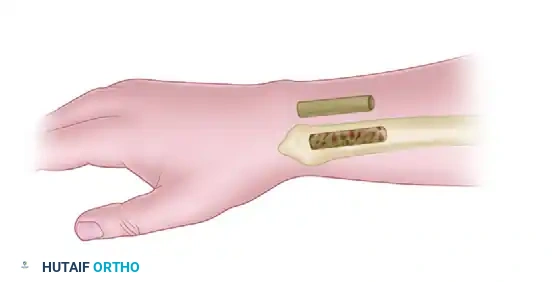
Definitive stabilization must execute the biomechanical goals established during preoperative planning. If absolute stability is required, interfragmentary compression is achieved using lag screws. The lag screw technique requires a gliding hole in the near cortex (equivalent to the outer thread diameter) and a thread hole in the far cortex (equivalent to the core diameter), allowing the screw head to compress the near fragment against the far fragment as the threads engage. If a plate is applied, it must be placed on the tension side of the bone to convert bending forces into compressive forces at the fracture site. For relative stability constructs, such as intramedullary nails or bridge plates, the hardware must possess sufficient fatigue life to withstand physiological loads until secondary bone healing occurs. The fixation is then rigorously assessed clinically for rotational alignment and fluoroscopically for hardware placement before proceeding to a meticulous, layered closure over a closed suction drain if necessary.
Complications, Incidence Rates, and Salvage Management
Despite meticulous planning and flawless execution, surgical fracture management carries an inherent risk of complications. The surgeon must be adept at recognizing these adverse events early and instituting appropriate salvage protocols. Complications can be broadly categorized into intraoperative, early postoperative, and late postoperative events. Intraoperative complications often involve iatrogenic neurovascular injury, malreduction, or hardware failure, such as broken drill bits or stripped screw heads. Iatrogenic nerve palsies (e.g., radial nerve palsy during humeral shaft plating) demand careful documentation; if the nerve was visualized and protected intraoperatively, expectant management is appropriate, whereas a loss of function after a closed reduction and nailing may necessitate early exploration.
Early postoperative complications are dominated by the threat of infection, hematoma formation, and compartment syndrome. Deep surgical site infections (SSI) occur in approximately 1-2% of closed fractures but can skyrocket to over 30% in severe Grade III open fractures. The presence of internal hardware complicates infection management, as bacteria rapidly form an impenetrable glycocalyx biofilm. Acute compartment syndrome is a surgical emergency characterized by tissue pressure exceeding capillary perfusion pressure, leading to irreversible muscle and nerve necrosis within hours. It requires immediate, aggressive four-compartment fasciotomies. Systemic complications, including deep vein thrombosis (DVT) and pulmonary embolism (PE), remain a significant cause of morbidity and mortality, necessitating stringent mechanical and chemoprophylaxis protocols.
Late complications primarily involve failures of the biological healing process or the mechanical construct. Delayed union is defined as a fracture that has not healed within the expected timeframe, while a nonunion represents a cessation of the healing process altogether. Hypertrophic nonunions are characterized by abundant callus formation that fails to bridge the gap, indicating adequate biology but insufficient mechanical stability. Salvage requires rigid fixation, typically via compression plating or exchange nailing. Atrophic nonunions exhibit no callus formation, indicating a biological failure due to devascularization or infection. Salvage requires debridement, optimization of stability, and the addition of osteoinductive and osteoconductive materials, such as autologous bone graft (frequently harvested via the Reamer-Irrigator-Aspirator or RIA system) or bone morphogenetic proteins (BMPs).
| Complication | Estimated Incidence | Etiology / Risk Factors | Salvage Management / Protocol |
|---|---|---|---|
| Deep Surgical Site Infection | 1-2% (Closed), 10-30% (Open) | Contamination, poor soft tissue envelope, prolonged OR time, diabetes, smoking. | Aggressive I&D, hardware retention if stable and early; hardware removal + external fixation if late/loose. Targeted IV antibiotics. |
| Hypertrophic Nonunion | 5-10% | Inadequate mechanical stability (excessive strain >10%), premature weight-bearing. | Enhance mechanical stability. Exchange to a larger IM nail or apply a compression plate. Bone graft is rarely needed. |
| Atrophic Nonunion | 5-10% | Biological failure, excessive periosteal stripping, smoking, NSAID use, infection. | Debride necrotic bone, rigid internal fixation, and biological augmentation (autograft, RIA, BMP-2/7). |
| Compartment Syndrome | 1-10% (Highest in tibial shaft) | High-energy crush, reperfusion injury, tight dressings, intramedullary reaming. | Emergent multi-compartment fasciotomies. Delayed primary closure or split-thickness skin grafting once swelling subsides. |
| Hardware Failure | 2-5% | Fatigue failure due to nonunion, short working length, patient non-compliance. | Revision osteosynthesis. Address the underlying cause of nonunion. Utilize load-sharing devices or dual plating. |
Phased Post-Operative Rehabilitation Protocols
The surgical procedure represents only the first phase of comprehensive fracture management. The AO-ASIF philosophy heavily emphasizes early, active mobilization to combat "fracture disease"—a debilitating cascade of joint stiffness, profound muscle atrophy, regional osteopenia, and complex regional pain syndrome (CRPS) resulting from prolonged immobilization. The postoperative rehabilitation protocol must be meticulously phased, balancing the biomechanical stability of the surgical construct with the biological progression of tissue healing. The surgeon must clearly communicate weight-bearing restrictions and range-of-motion goals to the physical therapy team.
Phase I: Immediate Postoperative Phase (Days 0-14). The primary objectives during this acute phase are edema control, pain management, and the prevention of deep vein thrombosis (DVT). The extremity must be strictly elevated above the level of the heart to facilitate venous and lymphatic drainage, thereby minimizing swelling and tension on the surgical incision. Passive and active-assisted range of motion of the adjacent, uninjured joints should commence immediately. Weight-bearing status is entirely dependent on the fracture pattern and the chosen fixation method. Diaphyseal fractures treated with load-sharing intramedullary nails often permit immediate weight-bearing as tolerated, as the nail biomechanically supports axial loads. Conversely, articular fractures treated with plate osteosynthesis typically require strict non-weight-bearing or toe-touch weight-bearing to prevent subsidence of the articular fragments before primary bone healing initiates.
Phase II: Early Healing and Mobilization (Weeks 2-6). Following suture removal and confirmation of primary wound healing, the rehabilitation focus shifts to restoring joint kinematics and preventing arthrofibrosis. For constructs providing absolute stability, active range of motion can be aggressively pursued, as the rigid fixation protects the fracture site. For constructs providing relative stability, the progression of weight-bearing and stress application is guided by radiographic evidence of early callus formation. Isometric strengthening exercises are introduced to mitigate muscle atrophy without applying deleterious shear forces across the fracture site. The physical therapist plays a critical role in monitoring the patient for signs of CRPS, initiating desensitization protocols if hyperalgesia or trophic changes are observed.
Phase III: Consolidation and Return to Function (Weeks 6-12+). As the fracture progresses toward clinical and radiographic union—defined as the presence of bridging callus on three out of four cortices on orthogonal radiographs and the absence of pain with weight-bearing—the rehabilitation protocol becomes more aggressive. Progressive resistance training is implemented to restore baseline muscular strength. Proprioceptive and neuromuscular control exercises are essential, particularly for lower extremity fractures, to restore balance and prevent secondary falls. The final progression to high-impact activities, heavy manual labor, or competitive sports is individually tailored, requiring full restoration of strength, painless range of motion, and mature radiographic consolidation.
Summary of Landmark Literature and Clinical Guidelines
The evolution of operative fracture management is deeply rooted in rigorous scientific inquiry and landmark clinical trials that have continuously refined the AO-ASIF principles. The foundational text, the Manual of Internal Fixation by Müller, Allgöwer, Schneider, and Willenegger, established the initial doctrine of rigid anatomical fixation. However, it was Stephan Perren’s seminal publications on the "Strain Theory" in the late 1970s that catalyzed the paradigm shift toward biological osteosynthesis, providing the mathematical and biological justification for relative stability and the preservation of the fracture hematoma.
In the realm of polytrauma, the concept of Damage Control Orthopedics (DCO) versus Early Total Care (ETC) has been extensively debated and refined. Landmark studies by Scalea, as well as the Hannover experience published by Pape and Giannoudis, delineated the physiological parameters of the "borderline" polytrauma patient. Their research demonstrated that aggressive, prolonged intramedullary nailing in patients with severe chest trauma or elevated inflammatory markers (IL-6) significantly increased the incidence of ARDS and mortality. This established the modern guideline that unstable or borderline patients must undergo rapid provisional external fixation, delaying definitive osteosynthesis until the physiological storm has abated.
The management of open fractures is governed by the Gustilo-Anderson classification and the subsequent guidelines regarding antibiotic prophylaxis and surgical debridement. The Lower Extremity Assessment Project (LEAP) study stands as a monumental, multicenter prospective trial that evaluated the outcomes of limb salvage versus early amputation in severe lower extremity trauma. The LEAP study profoundly impacted clinical practice by demonstrating that functional outcomes at two years were essentially equivalent between the salvage and amputation cohorts, emphasizing that the decision must be highly individualized, taking into account the patient's psychological resilience, economic support, and the presence of severe insensate foot or intractable soft tissue loss.
Modern advancements continue to push the boundaries of fracture management. The SPRINT trial (Study to Prospectively Evaluate Reamed Intramedullary Nails in Patients with Tibial Fractures) provided high-level evidence supporting the use of reamed over unreamed nails in closed tibial shaft fractures, demonstrating lower rates of nonunion and hardware failure. Furthermore, the integration of orthobiologics, such as the use of the Masquelet induced-membrane technique for massive bone defects, and the application of bone morphogenetic proteins (BMP-2), represent the cutting edge of biological augmentation. As technology advances with the introduction of robotic-assisted reduction and patient-specific 3D-printed implants, the core philosophy remains unchanged: the surgeon must flawlessly integrate mechanical stability with biological preservation to achieve optimal patient outcomes.
