Serious Complications of Fractures You Need to Know
Key Takeaway
In this comprehensive guide, we discuss everything you need to know about Serious Complications of Fractures You Need to Know. Complications of fractures encompass issues from major trauma, including hemorrhage, infection in open injuries, and metabolic responses. Other significant complications are acute respiratory distress syndrome (ARDS), fat embolism syndrome (FES), systemic inflammatory response syndrome (SIRS), and multiple organ dysfunction (MODS), all stemming from the body's systemic reaction to severe injury.
As academic orthopedic surgeons and medical educators, our commitment extends beyond technical proficiency to a deep understanding of the intricate pathophysiology and potential sequelae associated with musculoskeletal trauma. Fractures, while often appearing as isolated events, frequently initiate complex systemic and local responses that demand a comprehensive and nuanced approach to patient management. This review aims to distill high-yield information regarding the serious complications that can arise from fractures, guiding clinical decision-making from initial presentation through rehabilitation.
Introduction and Epidemiology
Fractures represent a significant global health burden, impacting individuals across all age groups and socioeconomic strata. Annually, millions of fractures occur worldwide, leading to substantial morbidity, functional impairment, and healthcare expenditure. The epidemiology of fractures is bimodal, with peaks in younger, active populations due to high-energy trauma, and in older adults, often precipitated by low-energy falls in the context of osteoporosis. The socioeconomic impact extends beyond direct medical costs to include lost productivity, long-term disability, and caregiver burden. Advances in trauma care, including early appropriate care and damage control orthopedics, have significantly improved outcomes, yet serious complications remain a formidable challenge, necessitating a proactive and multidisciplinary approach to their prevention, early detection, and management. A thorough understanding of these potential complications is paramount for every orthopedic surgeon, influencing treatment algorithms and ultimately patient prognosis.
Surgical Anatomy and Biomechanics
A profound understanding of surgical anatomy and biomechanics is foundational to effective fracture management and complication avoidance. Bone, as a dynamic living tissue, responds to mechanical stresses in accordance with Wolff's Law, adapting its architecture. Fractures disrupt this intricate system, altering normal load-bearing axes and stress distribution. The inherent biomechanical properties of bone—its viscoelasticity, anisotropic nature, and capacity for remodeling—dictate how it responds to injury and how fixation constructs must be designed to promote healing.
General Bone Biomechanics
Bone possesses distinct material properties. Cortical bone, dense and strong, primarily resists bending and torsional forces, while cancellous bone, porous and metabolically active, is crucial for load distribution and shock absorption. The periosteum, a vascularized connective tissue sheath, is critical for both cortical blood supply and osteoprogenitor cell recruitment, especially in pediatric fractures. Compromise of the periosteal blood supply, particularly after extensive surgical exposure or high-energy trauma, can significantly impair healing and increase the risk of nonunion. Fixation constructs must respect the bone's biomechanical environment, aiming for either absolute stability (e.g., interfragmentary compression for articular fractures) or relative stability (e.g., intramedullary nailing or bridging plates for diaphyseal fractures) to foster appropriate healing pathways.
Neurovascular Anatomy
Beyond the skeletal structure itself, the surrounding neurovascular bundles and soft tissue envelope are integral considerations. Major arterial and venous structures, as well as peripheral nerves, often lie in close proximity to long bones and joints. Understanding internervous planes is critical for surgical approaches, minimizing iatrogenic injury. For example, the radial nerve is intimately associated with the humeral shaft, the common peroneal nerve with the fibular neck, and the femoral artery and nerve with the proximal femur and pelvis. Preoperative assessment must meticulously evaluate neurovascular status, and intraoperative technique must prioritize their protection. The vascularity of bone fragments, particularly in complex intra-articular or avascular necrosis-prone regions (e.g., femoral head, scaphoid), directly influences healing potential and the risk of complications like avascular necrosis. Ischemic insult from direct trauma or extensive dissection can compromise healing and necessitate more complex reconstructive options.
Indications and Contraindications
The decision-making process for operative versus non-operative management of fractures is multifaceted, balancing fracture characteristics, patient physiology, and potential complications. This decision matrix is dynamic, often evolving with initial injury assessment and subsequent patient response.
Operative Indications
Operative intervention is typically indicated for fractures that are inherently unstable, irreducible, involve critical articular surfaces, or are associated with neurovascular compromise. Open fractures, by definition, require surgical debridement and often internal or external fixation to mitigate infection risk and promote soft tissue healing. Polytrauma patients often benefit from early appropriate care, which may involve surgical stabilization of major fractures to reduce systemic inflammatory burden, facilitate nursing care, and expedite rehabilitation. Relative indications include impending compartment syndrome, pathological fractures, and certain highly displaced fractures in the elderly where early mobilization is prioritized.
Non-Operative Indications
Non-operative management is appropriate for stable, non-displaced, or minimally displaced fractures that are unlikely to displace further or heal with unacceptable deformity. Many isolated upper extremity fractures, stress fractures, and certain stable pelvic injuries fall into this category. Patient factors such as severe comorbidities that preclude anesthesia or surgery, or a patient's informed refusal, can also guide a non-operative approach, albeit with careful consideration of the potential risks of delayed healing or malunion.
Operative Versus Non-Operative Indications Table
| Category | Operative Indications | Non-Operative Indications |
|---|---|---|
| Fracture Type | Open fractures (Gustilo-Anderson II-III) | Closed, stable, non-displaced/minimally displaced fractures |
| Stability | Unstable fractures (high risk of displacement) | Stable fractures (inherently or after reduction) |
| Articular Involvement | Displaced intra-articular fractures (step-off > 2mm) | Non-displaced intra-articular fractures |
| Neurovascular Status | Neurovascular compromise (e.g., arterial injury, nerve impingement) | Intact neurovascular status |
| Soft Tissue | Impending compartment syndrome | Healthy soft tissue envelope |
| Patient Factors | Polytrauma with need for early mobilization | Significant comorbidities precluding surgery |
| Pathological fractures requiring stabilization | Patient preference (after informed consent) | |
| Special Cases | Irreducible fractures (interposed soft tissue) | Stress fractures |
| Nonunions/Malunions requiring revision | Certain stable pediatric fractures |
Pre Operative Planning and Patient Positioning
Meticulous preoperative planning is paramount to surgical success and complication avoidance. It encompasses a comprehensive assessment of the patient, the fracture, and the surgical environment.
Preoperative Assessment and Imaging
Initial assessment involves a thorough history and physical examination, focusing on trauma mechanism, comorbidities, and neurovascular status. Standard orthogonal radiographs are the cornerstone of diagnosis, but computed tomography (CT) scans are invaluable for complex fractures, particularly intra-articular injuries, allowing for detailed assessment of fragment size, displacement, and joint congruence. Three-dimensional CT reconstructions are particularly useful for surgical planning, virtual reduction, and implant templating. Magnetic resonance imaging (MRI) may be indicated for suspected ligamentous injuries, occult fractures, or soft tissue pathology. Preoperative blood work, including a complete blood count, coagulation profile, and type and crossmatch, is essential, especially for anticipated blood loss. Medical optimization by internal medicine specialists or anesthesiologists is crucial for patients with significant comorbidities.
Surgical Templating and Contingency Planning
Detailed templating of implants (plates, nails, screws) onto contralateral limb radiographs or CT reconstructions allows for accurate size prediction and intraoperative efficiency. Understanding the fracture pattern thoroughly enables the surgeon to anticipate potential intraoperative challenges, such as comminution, bone loss, or difficult reduction maneuvers. Developing a contingency plan for alternative fixation strategies (e.g., from internal to external fixation) or potential complications (e.g., iatrogenic nerve injury, severe bleeding) is an integral part of robust preoperative preparation.
Patient Positioning
Correct patient positioning is critical for optimal surgical exposure, unimpeded image intensifier access, and prevention of iatrogenic injuries. Specific positions (supine, prone, lateral, beach chair) are dictated by the fracture location and chosen surgical approach. Key principles include:
* Protection of Pressure Points: Meticulous padding of bony prominences (heels, sacrum, elbows, ulnar nerve, peroneal nerve) to prevent skin breakdown, nerve palsies, and compartment syndrome.
* Airway and Vascular Access: Ensuring an unobstructed airway and secure intravenous access.
* Fracture Access and Reduction: Positioning must allow for unhindered access to the fracture site and facilitate reduction maneuvers, often requiring traction tables or specialized fracture beds.
* Image Intensifier Compatibility: The ability to obtain clear intraoperative fluoroscopic images in multiple planes is non-negotiable for accurate reduction and fixation.
* Surgeon Ergonomics: Positioning should also permit comfortable and efficient surgical technique.
Detailed Surgical Approach and Technique
While specific surgical approaches are dictated by individual fracture characteristics and anatomical location, universal principles govern successful open reduction and internal fixation (ORIF) or intramedullary nailing.
Principles of Surgical Access and Dissection
The chosen surgical approach must provide adequate exposure for reduction and fixation while minimizing soft tissue stripping, devascularization of bone fragments, and iatrogenic neurovascular injury. A deep understanding of internervous planes is paramount, allowing muscle splitting rather than muscle transection, thereby preserving muscle function and reducing postoperative pain. Meticulous hemostasis, gentle tissue handling, and precise dissection through tissue layers are crucial. For example, in tibial shaft fractures, the medial approach avoids major neurovascular structures, while lateral approaches must protect the superficial peroneal nerve. In proximal humeral fractures, the deltopectoral approach is commonly employed, carefully protecting the cephalic vein and axillary nerve.
Fracture Reduction Techniques
Fracture reduction aims to restore anatomical alignment, length, rotation, and articular congruity. Techniques vary based on fracture pattern and location:
* Direct Reduction: Involves directly manipulating fracture fragments under direct visualization. This is often necessary for intra-articular fractures to ensure precise joint reconstruction.
* Indirect Reduction: Utilizes traction, ligamentotaxis, or specialized instruments (e.g., large fragment clamps, distractor systems) to reduce fragments without extensive soft tissue stripping. This approach respects the biological environment and minimizes further vascular compromise, particularly favored in comminuted diaphyseal fractures.
* Percutaneous Reduction: Using instruments through small incisions under fluoroscopic guidance, often combined with minimally invasive plate osteosynthesis (MIPO) techniques.
Fixation Principles and Hardware Application
Once reduced, fragments are stabilized using appropriate internal or external fixation devices. The choice of implant depends on the fracture pattern, bone quality, and desired biomechanical environment:
* Plating: Plates provide stability by acting as neutralization plates, compression plates, buttress plates, or bridge plates. Locked plating systems are particularly beneficial in osteoporotic bone or multifragmentary fractures, providing angular stability independent of plate-bone interface friction.
* Intramedullary Nailing: Gold standard for many diaphyseal long bone fractures (femur, tibia, humerus). Nails provide load-sharing stability, preserving periosteal blood supply and promoting secondary bone healing. Reaming versus unreamed nailing remains a topic of debate, with reaming potentially enhancing healing but also increasing the risk of fat embolism.
* Screws: Used for interfragmentary compression (lag screws), neutralization, or antiglide constructs.
* External Fixation: A temporary or definitive solution for open fractures, highly comminuted fractures with significant soft tissue compromise, pelvic ring injuries, or as part of damage control orthopedics in critically ill patients. It allows for soft tissue monitoring and access while providing stability.
The goal is to achieve stable fixation that allows for early mobilization, minimizes the risk of hardware failure, and promotes union. Biologic fixation, emphasizing preservation of the soft tissue envelope and vascularity, often through indirect reduction and MIPO techniques, is increasingly favored to enhance healing outcomes.
Complications and Management
Complications following fractures can be broadly categorized into systemic (often life-threatening) and local (affecting the injured limb). Early recognition and aggressive management are critical to mitigate their impact.
Systemic Complications
The initial seed content highlighted critical systemic responses to major trauma. These complications are particularly relevant in polytrauma patients or those with high-energy fractures.
Hemorrhage and Hypovolemic Shock
Fractures, especially of the pelvis, femur, or multiple long bones, can lead to significant occult blood loss, culminating in hypovolemic shock. Pelvic fractures can result in life-threatening retroperitoneal hemorrhage.
* Incidence: Varies widely with fracture type (e.g., pelvic fractures up to 100% of cases require transfusion, femoral fractures average 1-2 units).
* Salvage Strategies: Early recognition, aggressive fluid resuscitation (crystalloids, blood products), tranexamic acid, and rapid hemorrhage control (external fixation, embolization, packing for pelvic trauma; definitive fixation for long bones). Damage control orthopedics principles are vital.
Infection in Open Fractures
Open fractures represent a surgical emergency due to direct communication between the fracture site and the external environment, carrying a high risk of infection.
* Incidence: Directly correlated with Gustilo-Anderson classification (Type I: 0-2%, Type II: 2-10%, Type III: 10-50%).
* Salvage Strategies: Urgent surgical debridement (within 6 hours if possible), copious irrigation, systemic broad-spectrum antibiotics, tetanus prophylaxis, stable wound coverage (primary closure, skin graft, flap), and appropriate fracture stabilization (often external fixation initially). Repeat debridement may be necessary.
Metabolic Responses of Trauma
Severe trauma triggers a systemic inflammatory response, leading to a hypermetabolic state.
* Electrolyte Shifts: Hypocalcemia, hyperkalemia (especially in crush injuries due to rhabdomyolysis), and imbalances in magnesium and phosphate.
* Protein Breakdown: Profound catabolism leading to negative nitrogen balance, muscle wasting, and impaired wound healing.
* Salvage Strategies: Meticulous fluid and electrolyte management, nutritional support (early enteral feeding preferred), and tight glycemic control. Monitoring for crush injury sequelae, including acute kidney injury.
Systemic Inflammatory Response Syndrome SIRS and Compensatory Anti-inflammatory Response Syndrome CARS
Major tissue trauma activates cell defense mechanisms which combat infection, remove damaged tissue and facilitate tissue repair. These processes may be affected by systemic mediators which might cause an imbalance. This may be towards a generalised pro-inflammatory state (systemic inflammatory response syndrome or SIRS) accompanied by cell damage with increased cell wall permeability, or to suppression of inflammation (compensatory anti-inflammatory response syndrome or CARS) which may lead to a susceptibility to infection. Such imbalances are said to be the result either of particularly severe trauma or a particular individual response. The acute respiratory distress syndrome (ARDS) is regarded as being a local manifestation of SIRS, and in those who survive, the cause of multiple organ dysfunction (MODS). This may include cardiac, gastrointestinal, renal, hepatic, haematological and cerebral failure.
Hypoxia and acute respiratory insufficiency are common after trauma, and the causes include upper airway obstruction, chest injury (e.g. due to pneumothorax) and circulatory failure. Most respond to treatment of the underlying cause and the administration of oxygen, but if this fails other reasons, ARDS or the fat embolism syndrome (FES) must be suspected.
Acute Respiratory Distress Syndrome ARDS
ARDS is a severe inflammatory lung injury characterized by diffuse alveolar damage, increased pulmonary capillary permeability, and non-cardiogenic pulmonary edema, leading to profound hypoxemia. It is a local manifestation of SIRS.
* Incidence: 10-20% in severe trauma, higher in polytrauma and associated chest injury.
* Pathophysiology: Release of inflammatory mediators (cytokines, chemokines) into the systemic circulation, leading to microvascular injury in the lungs, loss of alveolar-capillary membrane integrity, and surfactant dysfunction.
* Salvage Strategies: Supportive care, including mechanical ventilation with low tidal volumes, positive end-expiratory pressure (PEEP), prone positioning, and extracorporeal membrane oxygenation (ECMO) in refractory cases. Early definitive fracture stabilization in polytrauma may reduce the risk of ARDS by limiting secondary inflammatory insults.
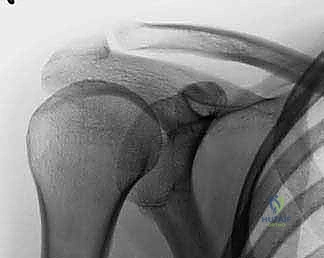
Fat Embolism Syndrome FES
After most fractures some fat is released into the circulation and causes no problem. In FES however, the situation is different, and this may be related to the quantity of fat involved. The presence of fat in the pulmonar
This image illustrates aspects related to the systemic response or a specific complication, often reflecting pulmonary involvement in major trauma.
FES is a serious, potentially fatal complication, most commonly associated with fractures of long bones (femur, tibia), pelvis, and multiple fractures. It results from the release of fat globules into the bloodstream, leading to mechanical obstruction and biochemical injury, primarily in the lungs and brain.
* Incidence: Clinical FES 1-10% of long bone fractures; subclinical fat emboli are common.
* Pathophysiology: Two main theories:
1. Mechanical Theory: Fat globules from bone marrow enter venous circulation, travel to the pulmonary capillaries, causing mechanical obstruction and hypoxia.
2. Biochemical Theory: Circulating free fatty acids and inflammatory mediators released from fat globules cause direct endothelial damage, leading to inflammation and increased capillary permeability in the lungs and brain.
* Clinical Presentation: The classic triad (Gurd and Wilson criteria) includes respiratory insufficiency (tachypnea, dyspnea, hypoxemia), cerebral dysfunction (confusion, delirium, seizures, coma), and petechial rash (over the neck, chest, axillae). Symptoms typically appear 24-72 hours post-injury.
* Salvage Strategies: Primarily supportive. Prevention includes early fracture stabilization (especially for long bone fractures, although intramedullary reaming may transiently increase embolic load), careful patient handling, and adequate fluid resuscitation. Once FES develops, management involves optimizing oxygenation and ventilation (mechanical ventilation if needed), maintaining hemodynamic stability, and general supportive care. Steroids are controversial but sometimes used.
Local Complications
Local complications directly affect the injured limb and surrounding tissues.
Nonunion and Delayed Union
Failure of a fracture to heal within the expected timeframe (delayed union) or complete cessation of healing (nonunion) is a significant morbidity.
* Incidence: Varies by bone and fracture pattern (e.g., tibial shaft: 5-10%, scaphoid: 5-15%). Increased risk with open fractures, severe soft tissue injury, smoking, diabetes, and infection.
* Salvage Strategies: Address underlying factors (e.g., infection, inadequate stability, poor biology). Surgical intervention often involves revision fixation (e.g., stiffer plates, larger nails), bone grafting (autograft, allograft), or biological adjuncts (e.g., bone morphogenetic proteins, platelet-rich plasma, pulsed electromagnetic fields).
Malunion
Healing of a fracture in an unacceptable anatomical position (e.g., significant angulation, rotation, or shortening).
* Incidence: Varies significantly based on fracture location and initial treatment.
* Salvage Strategies: May require corrective osteotomy if symptomatic or functionally limiting. Decision to intervene is based on patient age, functional demands, degree of deformity, and location.
Compartment Syndrome
Elevated pressure within a confined osteofascial compartment, leading to ischemia and potential necrosis of muscle and nerve tissue. Most common in the lower leg and forearm.
* Incidence: 1-9% of long bone fractures, higher in tibia and forearm.
* Salvage Strategies: Surgical fasciotomy is the only definitive treatment, performed urgently upon diagnosis (clinical suspicion, compartment pressure measurement). Delayed diagnosis leads to irreversible muscle damage and functional deficits (Volkmann's contracture).
Nerve and Vascular Injury
Direct injury to nerves or blood vessels can occur at the time of trauma or iatrogenically during surgery.
* Incidence: Variable, e.g., common peroneal nerve injury in fibular neck fractures (10-20%), brachial artery injury in supracondylar humerus fractures (5-10%).
* Salvage Strategies: Immediate assessment and documentation. Vascular injuries require urgent surgical repair or reconstruction. Nerve injuries may be observed for recovery (neuropraxia), repaired (neurorrhaphy, nerve graft), or managed with tendon transfers for long-term deficits.
Post-Traumatic Arthritis
Degenerative joint disease developing after intra-articular fractures, even with anatomical reduction, due to cartilage damage and altered joint mechanics.
* Incidence: High in displaced intra-articular fractures (e.g., tibial plateau, pilon, acetabular).
* Salvage Strategies: Early anatomical reduction and stable fixation are paramount. Symptomatic arthritis may require activity modification, bracing, intra-articular injections, or ultimately arthroplasty (joint replacement) or arthrodesis (fusion).
Heterotopic Ossification HO
Formation of mature lamellar bone in non-osseous soft tissues, typically muscles and ligaments around major joints, particularly after high-energy trauma, head injury, or burns.
* Incidence: Varies widely; common around the hip and elbow after trauma.
* Salvage Strategies: Prevention strategies include NSAIDs (indomethacin), radiation therapy, or early surgical excision once mature for symptomatic HO causing functional limitation.
Complex Regional Pain Syndrome CRPS
A chronic pain condition characterized by severe pain, swelling, autonomic dysfunction, and trophic changes, often disproportionate to the initial injury.
* Incidence: 2-5% of fractures, higher in distal radius fractures.
* Salvage Strategies: Early diagnosis and multidisciplinary management are crucial, involving physical therapy, pain management (nerve blocks, medications like gabapentinoids, tricyclic antidepressants), and psychological support.
Complications and Management Table
| Complication | Incidence (Approx.) | Salvage Strategies |
|---|---|---|
| Systemic Complications | ||
| Hemorrhage/Hypovolemic Shock | Pelvic: high; Femoral: 1-2 units | Aggressive fluid/blood resuscitation, TXA, hemorrhage control (fixation, embolization, packing). |
| Infection (Open Fractures) | Type I: 0-2%; Type III: 10-50% | Urgent debridement, copious irrigation, systemic antibiotics, tetanus, stable wound coverage, appropriate fixation. |
| Metabolic Derangements | High in severe trauma | Meticulous fluid/electrolyte management, nutritional support, glycemic control. |
| ARDS | 10-20% severe trauma | Supportive care (mechanical ventilation, low tidal volumes, PEEP, prone positioning), ECMO. Early definitive fracture fixation. |
| Fat Embolism Syndrome (FES) | Clinical 1-10% of long bone fractures | Supportive care (optimize oxygenation, ventilation, hemodynamics). Prevention: early fracture stabilization, careful handling. |
| Local Complications | ||
| Nonunion/Delayed Union | Tibia: 5-10%; Scaphoid: 5-15% | Address underlying factors (infection, stability, biology). Revision fixation, bone grafting, biological adjuncts. |
| Malunion | Highly variable by site | Corrective osteotomy if symptomatic/functional limitation. |
| Compartment Syndrome | 1-9% long bone fractures | Urgent fasciotomy. |
| Nerve Injury | Variable (e.g., radial nerve 5-10% humeral shaft) | Observation, surgical repair/graft, tendon transfers. |
| Vascular Injury | Variable | Urgent surgical repair/reconstruction. |
| Post-Traumatic Arthritis | High in displaced intra-articular fractures | Anatomical reduction, stable fixation. Activity modification, injections, arthroplasty, arthrodesis. |
| Heterotopic Ossification (HO) | Variable (high in TBI, burns, severe trauma) | Prophylaxis (NSAIDs, radiation). Surgical excision for symptomatic HO. |
| Complex Regional Pain Syndrome | 2-5% of fractures (higher in distal radius) | Multidisciplinary approach: PT, pain management (nerve blocks, meds), psychological support. |
Post Operative Rehabilitation Protocols
Postoperative rehabilitation is an integral component of comprehensive fracture management, aiming to restore strength, range of motion, and function while protecting the surgical repair. Protocols are tailored to the specific fracture, fixation stability, patient's bone quality, and overall health status.
Initial Phase Protection and Edema Control
Immediately post-surgery, the focus is on pain management, edema control, and wound care. Immobilization (splint, brace) is typically used to protect the repair. Early, gentle range of motion (ROM) is often initiated for adjacent joints not directly involved in the fracture or for stable intra-articular repairs (e.g., controlled passive ROM for stable elbow fractures). Weight-bearing status is critical and often progressive:
* Non-weight bearing (NWB): For unstable fractures, tenuous fixation, or significant soft tissue injury.
* Touch-down weight bearing (TDWB) / Partial weight bearing (PWB): Gradual loading based on fracture healing progression.
* Weight bearing as tolerated (WBAT): For stable fractures, good bone quality, or certain intramedullary nails.
Deep vein thrombosis (DVT) prophylaxis is crucial throughout this phase, typically with pharmacological agents (e.g., low molecular weight heparin) and mechanical devices (e.g., pneumatic compression stockings).
Intermediate Phase Controlled Motion and Strengthening
As pain subsides and radiographic signs of healing emerge (callus formation), rehabilitation progresses to controlled active ROM and early strengthening exercises. The goal is to gradually increase joint mobility and muscle strength without jeopardizing fracture healing. Isometric exercises are often initiated first, followed by light resistance training. Close communication between the surgeon and physical therapist is essential to ensure progression is appropriate and safe. Education on activity modification and avoidance of high-impact activities is reinforced.
Advanced Phase Progressive Loading and Functional Integration
Once fracture union is confirmed radiographically and clinically, the focus shifts to restoring full strength, endurance, and functional capacity. This phase involves progressive resistance training, proprioceptive exercises, and sport-specific or work-specific activities. The aim is to regain pre-injury levels of function and prevent long-term complications such as stiffness, weakness, or deconditioning. This often requires several months of dedicated therapy and patient commitment. For complex injuries or multi-trauma patients, a multidisciplinary team involving occupational therapists, pain specialists, and psychologists may be necessary to address all aspects of recovery.
Summary of Key Literature and Guidelines
The field of orthopedic trauma is continuously evolving, driven by clinical research and technological advancements. Several key concepts and guidelines underpin contemporary fracture management.
Damage Control Orthopedics DCO
The concept of DCO has revolutionized the management of critically ill polytrauma patients. Pioneered by Giannoudis and others, DCO advocates for initial temporary stabilization of major fractures (e.g., external fixation) in physiologically unstable patients, followed by definitive fixation once the patient is resuscitated and medically optimized. This approach minimizes the "second hit" of prolonged surgery in unstable patients, reducing systemic inflammatory burden, and ultimately improving survival and decreasing rates of ARDS and MODS.
Early Appropriate Care EAC
Building upon DCO, EAC emphasizes the importance of performing definitive fracture fixation within a specific physiological window (typically 24-72 hours post-injury) for stable polytrauma patients. This strategy balances the benefits of early stabilization (pain control, easier nursing, reduced inflammatory mediator release) against the risks of premature aggressive surgery in patients who are not yet fully resuscitated.
Biologic Osteosynthesis and MIPO
The shift towards biologic osteosynthesis, emphasizing preservation of the soft tissue envelope and periosteal blood supply, has been a major theme. Minimally Invasive Plate Osteosynthesis (MIPO) techniques, popularized by the AO Foundation, exemplify this philosophy. By utilizing indirect reduction and percutaneous plating, MIPO aims to achieve relative stability for secondary bone healing while minimizing additional soft tissue trauma, thus reducing rates of infection, nonunion, and enhancing healing.
Professional Guidelines
Numerous professional organizations, such as the American Academy of Orthopaedic Surgeons (AAOS), the Orthopaedic Trauma Association (OTA), and the AO Foundation, regularly publish evidence-based guidelines and best practice recommendations for specific fracture types and complications. These guidelines inform clinical practice, standardize care, and provide frameworks for quality improvement. For instance, guidelines on antibiotic prophylaxis for open fractures, DVT prevention, and management of nonunions are critical for reducing complication rates. Ongoing research continues to refine our understanding of optimal fracture management, from implant design to biological augmentation strategies, further emphasizing the academic orthopedic surgeon's role in translating these advancements into improved patient outcomes. Continuous engagement with peer-reviewed literature and participation in educational forums are imperative for staying abreast of these developments and delivering the highest standard of care.
