
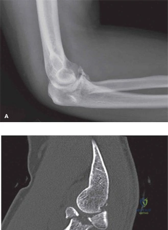
A 35-year-old patient presents to the Emergency Department following a fall onto an outstretched hand. The elbow is dislocated and visibly deformed. After immediate reduction, the elbow is noted to be grossly unstable in flexion and extension. What is your initial diagnostic workup, and what specific clinical entity must you exclude?
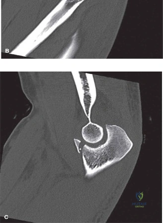
Candidate: I would examine the patient's neurovascular status, then order AP/Lateral X-rays. Since it is unstable, I would get a CT scan to look at the radial head and coronoid fractures. I am concerned about a "Terrible Triad" injury, which includes radial head, coronoid, and LUCL injury.
Failing to emphasize the "neurovascular" assessment prior to any imaging. A poor candidate often describes the imaging but misses the clinical necessity of documenting the status of the ulnar and median nerves, as well as checking for compartment syndrome in a high-energy injury.
Start with a structured ABCDE approach: Assess neurovascular status (median/ulnar nerves and perfusion) immediately post-reduction. Obtain plain radiographs (AP/Lateral) and, crucially, a high-resolution CT scan with 3D reconstructions to assess the coronoid process morphology (O'Driscoll classification) and the degree of radial head comminution. The diagnosis of "Terrible Triad" (Radial head fracture, Coronoid fracture, LUCL avulsion) must be presumed until proven otherwise, as these injuries are pathognomonic for gross posterolateral rotatory instability (PLRI).
You have decided to proceed to surgery. You are in the theatre. Describe your planned surgical approach and the sequence of your reconstruction for this Terrible Triad injury.
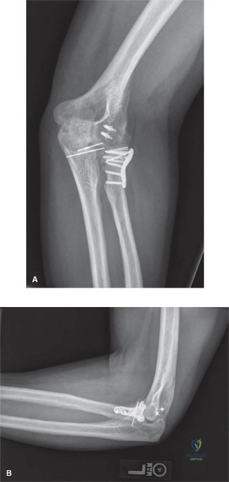
Candidate: I would use a posterolateral approach, specifically the Kocher interval between the anconeus and the ECU. I would fix the radial head first, then the coronoid, and finally repair the LUCL.
Getting the surgical sequence wrong. Fixing the radial head first is a common error. If you fix the radial head first, you may lose the ability to easily reduce the coronoid, which is the primary anterior buttress. Stability is restored from inside out.
The approach is a standard posterolateral approach (Kocher interval). The sequential reconstruction is: 1) Coronoid process (primary stabilizer), 2) Radial head (restore length and radio-capitellar stability via ORIF or arthroplasty), and 3) LUCL repair/reconstruction (to restore PLRI stability). This "inside-out" sequence ensures the foundational bony constraints are secured before ligamentous repair.
Post-operatively, the patient is at the 4-week mark. They are struggling with range of motion and you are concerned about potential complications. What are the key complications you must monitor for, and how would you manage the most common one?
Candidate: The biggest risk is elbow stiffness. I would also watch for heterotopic ossification, persistent instability, and ulnar nerve issues. For stiffness, I would advise physical therapy and perhaps manipulation under anesthesia.
Failing to mention the timeline for surgical interventions like arthrolysis. A candidate should know that we do not typically perform surgical releases for stiffness until at least 6-12 months post-op to allow for maturation.
The most common complication is elbow stiffness/loss of range of motion. Initial management involves aggressive, supervised physical therapy and dynamic splinting. If stiffness persists and creates functional limitation after 6-12 months, surgical arthrolysis (open or arthroscopic) is the gold standard. I must also monitor for Heterotopic Ossification (HO)—potentially prophylaxing with Indomethacin—and watch for ulnar neuropathy, which may require decompression if symptoms are progressive.
Detailed Chapters & Topics
Dive deeper into specialized chapters regarding terrible-triad-injury