Coronoid Fractures: Comprehensive Guide to Anatomy, Biomechanics, & Elbow Instability
Key Takeaway
Coronoid fractures are critical injuries to the ulna's coronoid process, often overlooked but vital for elbow stability. They prevent posterior displacement and resist varus/valgus stress, especially via the anteromedial facet and MCL attachment. Their involvement in complex injury patterns like Terrible Triads necessitates accurate diagnosis and treatment to prevent chronic instability.
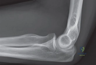
You are presented with a 35-year-old patient who sustained an elbow dislocation following a fall from a height. The patient was reduced in the A&E department. You are reviewing the patient in the clinic. Please evaluate the radiograph provided.
Candidate: The radiograph shows a reduced elbow with a fracture of the coronoid process. It appears to be a Regan-Morrey type 2 fracture. I would obtain a CT scan to fully characterize the fracture, specifically looking for anteromedial facet involvement, and assess for associated injuries like a radial head fracture or ligamentous instability.
Focusing only on the Regan-Morrey classification. This classification is size-dependent and ignores the critical biomechanical importance of the anteromedial facet. Failing to mention the need for a CT scan in all coronoid fractures is a major oversight, as plain radiographs frequently miss the extent of comminution or the specific involvement of the sublime tubercle.
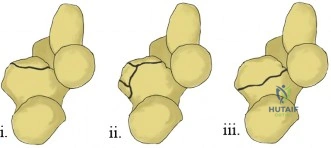
The candidate identifies the fracture and immediately emphasizes the need for high-resolution CT imaging. They classify the fracture using the O'Driscoll system rather than just Regan-Morrey, noting the risk of Varus Posteromedial Rotatory Instability (VPMRI). They explain that the coronoid is a primary anterior buttress and that the sublime tubercle is the site of the MCL attachment, necessitating a high index of suspicion for associated 'Terrible Triad' or trans-olecranon injuries.
Based on your assessment of the CT scan below, this is an O'Driscoll Type 2 anteromedial facet fracture associated with a radial head fracture. What is your surgical strategy for this "Terrible Triad" presentation?
Candidate: The strategy follows the "inside-out" approach. First, I would address the coronoid via a medial or anterior approach to stabilize the anteromedial facet. Then, I would address the radial head fracture—either ORIF or replacement if comminuted—and finally repair the lateral collateral ligament (LCL) complex. Intraoperative fluoroscopy is essential to ensure a concentric reduction through a functional arc of motion.
Starting with the radial head or the lateral side. In a terrible triad, stability is often contingent upon the coronoid buttress. Failing to mention the systematic "Horii circle" approach or neglecting to explicitly state that the goal is a stable concentric reduction through the range of motion before closure.
The candidate outlines a structured, step-by-step approach: 1) Coronoid fixation (to restore the anterior buttress), 2) Radial head reconstruction (to restore lateral stability), and 3) LCL repair. They specify using either a suture lasso/anchor for small fragments or a buttress plate for larger anteromedial facets. They highlight the importance of ulnar nerve protection during the medial approach and confirm that final stability must be tested via fluoroscopy in the supine position.
