Terrible Triad Injury of the Elbow: A Detailed Clinical Case Study & Diagnostic Imaging Analysis
Key Takeaway
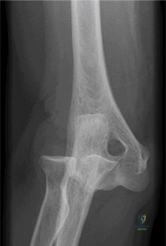
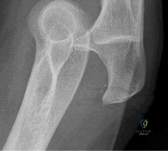
A terrible triad elbow injury involves posterior elbow dislocation, radial head fracture, and coronoid process fracture, typically from a FOOSH mechanism. Diagnosis relies on clinical examination revealing deformity and pain, confirmed by X-rays showing dislocation and fractures, followed by CT for detailed surgical planning and assessment of fracture morphology.
A 44-year-old male presents with a high-energy FOOSH injury. Clinical assessment shows gross deformity and skin tension over the olecranon. Describe your immediate systematic approach to this patient in the Emergency Department and identify the key clinical classification of this injury.
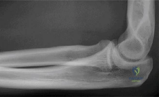
Candidate: I would start with ATLS primary survey to ensure hemodynamic stability. Locally, I need to assess the neurovascular status, specifically the ulnar and median nerves, and the brachial artery. I would assess the soft tissue using Tscherne classification due to the tension over the olecranon. Urgent reduction is required. Based on the radiographs showing posterior dislocation with radial head and coronoid fractures, this is a "Terrible Triad" injury of the elbow.
Failing to emphasize the "urgent" nature of the reduction due to skin tenting. Candidates often forget to document the pre-reduction neurovascular status, which is medicolegally catastrophic. Some also fail to link the "Horii circle" concept to explain why this injury pattern occurs.
A structured response: 1. ATLS/Systemic stability; 2. Neurovascular integrity (documenting ulnar/median/AIN/radial nerve and pulse); 3. Soft tissue assessment (Tscherne grade) acknowledging the need for urgent reduction to salvage the envelope; 4. Radiographic diagnosis ("Terrible Triad") and biomechanical justification (O’Driscoll’s Horii circle: lateral-to-medial soft tissue disruption plus loss of osseous buttresses).
The patient has been reduced. The CT scan confirms a comminuted radial head fracture and a coronoid tip fracture. Explain your decision-making regarding the radial head: Why might you choose arthroplasty over ORIF in this specific scenario?
Candidate: The radial head is critical for valgus and longitudinal stability, especially in a Triad injury where the MCL is often compromised. If the fracture is highly comminuted (Mason III), ORIF is prone to non-union or post-traumatic arthritis. Arthroplasty (RHA) provides a reliable mechanical block to posterior subluxation and restores joint congruity without the risk of hardware prominence or osteonecrosis.
Suggesting that "radial head excision" is an option. It is absolutely contraindicated in a Terrible Triad injury due to resulting proximal migration of the radius and severe valgus/longitudinal instability.
Highlight the role of the radial head as a "secondary stabilizer" that becomes a "primary stabilizer" once the MCL is disrupted. Emphasize that Mason III comminution (>3 fragments) precludes stable ORIF. Mention the importance of avoiding "overstuffing" the joint with the prosthesis, which leads to capitellar cartilage wear and stiffness.
During surgery, you are fixing the coronoid process. What is the rationale for the "suture lasso" technique, and how do you ensure the stability of the elbow after your reconstruction?
Candidate: The coronoid is the anterior buttress against posterior subluxation. When the fracture is small or comminuted, screw fixation is difficult. The suture lasso technique secures the anterior capsule to the ulnar shaft, restoring the anterior soft-tissue restraint. After internal fixation, I perform the "Hang Test"—extending the arm under fluoroscopy. If it remains reduced, no further steps are needed.
Forgetting to mention the specific importance of the "anterior capsule" as a stabilizer, or failing to acknowledge that if the elbow remains unstable after RHA and coronoid repair, a hinged external fixator is the next line of defense.
Structure the answer using the Pugh protocol: (1) Anterior buttress (coronoid/capsule), (2) Radial head (RHA), (3) Lateral tension band (LUCL repair). Explicitly mention the "Hang Test" as the intraoperative gold standard for assessing stability in the supine, anesthetized patient.
