Mastering Arthrodesis of the First MTP Joint: Key Insights
Key Takeaway
Looking for accurate information on Mastering Arthrodesis of the First MTP Joint: Key Insights? Hallux rigidus is painful degenerative arthritis of the first metatarsophalangeal (MTP) joint, marked by restricted motion and bone formation. Initially, it causes pain and swelling, progressing to bony osteophytes and potential bony ankylosis. For advanced cases of hallux rigidus, **arthrodesis of the first** MTP joint is a surgical treatment that permanently fuses the joint, effectively eliminating motion to relieve severe pain.
Introduction and Epidemiology
The term hallux rigidus refers to a painful condition of the first metatarsophalangeal joint of the great toe that is characterized by restricted motion, mainly dorsiflexion, and periarticular bone formation. The basic pathologic entity is that of degenerative arthritis. Initially, hallux rigidus is characterized by pain, swelling, and metatarsophalangeal joint synovitis. As the degenerative process proceeds, proliferation of bony osteophytes on the dorsal and dorsolateral aspect of the first metatarsal head develop. With advanced disease, near-complete bony ankylosis may occur.
The cause of hallux rigidus has not been definitively established, but joint trauma is frequently cited as a major predisposing factor. This may occur as a single episode, such as an intra-articular fracture, as a crush injury, or secondary to repetitive microtrauma. In a patient who sustains an acute injury to the metatarsophalangeal joint, forced hyperextension or plantarflexion may lead to an acute chondral or osteochondral injury. The documented factors associated with the cause of hallux rigidus include a flat or chevron-shaped metatarsal articular surface, bilaterality in those with a positive family history, and female gender. Metatarsus primus elevatus typically is a secondary phenomenon related to the severity of the disease and restricted joint motion, rather than a primary cause of hallux rigidus.
A patient with hallux rigidus typically complains of stiffness with ambulation and pain localized to the dorsal aspect of the first metatarsophalangeal joint that is aggravated by walking, especially during the toe-off phase of the gait cycle. Patients tend to ambulate with an inverted foot posture to prevent stress on the first ray. With time and further osteophyte formation, increased bulk around the joint periphery can lead to substantial discomfort with constricting footwear. More than 80% of patients, if followed long enough, will develop bilateral symptoms. Ninety-five percent of patients with bilateral symptoms have a positive family history.

Figure: Radiographic and clinical progression of osteophyte formation in hallux rigidus.

Figure: Metatarsus primus elevatus, often a secondary phenomenon in advanced disease.

Figure: Clinical manifestation of advanced hallux rigidus with dorsal prominence.
Surgical Anatomy and Biomechanics
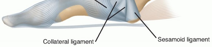
The round, cartilage-covered first metatarsal head articulates with the somewhat smaller, concave base of the proximal phalanx. Articulating on the plantar surface of the metatarsal head are the two sesamoids, which are contained in the tendon of the flexor hallucis brevis. Distally, the two sesamoids are attached by the plantar plate to the base of the proximal phalanx. The sesamoids are connected by the intersesamoidal ligament and protect, on their plantar surface, the tendon of the flexor hallucis longus within its tendon sheath.
Dorsally, the extensor hallucis longus is anchored medially and laterally by the dorsal capsule and metatarsophalangeal joint hood ligaments. The tendons of the abductor and adductor hallucis pass medially and laterally but much closer to the plantar surface of the joint.

Figure: Axial drawing of the first MTP joint highlighting the sesamoid complex and capsular attachments.

Figure: Lateral drawing of the first MTP joint.
Biomechanical Considerations
The first metatarsophalangeal joint plays a critical role in the windlass mechanism described by Hicks. During the propulsive phase of gait, dorsiflexion of the hallux tightens the plantar fascia, elevating the medial longitudinal arch and rigidifying the midfoot for efficient load transfer. In hallux rigidus, the loss of dorsiflexion disrupts this mechanism, leading to compensatory kinematics such as premature heel lift, decreased stride length, and lateral weight transfer.
Arthrodesis of the first metatarsophalangeal joint aims to restore a stable medial column while eliminating pain. The biomechanical success of the procedure relies heavily on the precise positioning of the fused joint. Excessive dorsiflexion leads to dorsal shoe wear and transfer metatarsalgia, while insufficient dorsiflexion impairs rollover during toe-off and increases stress on the interphalangeal joint.

Figure: Deep anatomical dissection of the first ray.

Figure: Biomechanical axes of the first metatarsophalangeal joint.

Figure: Plantar plate and sesamoid complex.
Indications and Contraindications
Arthrodesis of the first metatarsophalangeal joint remains the gold standard for end-stage hallux rigidus (Coughlin and Shurnas Grade 3 and 4). It provides reliable pain relief, restores medial column stability, and yields high patient satisfaction rates. It is also indicated for severe hallux valgus deformities, rheumatoid arthritis, neuromuscular disorders, and as a salvage procedure for failed prior forefoot surgeries, such as failed Keller arthroplasty or failed silastic implants.
Contraindications are primarily related to general surgical risks, active infection, and severe vascular compromise. Relative contraindications include pre-existing severe osteoarthritis of the hallux interphalangeal joint, as arthrodesis of the metatarsophalangeal joint will inherently increase the mechanical demands placed upon the interphalangeal articulation.
| Indication Category | Specific Conditions |
|---|---|
| Operative Indications | Coughlin/Shurnas Grade 3 or 4 Hallux Rigidus |
| Severe Hallux Valgus (IM angle > 20 degrees) | |
| Rheumatoid Arthritis with severe forefoot deformity | |
| Salvage of failed Keller arthroplasty | |
| Salvage of failed first MTP joint arthroplasty / implant | |
| Neuromuscular instability (e.g., Charcot-Marie-Tooth) | |
| Non-Operative Indications | Coughlin/Shurnas Grade 1 or 2 Hallux Rigidus |
| High surgical risk / severe medical comorbidities | |
| Patient preference for conservative management | |
| Absolute Contraindications | Active local or systemic infection |
| Severe peripheral arterial disease (inadequate perfusion) | |
| Relative Contraindications | Severe, symptomatic hallux interphalangeal joint arthritis |
| Non-ambulatory status |

Figure: Preoperative radiograph demonstrating end-stage hallux rigidus suitable for arthrodesis.

Figure: Severe hallux valgus deformity, an excellent indication for MTP arthrodesis.

Figure: Rheumatoid arthritis affecting the first ray.
Pre Operative Planning and Patient Positioning
Comprehensive clinical and radiographic evaluation is mandatory. The interdigital spaces should be palpated for any tenderness, and a Mulder test should be performed to rule out concomitant interdigital neuroma, most commonly located in the second and third interspaces. The patient should be examined while standing to evaluate for the presence of hammer or mallet toe deformities, as well as to assess the overall alignment of the foot and ankle.
Radiographic Evaluation
Standard weight-bearing anteroposterior, lateral, and sesamoid axial radiographs of the foot are required. The surgeon must evaluate the degree of joint space narrowing, the size and location of osteophytes, the presence of subchondral cysts, and the overall bone stock. In cases of revision surgery, assessment of prior hardware and bone loss is critical. The interphalangeal joint must be scrutinized for pre-existing arthritic changes.
Patient Positioning and Anesthesia
The procedure is typically performed under regional anesthesia (ankle block or popliteal block) combined with intravenous sedation, or under general anesthesia depending on patient factors and surgeon preference. The patient is positioned supine on the operating table. A small bump may be placed under the ipsilateral hip to internally rotate the leg to a neutral position, ensuring the patella is pointing directly toward the ceiling. A calf or thigh tourniquet is applied to provide a bloodless surgical field.

Figure: Weight-bearing AP radiograph used for preoperative templating.

Figure: Weight-bearing lateral radiograph assessing bone stock and elevation.

Figure: Supine positioning with appropriate draping and tourniquet placement.
Detailed Surgical Approach and Technique
The surgical technique for first metatarsophalangeal joint arthrodesis requires meticulous soft tissue handling, precise joint preparation, accurate positioning, and rigid internal fixation.
Incision and Soft Tissue Dissection
A dorsal longitudinal incision is typically utilized, centered over the first metatarsophalangeal joint. The incision extends from the mid-shaft of the first metatarsal to the mid-shaft of the proximal phalanx. The internervous plane lies between the medial dorsal cutaneous nerve (a branch of the superficial peroneal nerve) and the terminal branches of the deep peroneal nerve. These sensory branches must be carefully identified and retracted using blunt dissection to prevent postoperative neuromas.
The extensor hallucis longus tendon is identified and mobilized. It can be retracted either medially or laterally, though lateral retraction is most common to expose the dorsal capsule. A longitudinal capsulotomy is performed, and full-thickness flaps are elevated medially and laterally to expose the metatarsal head and the base of the proximal phalanx. Collateral ligaments are released to allow for adequate plantarflexion of the proximal phalanx, providing comprehensive exposure of the articular surfaces.

Figure: Dorsal longitudinal incision over the first MTP joint.

Figure: Identification and retraction of the extensor hallucis longus tendon.

Figure: Longitudinal capsulotomy exposing the degenerative joint.
Joint Preparation
Extensive dorsal, medial, and lateral osteophytes are excised using a rongeur or an oscillating saw. This not only improves exposure but also prevents dorsal impingement postoperatively.
Preparation of the articular surfaces can be performed using either flat cuts or a cup-and-cone reaming technique. The cup-and-cone technique is widely favored because it preserves bone stock, allows for infinite multiplanar adjustment of the fusion angle prior to fixation, and provides excellent cancellous bone contact.
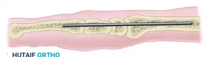
A guidewire is driven centrally into the metatarsal head, and the convex reamer is used to remove the remaining cartilage and subchondral bone until bleeding cancellous bone is exposed. A corresponding concave reamer is then used on the base of the proximal phalanx. If severe deformity or bone loss is present, flat cuts using an oscillating saw may be necessary, though this shortens the first ray and reduces the surface area for healing.

Figure: Excision of the dorsal osteophyte using a rongeur.

Figure: Central placement of the guidewire in the metatarsal head.

Figure: Application of the convex reamer to the metatarsal head.

Figure: Preparation of the proximal phalanx base.

Figure: Bleeding cancellous bone surfaces ready for apposition.
Positioning the Arthrodesis
Positioning is the most critical step of the procedure. The ideal position for the fused first metatarsophalangeal joint is:
1. Valgus: 10 to 15 degrees of valgus relative to the first metatarsal shaft, allowing the hallux to rest parallel to the second toe without impingement.
2. Dorsiflexion: 10 to 15 degrees of dorsiflexion relative to the floor, or approximately 20 to 25 degrees relative to the longitudinal axis of the first metatarsal. This allows for clearance of the toe during the swing phase and appropriate loading during toe-off.
3. Rotation: Neutral rotation, ensuring the toenail faces directly dorsal.
Intraoperative fluoroscopy and clinical simulation (loading the foot with a flat plate to simulate weight-bearing) are utilized to confirm optimal alignment.

Figure: Clinical assessment of the hallux position using a simulated weight-bearing plate.

Figure: Intraoperative fluoroscopy confirming alignment and provisional fixation.
Internal Fixation
Once the optimal position is achieved, provisional fixation is obtained with smooth Kirschner wires. Multiple fixation constructs are biomechanically sound and clinically utilized.
The most common contemporary construct is a low-profile dorsal titanium plate combined with an independent interfragmentary lag screw. The lag screw is typically placed from the medial aspect of the metatarsal head, directing distally and laterally into the proximal phalanx, providing robust compression across the arthrodesis site. Following lag screw insertion, a pre-contoured dorsal plate is applied. Locking screws are placed into the metatarsal and phalanx to provide a neutralization construct that resists bending and torsional forces.
Alternative methods include crossed interfragmentary lag screws (which require excellent bone stock and precise technique), or intramedullary memory-metal staples.

Figure: Provisional K-wire fixation maintaining the desired alignment.

Figure: Insertion of the independent interfragmentary lag screw.

Figure: Application of the low-profile dorsal locking plate.

Figure: Final fixation construct demonstrating compression and stability.

Figure: Final intraoperative AP fluoroscopic image.

Figure: Final intraoperative lateral fluoroscopic image.
Closure
The surgical site is irrigated copiously. The capsule and extensor retinaculum are repaired over the hardware using absorbable sutures to provide a soft tissue envelope that minimizes hardware prominence. Subcutaneous tissues and skin are closed in a standard layered fashion. A sterile, bulky compressive dressing is applied.

Figure: Meticulous capsular closure over the dorsal plate.

Figure: Final skin closure.
Complications and Management
While first metatarsophalangeal joint arthrodesis is highly successful, complications can occur. Meticulous surgical technique and adherence to biomechanical principles minimize these risks.
| Complication | Estimated Incidence | Etiology / Presentation | Salvage Strategy |
|---|---|---|---|
| Nonunion (Pseudoarthrosis) | 5% - 10% | Inadequate compression, poor bone stock, smoking, premature weight-bearing. Often asymptomatic. | If symptomatic: Revision arthrodesis with structural bone graft (iliac crest) and robust locking plate fixation. |
| Malunion | 5% - 8% | Incorrect intraoperative positioning. Excessive dorsiflexion causes shoe impingement; excessive plantarflexion causes IP joint arthritis. | Corrective closing or opening wedge osteotomy through the fusion mass. |
| Hardware Prominence | 10% - 15% | Thin dorsal soft tissue envelope over plates/screws. | Hardware removal after radiographic confirmation of solid fusion (typically > 6 months postop). |
| Transfer Metatarsalgia | 5% - 12% | Shortening of the first ray or excessive dorsiflexion of the hallux. | Custom orthotics with metatarsal pads. Rarely, lesser metatarsal shortening osteotomies (Weil osteotomy). |
| IP Joint Degeneration | 10% - 20% | Increased mechanical stress on the IP joint post-fusion. | Conservative management (stiff sole shoe). Rarely requires IP joint arthrodesis. |
| Infection | 1% - 3% | Superficial or deep surgical site infection. | Oral/IV antibiotics, surgical debridement, hardware removal if fusion is solid or if biofilm cannot be eradicated. |

Figure: Radiographic evidence of atrophic nonunion at the arthrodesis site.

Figure: Clinical presentation of a malunion with excessive valgus and pronation.

Figure: Hardware prominence necessitating removal after solid bony union.

Figure: Plantar callosities indicative of transfer metatarsalgia following first ray shortening.
Post Operative Rehabilitation Protocols
Postoperative rehabilitation must balance the need for rigid immobilization to achieve bony union with the patient's functional mobility requirements.
Immediate Postoperative Phase
During the first two weeks, the patient is placed in a rigid postoperative shoe or a controlled ankle motion (CAM) boot. Weight-bearing is restricted to heel-touch or strict heel-weight-bearing. Elevation of the operative extremity is critical to minimize edema and promote wound healing. Sutures are typically removed at 10 to 14 days postoperatively.
Intermediate Phase
From weeks 2 to 6, the patient continues to wear the CAM boot or stiff-soled surgical shoe. Weight-bearing is progressively advanced to full weight-bearing as tolerated, provided the patient maintains a flat-foot or heel-strike gait pattern to avoid overloading the forefoot. Radiographs are obtained at 6 weeks to assess early trabecular bridging across the arthrodesis site.
Late Phase
Between 6 and 12 weeks, upon radiographic confirmation of progressive union, the patient is transitioned into standard, supportive footwear. A shoe with a stiff sole or a slight rocker bottom is highly recommended to decrease stress on the midfoot and the hallux interphalangeal joint. High-impact activities and running are generally restricted until 4 to 6 months postoperatively, when solid bony consolidation is unequivocally evident.

Figure: Typical controlled ankle motion (CAM) boot utilized in the postoperative phase.
Summary of Key Literature and Guidelines
The academic consensus strongly supports first metatarsophalangeal joint arthrodesis as the definitive management for severe hallux rigidus.
Coughlin and Shurnas, in their landmark series, demonstrated that arthrodesis provides reliable, long-lasting pain relief with a union rate exceeding 90%. Their classification system remains the standard for determining operative indications, with Grade 3 (severe loss of motion, extensive osteophytes) and Grade 4 (stiff, painful joint throughout the range of motion) being the primary indications for fusion.
Biomechanical studies comparing fixation techniques have shown that the combination of an interfragmentary lag screw and a dorsal neutralization plate provides superior construct stiffness and load-to-failure characteristics compared to crossed screws or dorsal plates alone. Politi et al. demonstrated that this construct minimizes micromotion at the arthrodesis site, which is critical for primary
Clinical & Radiographic Imaging
You Might Also Like