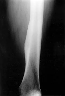
Comprehensive Master Guide · Medically Reviewed

Orthopedic Board Prep: Interactive Viva Exam Practice for Trauma & Surgical Cases

Master your orthopedic board exams with interactive viva practice. Switch between study and exam modes to conquer trauma and surgical cases today!

22 Detailed Chapters
150 min read
Updated: Apr 2026
Dr. Mohammed Hutaif
Medically Reviewed by
Prof. Dr. Mohammed Hutaif
Verified Content Expert Reviewed

Quick Medical Answer

To effectively prepare for your Orthopedic Board Viva, engage with interactive practice questions focusing on trauma and complex surgical cases. Utilize MCQ engines to hone clinical reasoning and adaptive problem-solving skills. Consistent review of evidence-based guidelines, coupled with mock viva sessions, enhances preparedness for the rigorous oral examination.

Practicalities of the oral exam

Comprehensive Exam


00:00

Start Quiz

Question 1

A candidate is preparing for an orthopedic viva focusing on complex trauma cases. Which of the following preparation strategies is LEAST effective for developing the necessary critical thinking and adaptive problem-solving skills crucial for success in an oral examination?


Explanation

While knowing algorithms can be helpful, solely relying on memorized, rigid algorithms without understanding the underlying principles or adaptability to variations is a significant pitfall in a viva. Examiners seek evidence of critical thinking, the ability to justify decisions, and to manage deviations from standard protocols. The other options promote active learning, evidence integration, and flexible problem-solving, which are essential for navigating the dynamic nature of an oral exam.

Question 2

During an orthopedic viva, an examiner asks a candidate to describe the surgical management of a complex pilon fracture. The candidate begins with a well-structured introduction. The examiner then interjects, 'Tell me the two most critical principles guiding your approach to the soft tissues in this scenario.' What is the MOST appropriate immediate response?


Explanation

When an examiner interjects with a specific question, it is crucial to address it directly and concisely before attempting to revert to a broader discussion. Stating the principles (e.g., respect for soft tissues, staged management) immediately demonstrates responsiveness and understanding of the examiner's focus. Briefly elaborating shows depth of knowledge. Asking for clarification can be interpreted as uncertainty, and continuing with the original plan disregards the examiner's direction. Jumping to specific techniques without principles lacks structure.

Question 3

You are presenting a case of a delayed presentation of a scaphoid nonunion in your viva. The examiner queries, 'What is your threshold for offering surgical intervention versus continued conservative management in this specific patient?' Which of the following responses BEST demonstrates a nuanced and patient-centered approach?


Explanation

A nuanced, patient-centered approach to surgical decision-making considers a comprehensive array of factors beyond just imaging or age. Option C encompasses key patient-specific variables (functional demands, occupation, willingness to accept risk), pain, and radiographic evidence, reflecting a holistic understanding of shared decision-making. Options A, B, and E are too rigid and fail to account for the complexity of patient preferences and functional outcomes. Option D, while a reasonable step, doesn't answer the 'your threshold' question and defers responsibility.

Question 4

An examiner asks you about the indications for total hip arthroplasty in a 35-year-old patient. After you list the common indications, the examiner presses, 'What are the two most common reasons for early revision (<5 years) in this specific demographic?' Which pair of reasons is most accurate?


Explanation

For young, active patients undergoing total hip arthroplasty, the most common reasons for early revision (within 5 years) are periprosthetic joint infection (PJI) and instability/dislocation. While aseptic loosening and polyethylene wear are significant long-term failure modes, they typically manifest later. Osteolysis is a long-term consequence of wear. Femoral component fracture, nerve injury, vascular injury, and DVT are less common causes for early revision compared to PJI and dislocation.

Question 5

During a viva, you are asked to describe the approach to a suspected compartment syndrome in the forearm. You correctly outline the clinical assessment and indications for fasciotomy. The examiner then probes, 'What specific pitfalls might you encounter in diagnosing compartment syndrome in an intubated, sedated patient in the ICU?' Which pitfall is MOST critical to emphasize?


Explanation

While all options represent challenges, the absence of the 'pain out of proportion' hallmark symptom, due to sedation and analgesia, is arguably the most critical pitfall. This key diagnostic criterion is negated, requiring a heightened index of suspicion and reliance on other, often subtle, signs and frequent compartment pressure measurements. Difficulty assessing motor/sensory (A) is also true, but pain is often the earliest and most reliable subjective sign. Pressure measurement challenges (C) can be overcome. Visible swelling (D) is often late. SIRS (E) is a systemic issue, not a direct diagnostic pitfall for compartment syndrome itself.

Question 6

When discussing the management of metastatic bone disease in the spine during a viva, the examiner asks, 'Beyond direct oncological treatment, what is your primary goal as an orthopedic surgeon in managing these patients, and how do you achieve it?' Which statement best encapsulates this primary goal and its achievement?


Explanation

The primary goal of orthopedic surgery in metastatic spinal disease is palliative: to improve the patient's quality of life by addressing pain, preventing or treating neurological compromise, and stabilizing the spine. This requires a multidisciplinary approach, carefully balancing the risks and benefits of intervention against the patient's overall prognosis and wishes. Eradication of disease (A) is rarely the primary goal for the orthopedic surgeon in this context. Prolonging survival (B) is usually a systemic oncology goal, not necessarily the orthopedic primary aim at all costs. Preventing all fractures (D) can be overly aggressive. Referring to palliative care (E) without considering surgical interventions that can significantly improve quality of life is inappropriate.

Question 7

A candidate is discussing surgical consent for an elective total knee arthroplasty. The examiner asks, 'How do you ensure truly informed consent, especially regarding less common but significant risks, without unduly alarming the patient?' Which approach is most ethically sound and practically effective?


Explanation

Truly informed consent involves more than just a checklist. It requires a tailored discussion focusing on risks relevant to the specific patient and procedure. Grouping less common but serious risks allows for efficient communication without overwhelming the patient, while still fulfilling ethical and legal obligations. Option A can cause undue anxiety. Option B is inadequate for informed consent. Option D is legally and ethically unsound. Option E is a delegation of a core responsibility and inappropriate.

Question 8

In a trauma scenario, you are asked about managing an open tibia fracture with significant soft tissue injury. The examiner interjects, 'Before you even consider fixation, what is your absolute priority in the immediate management of this limb?'


Explanation

While antibiotics (A) are critical for open fractures and radiographs (D) are necessary, the absolute immediate priority in any limb trauma, especially with significant soft tissue involvement, is to assess for and address neurovascular compromise (e.g., check pulses, sensation, capillary refill) and provide temporary stabilization/splinting. This ensures limb viability and prevents further injury. Debridement (B) is urgent but comes after initial assessment and temporary stabilization. Definitive fixation (E) is much later.

Question 9

You are presenting a case of recurrent shoulder instability in an overhead athlete. The examiner asks, 'What specific factors would lead you to favor a bony reconstructive procedure (e.g., Latarjet) over an arthroscopic soft-tissue repair (e.g., Bankart) in this patient?'


Explanation

Significant glenoid bone loss (typically >20-25% of the inferior glenoid) or an engaging Hill-Sachs lesion (where the humeral head defect engages the anterior glenoid rim) are the primary indications to favor a bony reconstructive procedure like the Latarjet over an arthroscopic soft-tissue repair for recurrent shoulder instability. These factors are associated with higher failure rates after isolated soft tissue repair. Patient preference (A) is secondary to biomechanical stability. A single prior dislocation (C) without bone loss would typically favor soft tissue repair. Age (D) is a risk factor for recurrence, but not a direct indication for bony procedure without bone loss. WOSI scores (E) reflect the severity of instability symptoms but not necessarily the underlying pathology dictating surgical choice.

Question 10

In an oral exam, you are discussing the management of a displaced intra-articular calcaneal fracture. The examiner interrupts, 'Before you outline your surgical approach, how do you manage the critical 'window' period between injury and definitive fixation?'


Explanation

The management of the critical 'window' period for displaced intra-articular calcaneal fractures is crucial due to the significant associated soft tissue swelling. Immediate surgery is generally contraindicated as it drastically increases the risk of wound complications. The most appropriate management involves strict elevation, ice, compression, and non-weight bearing until the soft tissue envelope has recovered sufficiently, typically indicated by the 'wrinkle sign' or resolution of significant edema. This approach optimizes conditions for eventual surgical intervention. An external fixator (C) can be used, but the core principle is still soft tissue management, and it's not the only immediate management. Early mobilization (D) is inappropriate for a displaced fracture. Immediate discharge (E) is irresponsible.

Question 11

You are asked about the differential diagnosis of hip pain in a young adult. After listing common causes, the examiner asks, 'What specific signs or symptoms would raise your suspicion for a rare but critical diagnosis like avascular necrosis (AVN) of the femoral head in this demographic?'


Explanation

Avascular necrosis (AVN) of the femoral head typically presents with insidious onset of deep, aching groin pain, often made worse with weight-bearing, in the absence of a clear acute traumatic event. The presence of risk factors (e.g., steroid use, excessive alcohol, sickle cell disease, lupus, trauma with vascular disruption) significantly increases suspicion. Acute pain with external rotation/shortening (B) is classic for hip fracture/dislocation. Buttock pain radiating down the posterior thigh (C) suggests piriformis syndrome or sacroiliac joint dysfunction. Clicking/catching with FADIR (D) is indicative of femoroacetabular impingement (FAI) or labral tear. Lateral hip pain (E) points to trochanteric bursitis.

Question 12

In a discussion about the management of osteomyelitis, the examiner asks, 'When considering surgical debridement for chronic osteomyelitis, what is the 'surgical goal' and why is it so critical?'


Explanation

The cornerstone of chronic osteomyelitis management is aggressive surgical debridement. The goal is to remove all non-viable, infected bone and soft tissue, foreign bodies (implants if infected), and any sequestra or involucrum, creating a clean, well-vascularized environment. This is critical because necrotic bone acts as a sanctuary for bacteria, impenetrable to systemic antibiotics and host immune defenses. Antibiotic penetration and immune cell access are only effective in viable tissue. Options A, C, D, and E represent incomplete or incorrect surgical goals for chronic osteomyelitis.

Question 13

You are discussing the indications for spinal fusion in degenerative conditions. The examiner asks, 'What is the MOST compelling indication for adding fusion to decompression in a patient with lumbar spinal stenosis?'


Explanation

The most compelling indication for adding spinal fusion to decompression in lumbar stenosis is the presence of pre-existing spinal instability, such as degenerative spondylolisthesis (especially grades I and II) that is symptomatic, or the potential for iatrogenic instability created by a necessary extensive decompression (e.g., bilateral facetectomy, significant removal of stabilizing posterior elements). In these scenarios, decompression alone can worsen instability or lead to future instability, justifying fusion to maintain stability. While the other options can be contributing factors, they are not as direct or compelling indications for fusion as instability.

Question 14

During a viva, the examiner presents a case of a patient with knee pain after a twisting injury. You diagnose an ACL tear. The examiner then asks, 'What are the key patient factors you would consider when deciding between operative (ACL reconstruction) and non-operative management in this patient?'


Explanation

The decision between operative and non-operative management for an ACL tear is highly individualized and multifactorial. Key patient factors include age (younger patients, especially those returning to high-demand sports, tend to have better outcomes with surgery), activity level and functional demands (pivoting/cutting sports highly recommend surgery), presence of concomitant injuries (meniscal tears, collateral ligament injuries often push towards surgery), and the patient's psychological readiness and commitment to rehabilitation. BMI (A) and graft choice (D) are considerations for surgical technique or outcomes but not primary drivers for operative vs. non-operative. Socioeconomic status (C) is a practical consideration, but not a primary clinical indication. Crutch preference (E) is trivial.

Question 15

You are asked about the management of a displaced midshaft clavicle fracture in an adult. The examiner then asks, 'What specific patient or fracture characteristics would lead you to strongly consider surgical fixation over non-operative management?'


Explanation

While many midshaft clavicle fractures heal with non-operative management, specific factors indicate a higher risk of nonunion or poorer functional outcomes, necessitating surgical fixation. These include significant shortening (>2 cm), marked displacement (especially >100% or override), significant comminution, skin tenting (indicating impending open fracture), and neurovascular compromise. These factors often lead to higher rates of nonunion, malunion, and poorer functional results with conservative management. Swelling (A) is common. Age over 60 with low demands (B) might lean towards non-op. Simple transverse fracture (D) is ideal for non-op. Patient preference (E) is important but doesn't override clear surgical indications.

Question 16

In a viva setting, an examiner presents a patient with chronic low back pain and significant leg pain, suspected to be lumbar radiculopathy. You've outlined your diagnostic approach. The examiner asks, 'What is the role of an MRI scan in your initial management plan for this patient, and when would you order it?'


Explanation

For most patients with acute low back pain and radiculopathy, an MRI is not indicated in the initial 4-6 weeks unless 'red flag' symptoms (e.g., cauda equina syndrome, progressive motor weakness, signs of infection or malignancy) are present, or if symptoms are severe and debilitating and persist beyond this period despite appropriate conservative management. Early imaging often reveals asymptomatic disc bulges or herniations, leading to over-investigation and potentially unnecessary interventions. Option A is incorrect due to the 'always' clause and potential for over-diagnosis. Option C delays necessary diagnostics. Options D and E are fundamentally incorrect about MRI's utility and ordering practices.

Question 17

You are describing the technique for a total shoulder arthroplasty. The examiner abruptly asks, 'What is the most common and often devastating complication unique to reverse total shoulder arthroplasty (rTSA) compared to anatomic TSA, and how do you mitigate it?'


Explanation

Scapular notching is a complication unique and relatively common to reverse total shoulder arthroplasty, where the humeral polyethylene liner impinges on the inferior aspect of the scapular neck during adduction and internal rotation, leading to erosion of the bone. It can lead to pain, reduced range of motion, and implant loosening. Mitigation strategies include lateralizing the glenosphere, inferomedial placement of the glenosphere, and achieving appropriate soft tissue tension. Axillary nerve palsy (B) can occur in both, but scapular notching is unique to rTSA. Aseptic loosening (A) is a general arthroplasty complication, not unique. Infection (D) is also a general complication. Rotator cuff failure (E) is the reason for rTSA in many cases, not a unique complication of rTSA in the context of anatomic TSA comparison.

Question 18

In a discussion about post-operative rehabilitation following knee surgery, the examiner asks, 'What is the primary rationale for early weight-bearing and mobilization protocols after many lower extremity orthopedic procedures, especially those involving articular cartilage?'


Explanation

The primary rationale for early weight-bearing and mobilization, particularly after procedures involving articular cartilage (e.g., microfracture, cartilage repair, meniscal repair, even arthroplasty), is to promote chondrocyte health through cyclical loading, enhance synovial fluid circulation for cartilage nutrition, and prevent deleterious effects like joint stiffness, adhesions, and disuse atrophy. While DVT prevention (A) is a benefit, it's not the primary rationale for articular cartilage health. Accelerating bone healing (B) is true for some fractures but not the universal primary reason across all lower extremity procedures involving cartilage. Reducing dependence (D) and pain (E) are secondary benefits.

Question 19

You're asked to discuss a complex case of osteosarcoma in a young patient. The examiner probes, 'Beyond oncologic resection, what is the single most critical consideration for the orthopedic surgeon in the pre-operative planning phase?'


Explanation

The single most critical consideration in pre-operative planning for osteosarcoma is accurate tumor staging and meticulously planning the surgical margins to achieve a wide oncologic resection. This dictates whether limb salvage is feasible and, if so, which reconstructive options are appropriate, always prioritizing oncologic control over other factors. Functional limb preservation (B) and cosmetic preferences (B, E) are important but secondary to achieving adequate oncologic margins. Financial resources (A) are practical but not the 'most critical' surgical consideration. Rehab coordination (D) is important but comes after definitive surgical planning.

Question 20

An examiner asks about the management of adolescent idiopathic scoliosis. They then ask, 'What is the primary indication for surgical correction in adolescent idiopathic scoliosis?'


Explanation

The primary indication for surgical correction of adolescent idiopathic scoliosis is typically curve progression to greater than 40-45 degrees (depending on the specific curve type and surgeon preference), especially in skeletally immature patients. Surgical consideration also involves the potential for future progression, spinal balance, and the degree of cosmetic deformity affecting the patient's quality of life. Curves >20 degrees (A) might warrant bracing, but not necessarily surgery. Cosmetic concerns (C) alone are generally not an indication without significant curve magnitude. Back pain (D) is not typically an indication for surgery in AIS unless associated with significant neurological deficit or instability. Failure of physical therapy (E) does not apply to structural scoliosis.

Question 21

During a viva, you are asked to discuss a patient presenting with an acute cervical radiculopathy. You've outlined your initial non-operative approach. The examiner asks, 'What are the 'red flag' symptoms in cervical radiculopathy that would prompt immediate advanced imaging and potentially urgent surgical referral?'


Explanation

Red flag symptoms in cervical radiculopathy necessitating urgent evaluation include progressive motor weakness (indicating impending neurological compromise), signs of myelopathy (which suggests spinal cord compression, a much more serious condition than radiculopathy), or intractable pain that is severely debilitating and completely refractory to aggressive conservative measures. Persistent neck pain (A), gradual numbness (B), sleep difficulty (D), and unilateral arm pain (E) are common features of radiculopathy, but without progression or myelopathic signs, they typically fall within the scope of initial conservative management.

Question 22

An examiner presents a scenario where you've operated on a complex tibial plateau fracture, and post-operatively, the patient develops increasing pain, swelling, and neurological deficits. You suspect compartment syndrome. What is your MOST immediate and critical action?


Explanation

In a suspected compartment syndrome, time is critical. The MOST immediate and critical action is to measure compartment pressures. If pressures are elevated (typically within 30 mmHg of diastolic blood pressure, or absolute pressure >30-40 mmHg, depending on protocols), emergent fasciotomy is indicated. Delay in diagnosis and treatment can lead to irreversible muscle and nerve damage. While CT (A) might be used in some contexts, it delays definitive diagnosis of compartment syndrome. Analgesia and elevation (B) are contraindicated if compartment syndrome is suspected as they can mask symptoms or worsen perfusion. Vascular consultation (D) might be needed but only after addressing compartment syndrome. Reassurance (E) is dangerous.

Question 23

You are discussing the post-operative management of a patient who underwent Achilles tendon repair. The examiner asks, 'What is the rationale behind early functional rehabilitation (e.g., controlled ankle motion, early weight-bearing) versus traditional prolonged immobilization after Achilles repair?'


Explanation

The rationale for early functional rehabilitation after Achilles tendon repair is multifactorial and evidence-based. It promotes better collagen fiber alignment and maturation, improves tendon strength by stimulating mechanoreceptors, reduces the formation of adhesions that can restrict motion, and facilitates a faster return to activity. Modern protocols show that controlled early motion and weight-bearing, when carefully implemented, do not increase the risk of re-rupture compared to prolonged immobilization, and often lead to superior functional outcomes. While DVT prevention (A) is a benefit, it's not the primary rationale for tendon healing. Option B is incorrect; prolonged immobilization can lead to tendon weakening, but not necessarily higher re-rupture rates if protected. Options D and E are incomplete or incorrect.

Question 24

An examiner asks you to describe your approach to a chronic non-healing ulcer on the foot of a diabetic patient. You outline history, examination, and initial investigations. The examiner then asks, 'What is the most critical first-line investigation to perform in ALL diabetic foot ulcers that will significantly guide further management?'


Explanation

For any chronic non-healing diabetic foot ulcer, assessing peripheral arterial disease (PAD) is paramount as adequate blood supply is essential for wound healing. An Ankle-Brachial Index (ABI) and/or toe pressures are critical first-line investigations. Without adequate perfusion, aggressive wound care, antibiotics, or even surgery will likely fail. While radiographs (A) for osteomyelitis, blood tests (B) for glycemic control and infection, cultures (D) for targeted antibiotics, and EMG (E) for neuropathy are all important, addressing perfusion (C) is often the most critical initial step impacting the entire management pathway.

Question 25

In a viva, you are presenting a case of a femoral shaft fracture. The examiner interjects, 'Describe the principles of damage control orthopedics (DCO) and when it would be specifically indicated for this patient.'


Explanation

Damage control orthopedics (DCO) is a strategy employed in the management of multiply injured patients or those in physiological extremis (e.g., hemorrhagic shock, severe head injury, severe chest injury). Its principle is to provide temporary stabilization of long bone fractures (often with external fixation) to control hemorrhage, pain, and prevent further tissue damage, deferring definitive fixation until the patient's systemic physiological status has stabilized. This avoids a 'second hit' inflammatory response that can worsen outcomes in already compromised patients. Options A, B, D, and E do not accurately describe the core principles or indications of DCO.

Question 26

You are asked about the non-operative management of osteoarthritis of the knee. After outlining initial steps, the examiner asks, 'What is the evidence-based role of intra-articular corticosteroid injections in the long-term management of knee OA?'


Explanation

Intra-articular corticosteroid injections can provide effective short-to-medium term pain relief (typically weeks to a few months) by reducing inflammation in an osteoarthritic joint. They can be a valuable adjunct in a comprehensive management plan, especially to bridge a patient to other interventions or alleviate acute flares. However, they do not alter the underlying disease progression or provide long-term structural modification. Frequent injections carry risks (e.g., infection, cartilage damage, systemic effects), thus judicious use is recommended. Options A, B, D, and E are incorrect or overstate/understate their role.

Question 27

In an orthopedic viva, you are discussing a case of suspected septic arthritis of the knee. You correctly identify the need for aspiration. The examiner then asks, 'What is the MOST critical test to send from the aspirated synovial fluid for rapid diagnosis and initial management guidance?'


Explanation

While all listed tests can be helpful, the Gram stain and urgent cell count with differential are the MOST critical for rapid diagnosis and initial management guidance in suspected septic arthritis. The cell count (specifically polymorphonuclear cell count) helps confirm inflammation and likelihood of infection, while the Gram stain can rapidly identify the causative organism (Gram-positive cocci vs. Gram-negative rods), allowing for initiation of appropriate empiric antibiotic therapy before culture results are available. Cultures (B) are essential for definitive diagnosis and sensitivity, but take longer. Glucose/protein (C) and LDH (D) are less specific. Crystal analysis (E) is important to rule out inflammatory arthropathy, but septic arthritis remains the priority until ruled out.

Question 28

You are asked about the surgical management of carpal tunnel syndrome. The examiner asks, 'What is the primary goal of carpal tunnel release surgery, and how does it achieve this?'


Explanation

The primary goal of carpal tunnel release surgery is to decompress the median nerve within the carpal tunnel. This is achieved by transecting (cutting) the transverse carpal ligament, which forms the roof of the tunnel. This increases the volume of the carpal tunnel, relieving pressure on the median nerve and allowing for restoration of its function and resolution of symptoms (pain, numbness, tingling). Excising the entire ligament (A) is an overstatement. Removing osteophytes/synovium (C) is not the primary goal but can be an adjunct in some cases. Relocating the nerve (D) is not the standard procedure. Neurolysis (E) is a separate procedure, usually only if intrinsic nerve pathology persists or is suspected.

Question 29

In a viva discussing total hip arthroplasty, the examiner asks, 'What is the primary advantage of a direct anterior approach (DAA) compared to posterior or lateral approaches, and what is its main technical challenge?'


Explanation

The primary advantage of the direct anterior approach (DAA) for total hip arthroplasty is its potential for a lower dislocation rate post-operatively due to preservation of the posterior soft tissue structures (capsule, external rotators). It is often associated with less post-operative pain and faster initial rehabilitation and fewer post-operative restrictions. However, its main technical challenges include a steeper learning curve, increased risk of lateral femoral cutaneous nerve (LFCN) injury (resulting in meralgia paresthetica), and potentially more difficult femoral exposure, especially in obese or muscular patients, or those with significant deformity. Higher risk of sciatic nerve injury (C) is generally associated with posterior approaches. Dislocation is a concern for posterior/lateral approaches not DAA. The other options are either less accurate or not the primary advantage/challenge.

Question 30

You are asked to describe the initial management of a suspected acute deep vein thrombosis (DVT) in a post-operative orthopedic patient. The examiner focuses on diagnostics. What is the MOST appropriate first-line diagnostic investigation?


Explanation

The most appropriate first-line diagnostic investigation for suspected acute DVT is a Doppler ultrasound of the affected limb. It is non-invasive, widely available, and highly sensitive and specific for proximal DVT. While a D-dimer test (D) can be used to rule out DVT in low-probability patients, it is not sufficiently specific in post-operative orthopedic patients (who often have elevated D-dimer due to surgery itself) to be the most appropriate first-line diagnostic when DVT is suspected. CTPA (B) is for suspected PE. MRI venography (A) and ascending venography (E) are more invasive or costly and typically reserved for complex cases or when ultrasound is inconclusive.

Question 31

During a viva, an examiner presents a patient with a proximal humerus fracture. You outline your management options. The examiner then asks, 'What are the two most common and distinct complications associated with the use of a hemiarthroplasty for complex proximal humerus fractures?'


Explanation

For hemiarthroplasty in complex proximal humerus fractures, the primary goal is often pain relief, but functional outcomes can be limited. The two most common and distinct complications are malposition or nonunion of the tuberosities, which compromises rotator cuff function and leads to poor active elevation, and progressive glenoid erosion due to articulation of the prosthetic humeral head with the native glenoid cartilage, leading to pain and often requiring revision to rTSA. PJI and DVT (A) are general surgical complications. Aseptic loosening of the glenoid and poly wear (B) are specific to total shoulder arthroplasty. Heterotopic ossification (D) and nerve injury (D) can occur but are less common or less unique to hemiarthroplasty for fracture than the tuberosity and glenoid issues. Stiffness (E) is a common outcome, but tuberosity malposition is the underlying reason, and implant fracture is less common than glenoid erosion or tuberosity issues.

Question 32

You are discussing the non-operative management of a stable intertrochanteric hip fracture in a very frail, non-ambulatory patient. The examiner asks, 'What is the MOST critical aspect of their management plan to prevent significant morbidity and mortality?'


Explanation

For a very frail, non-ambulatory patient with a stable intertrochanteric hip fracture managed non-operatively, the MOST critical aspect is to focus on comfort and preventing the devastating complications of immobility. This includes aggressive pain management to facilitate movement, early mobilization out of bed to a chair (even if not weight-bearing on the limb), meticulous skin care for pressure ulcer prevention, and adequate nutritional support. Strict bed rest (A, E) significantly increases the risk of pneumonia, DVT/PE, pressure ulcers, and functional decline. Spica cast (C) is inappropriate for intertrochanteric fractures in this demographic. Antibiotic prophylaxis (D) is not indicated.

Question 33

In a viva, you are asked about the ethical considerations in treating a Jehovah's Witness patient who requires urgent surgical intervention for a major orthopedic trauma but refuses blood transfusion. How would you proceed?


Explanation

This is a common and critical ethical scenario. The most appropriate approach is to respect the patient's autonomy and religious beliefs while providing the best possible care within those constraints. This involves a thorough discussion with the patient about the risks, exploring all blood conservation strategies, and involving an ethics committee for guidance if the situation allows. Overriding patient wishes (B) is ethically problematic, and refusing to operate (A) or delaying surgery (D) can lead to worse outcomes. Transferring (E) might be an option if specialist facilities exist, but the initial local management should follow option C.

Question 34

You are presenting a case of recurrent patellar dislocation in an adolescent. The examiner asks, 'What specific anatomical or biomechanical factors predispose a patient to recurrent patellar instability, and how do you assess them?'


Explanation

Recurrent patellar instability is typically multifactorial. Key predisposing factors include osseous abnormalities such as trochlear dysplasia (a shallow or flat trochlear groove), patella alta (high-riding patella), and an increased tibial tuberosity-trochlear groove (TT-TG) distance, which indicates lateralization of the patellar tendon insertion. Ligamentous laxity, especially medial patellofemoral ligament (MPFL) insufficiency, is also critical. These are assessed through clinical examination (e.g., patellar apprehension test, J-sign) and advanced imaging (MRI for trochlear morphology and MPFL integrity, CT for accurate TT-TG measurement). Quadriceps weakness (A) and retinacular tightness (D) are usually secondary or less dominant factors. Genu varum (B) is less common, and meniscal tears (E) are not a primary predisposing factor for patellar instability.

Question 35

In a viva, the examiner observes you are using jargon. They interrupt and ask, 'If you were explaining 'avascular necrosis of the femoral head' to an educated but non-medical patient and their family, how would you describe it simply but accurately?'


Explanation

Effective communication with patients involves using clear, understandable language. Option A accurately describes avascular necrosis by explaining the core pathology (interrupted blood supply, bone cell death) and its consequence (bone collapse, arthritis) in simple terms. Options B, C, D, and E are inaccurate or misleading explanations for a non-medical audience, confusing AVN with infection, osteoporosis, osteoarthritis, or autoimmune conditions.

Question 36

You are presenting a case of a femoral neck fracture in an elderly patient. The examiner asks, 'What is the most significant factor influencing the choice between internal fixation (e.g., cannulated screws) and arthroplasty (e.g., hemiarthroplasty or THR) for this fracture?'


Explanation

The most significant factor influencing the treatment choice for femoral neck fractures in the elderly is the patient's pre-injury functional status and cognitive ability, along with fracture displacement and bone quality. Active, cognitively intact patients with non-displaced fractures might be candidates for internal fixation, whereas displaced fractures in less active or cognitively impaired patients often fare better with arthroplasty (hemi or total). BMI (A) and exact location (B) are secondary considerations. Implant availability (D) should not dictate the best medical decision. Osteoporosis (E) is a common comorbidity, influencing bone quality, but not the sole determinant.

Question 37

In a viva, you've explained your reasoning for using a particular surgical approach. The examiner challenges, 'But why would you choose that approach when XYZ approach has a demonstrably lower infection rate in some series?' What is the BEST way to respond to this challenge?


Explanation

When challenged, a confident and knowledgeable candidate acknowledges the validity of the examiner's point (if appropriate) but then provides a reasoned, case-specific justification for their decision. This demonstrates critical thinking, awareness of controversies, and the ability to apply evidence to individual patients. Avoiding defensiveness (B) or immediate retraction (A) is crucial. Changing the topic (D) is a significant viva error. Simply quoting institutional data (E) without explanation is insufficient.

Question 38

You are discussing the management of a stress fracture in a professional athlete. The examiner asks, 'What is the most critical factor to identify and address in a non-healing or recurrent stress fracture in an athlete, beyond just rest and immobilization?'


Explanation

Beyond initial rest and immobilization, the most critical factors for non-healing or recurrent stress fractures, especially in athletes, are underlying physiological and biomechanical imbalances. This includes assessing bone mineral density (for conditions like osteoporosis), nutritional status (e.g., vitamin D, calcium, energy availability in female athlete triad), hormonal balance (e.g., amenorrhea), and meticulous review of training errors (e.g., overtraining, rapid increase in load, inadequate recovery, poor technique). Addressing these systemic and biomechanical factors is crucial for successful long-term management and prevention of recurrence. Other factors are less critical or directly modifiable.

Question 39

During your orthopedic viva, the examiner presents an X-ray of a child with a Salter-Harris Type II physeal fracture of the distal tibia. They ask, 'What are the two MOST important considerations in the management of any physeal fracture in a child?'


Explanation

For any physeal fracture in a child, the two most important considerations are: 1) Preserving the integrity and function of the physis (growth plate) to prevent growth arrest (shortening) or angular deformity, and 2) Achieving an adequate, typically anatomical or near-anatomical reduction to prevent malunion which can also lead to growth disturbances. Overly aggressive fixation (D) can damage the physis. Absolute anatomical reduction (A) is important but less critical than preserving the physis, and immediate weight-bearing is inappropriate. DVT prevention (B) and pain management are general post-op considerations, not specific to physeal fractures. Strict bed rest (E) is often detrimental.

Question 40

You are asked about the approach to patients with a suspected pathological fracture. The examiner then asks, 'What is the most appropriate initial imaging study to help determine the nature and extent of the underlying pathology causing the fracture?'


Explanation

The most appropriate initial imaging study for a suspected pathological fracture is plain radiographs (orthogonal views) of the affected bone, and often the entire bone and adjacent joint. This is fundamental for assessing the fracture pattern, characteristics of the lesion (lytic, blastic, mixed, periosteal reaction, cortical destruction), and overall bone quality. These findings guide subsequent advanced imaging (MRI, CT, bone scan, PET-CT) and potential biopsy. A bone scan (A), MRI (C), or PET-CT (D) are typically performed after initial plain films and further clinical correlation. Biopsy (E) is invasive and should be guided by prior imaging.

Question 41

In a viva discussing total hip arthroplasty, the examiner presents a scenario where a patient develops a leg length discrepancy post-operatively. What is the MOST crucial initial step in managing this complaint?


Explanation

The MOST crucial initial step in managing post-operative leg length discrepancy (LLD) is a thorough assessment. This involves eliciting the patient's symptoms (pain, gait disturbance, low back pain), determining the functional impact, and objectively measuring the LLD both clinically and radiographically. It's important to distinguish between true LLD and perceived LLD (often due to pelvic obliquity or soft tissue imbalances). Only after a comprehensive assessment can an appropriate management plan (e.g., shoe lift, therapy, or rarely, revision) be formulated. Immediate revision (A) or prescribing orthotics (B) without assessment is premature. Reassurance (E) is dismissive without a proper evaluation.

Question 42

You are asked about the management of a patient with chronic shoulder pain, and you suspect rotator cuff tendinopathy. The examiner asks, 'What is the most important component of conservative management for this condition?'


Explanation

The most important component of conservative management for rotator cuff tendinopathy is a structured, progressive physical therapy program. This focuses on improving rotator cuff strength and endurance, enhancing scapular mechanics and stability, and addressing any postural imbalances. This approach aims to restore proper shoulder biomechanics and reduce impingement. While NSAIDs (D) can help with pain, and injections (A) can offer short-term relief, they are adjuncts, not the primary management. Complete immobilization (B) is detrimental. Immediate surgical referral (E) is not indicated for tendinopathy unless it progresses to a tear failing conservative management.

Question 43

In a viva, you are discussing the management of a critically ill, elderly patient who has sustained a periprosthetic hip fracture. The examiner asks, 'What is the absolute priority in the initial assessment and stabilization of this patient, prior to surgical planning?'


Explanation

For a critically ill, elderly patient with a periprosthetic hip fracture, the absolute priority in the initial phase is to medically optimize their physiological status. This involves a comprehensive assessment and management of pre-existing comorbidities (cardiac disease, pulmonary issues, renal insufficiency, diabetes) and ensuring adequate pain control. Making the patient medically fit for surgery significantly reduces perioperative risks and improves outcomes. While imaging (A) is necessary for planning, and family consultation (B) and OR availability (E) are important, they are secondary to medical stabilization. Antibiotics (D) are part of perioperative care but not the absolute priority for overall patient stabilization.

Question 44

You are asked about the principles of managing open fractures. The examiner probes, 'What is the primary role of a staged approach (e.g., initial debridement, temporary stabilization, delayed definitive fixation) in the management of severe open fractures?'


Explanation

The primary role of a staged approach for severe open fractures is to prioritize soft tissue management and infection control. This involves urgent, thorough debridement and irrigation to remove contaminated and devitalized tissue, provisional stabilization (often with external fixation) to prevent further soft tissue injury and facilitate nursing care, and potentially early soft tissue coverage. Definitive fixation is then delayed until the soft tissue envelope is healthy and the risk of infection is minimized. This approach aims to reduce complications like infection, nonunion, and chronic osteomyelitis. Options A, B, D, and E are either partially correct, secondary, or incorrect interpretations of the primary rationale.

Question 45

In a discussion about pediatric fractures, the examiner shows you an X-ray of a displaced supracondylar humerus fracture in a 7-year-old. They ask, 'What is the most important clinical assessment to perform immediately in the emergency department, prior to any reduction or immobilization?'


Explanation

For a displaced supracondylar humerus fracture, neurovascular compromise is a significant and potentially devastating complication. Therefore, the MOST important clinical assessment to perform immediately is a detailed neurovascular examination (checking for radial pulse presence/quality, capillary refill, and function of the median, radial, and ulnar nerves) and thorough documentation. This guides immediate management decisions (e.g., urgent reduction for pulseless limb) and serves as a baseline for future comparison. Range of motion (A) is contraindicated initially. Pain assessment (C) is important but secondary. Vaccination history (D) and mechanism of injury (E) are important but not the immediate priority over limb viability.

Question 46

You are presenting a case of failed primary anterior cruciate ligament (ACL) reconstruction. The examiner asks, 'What are the most common identifiable reasons for failure of a primary ACL reconstruction?'


Explanation

The most common identifiable reasons for failure of a primary ACL reconstruction are technical errors, particularly malpositioning of the femoral or tibial tunnels, which leads to non-anatomic graft placement and persistent instability. Other factors include significant concomitant injuries (meniscal tears, collateral ligament injuries) that were not adequately addressed, and biological factors like graft healing or tensioning. Early return to sports without adequate rehabilitation is also a factor, but tunnel malposition is often a primary cause. Inadequate pain control (A) is not a direct cause of failure. Graft rejection (C) is rare. Allograft (D) may have a higher failure rate than autograft in some populations but is not the most common overall reason. Excessive rehabilitation (E) is generally not the cause; inadequate or poor rehabilitation is.

Question 47

In a viva, you are asked about the surgical management of rotator cuff tears. The examiner asks, 'What is the primary rationale for repairing a symptomatic, full-thickness rotator cuff tear in an active individual, even if symptoms have been long-standing?'


Explanation

The primary rationale for repairing a symptomatic, full-thickness rotator cuff tear in an active individual is to restore the normal biomechanics and function of the rotator cuff, alleviate pain, prevent the tear from propagating (enlarging), and, critically, to avert the long-term development of rotator cuff arthropathy (cuff tear arthropathy), which is a much more debilitating condition. Repair allows for better long-term functional outcomes. Preventing adhesive capsulitis (A) is not the primary reason. Lifting heavy weights (C) is an unrealistic immediate goal. Avoiding physical therapy (D) is incorrect; rehabilitation is vital. DVT risk reduction (E) is a general surgical benefit.

Question 48

You are discussing the assessment of a patient with suspected cauda equina syndrome. The examiner asks, 'What is the MOST critical and specific symptom that would raise your suspicion for cauda equina syndrome and necessitate immediate investigation?'


Explanation

The MOST critical and specific symptoms for cauda equina syndrome (CES) are sacral nerve root involvement leading to saddle anesthesia (loss of sensation in the S2-S5 dermatomes), new-onset bladder dysfunction (especially urinary retention with overflow incontinence or true incontinence), and/or bowel incontinence. These, combined with varying degrees of lower extremity motor weakness and sensory changes, necessitate immediate investigation and often urgent surgical decompression. Bilateral leg pain/weakness (A) and absent ankle reflexes (E) can be present but are less specific to CES alone. Unilateral foot drop (B) usually indicates a single nerve root or peripheral nerve issue. Progressive low back pain (C) is common in many spinal conditions.

Question 49

In an orthopedic viva, you are asked about the management of osteochondritis dissecans (OCD) of the knee. The examiner asks, 'What is the most significant prognostic factor in determining the outcome and guiding management for an OCD lesion?'


Explanation

The most significant prognostic factors in osteochondritis dissecans (OCD) of the knee are the skeletal maturity of the patient (whether growth plates are open or closed), the size of the lesion, and its stability (stable vs. unstable or displaced). In skeletally immature patients, stable lesions often have a higher chance of healing with non-operative management. Larger, unstable, or displaced lesions, especially in skeletally mature patients, typically require surgical intervention and have a poorer prognosis for complete healing. Other factors are less dominant. Duration of symptoms (B) is related to stability but less direct. BMI (A), location (D), and activity level (E) are secondary considerations.

Question 50

You are discussing the indications for total ankle arthroplasty (TAA) versus ankle fusion. The examiner asks, 'What is the primary contraindication for total ankle arthroplasty that would strongly favor ankle fusion?'


Explanation

The primary contraindications for total ankle arthroplasty (TAA) that would strongly favor ankle fusion include active or recurrent infection, severe deformity that cannot be corrected, significant bone loss that precludes stable implant placement, severe peripheral vascular disease (compromising healing), Charcot arthropathy, and neuropathic joints. These conditions significantly increase the risk of TAA failure, infection, and poor outcomes. Age (A) is a relative, not absolute, contraindication. Patient preference (B) is important but doesn't override absolute contraindications. Prior sprain (D) is irrelevant. Moderate obesity (E) is a relative contraindication, not absolute.

Question 51

In a viva, you are asked about the non-operative management of lumbar disc herniation with radiculopathy. You outline rest, NSAIDs, and physical therapy. The examiner then asks, 'What is the evidence-based role of epidural corticosteroid injections in this management plan?'


Explanation

Epidural corticosteroid injections can be a valuable adjunct in the non-operative management of lumbar disc herniation with radiculopathy. They aim to reduce local inflammation around the nerve root, providing short-to-medium term pain relief (weeks to a few months). This pain reduction can facilitate the patient's participation in physical therapy and rehabilitation. However, they are not curative, do not alter the natural history of disc herniation, and should be used judiciously due to potential risks and limited long-term efficacy. Repeated, frequent injections are generally not recommended. Options A, B, D, and E are incorrect or misrepresent their role and efficacy.

Question 52

You are asked to describe the surgical management of a common pediatric condition, such as developmental dysplasia of the hip (DDH). The examiner interjects, 'How do you ensure you are communicating effectively and building rapport with the child's parents, who are clearly anxious about surgery?'


Explanation

Effective communication and rapport-building with anxious parents are crucial in pediatric orthopedics. This involves active listening to their specific concerns, using clear, jargon-free and empathetic language, providing simple explanations with visual aids, involving them in shared decision-making, and patiently addressing all their questions. This approach builds trust and ensures informed consent. Overloading with literature (A) or jargon (B) can overwhelm and confuse. Delegating (D) or assuming understanding (E) are poor communication practices.

Question 53

In a viva, you are discussing the management of a stress fracture of the femoral neck. The examiner asks, 'What specific type of femoral neck stress fracture requires urgent surgical fixation, and why?'


Explanation

Tension-sided femoral neck stress fractures (typically located on the superior aspect of the femoral neck) require urgent surgical fixation. This is because they are at a very high risk of propagating to a complete, displaced femoral neck fracture, which carries a significant risk of avascular necrosis of the femoral head and nonunion, especially in active individuals. Compression-sided stress fractures (inferior aspect) generally have a lower risk of displacement and can often be managed non-operatively with protected weight-bearing. Professional athlete status (C) influences management but is secondary to fracture type. Trochanteric stress fractures (D) are different. Healed fractures (E) don't require urgent fixation.

Question 54

You are presenting a case of recurrent instability of the distal tibiofibular syndesmosis after initial operative fixation. The examiner asks, 'What are the two most common reasons for recurrent instability after syndesmotic fixation?'


Explanation

The two most common reasons for recurrent instability of the distal tibiofibular syndesmosis after initial operative fixation are malreduction of the syndesmosis during the primary surgery (often leading to persistent diastasis or impingement) and/or hardware failure or loosening (e.g., screw breakage, loosening of suture button). Correct anatomical reduction is paramount for long-term stability. While inadequate rehab (A) and non-compliance (B) can contribute, technical errors in reduction or hardware problems are often the primary culprits. DVT (D) is a complication, not a cause of instability. Over-tightening (E) can cause stiffness and pain but typically doesn't lead to recurrent instability as the primary issue.

Question 55

In a viva, an examiner states, 'You've just explained the indications for surgical intervention. Now, tell me, how do you manage a patient who, despite meeting all your surgical indications, steadfastly refuses surgery?'


Explanation

Patient autonomy is a fundamental ethical principle. When a patient refuses indicated surgery, even if it seems suboptimal, the appropriate response is to respect their decision. This involves thorough documentation, re-education on risks/benefits in a non-coercive manner, actively listening to understand their concerns, offering alternatives like a second opinion, and continuing to provide appropriate conservative care while maintaining an open-door policy for future discussions. Refusing care (A, D), forcing the issue (E), or immediate referral (C) are inappropriate and breach ethical principles.

Question 56

You are discussing the management of a patient with chronic lateral epicondylitis (tennis elbow) who has failed extensive conservative management. The examiner asks, 'What is the most appropriate next step in management for this patient?'


Explanation

For chronic lateral epicondylitis refractory to initial conservative care, the most appropriate next step is a comprehensive re-evaluation to confirm the diagnosis and rule out other causes of lateral elbow pain (e.g., radial tunnel syndrome, cervical radiculopathy). Following this, advanced conservative options such as platelet-rich plasma (PRP) injections, dry needling, or focused extracorporeal shockwave therapy can be considered, as evidence supports their use in some cases. Surgical release (B) is usually a last resort after exhausting all non-operative options. Oral steroids (A) have limited long-term benefit. Opioid management (D) is inappropriate. Complete cessation (E) is rarely practical or beneficial long-term.

Question 57

In a viva, you are asked about the surgical management of scoliosis. The examiner suddenly asks, 'What are the 'wake-up test' and 'somatosensory evoked potentials (SSEPs)' used for during scoliosis surgery, and what is their primary benefit?'


Explanation

The 'wake-up test' and somatosensory evoked potentials (SSEPs) are critical intraoperative neurophysiological monitoring techniques used during scoliosis correction surgery. Their primary benefit is to detect real-time changes in spinal cord function that might indicate ischemia or injury during spinal manipulation and instrumentation. This allows the surgical team to immediately adjust traction, instrumentation, or even remove implants to avert permanent neurological deficits, such as paraplegia. They are not for assessing anesthesia depth (A), vital signs (B), post-op success (D), or pre-op function (E).

Question 58

You are presenting a case of a patient with hip pain and functional limitations, and you are considering total hip arthroplasty. The examiner asks, 'What are the two most common complications leading to early reoperation or revision within the first few years after total hip arthroplasty?'


Explanation

While all listed options can be complications, the two most common reasons for early reoperation or revision within the first few years following total hip arthroplasty are periprosthetic joint infection (PJI) and instability/dislocation. These complications are typically acute or subacute. Aseptic loosening and osteolysis (B) are generally long-term failure mechanisms. DVT/PE (C) are serious but usually medical complications, not typically leading to reoperation unless the PE is fatal. Heterotopic ossification and nerve injury (D) are less common causes for early reoperation. Implant fracture (E) is rare, and LLD (E) often managed non-surgically unless severe and symptomatic.

Question 59

In a viva, you are discussing the management of a patient with a non-displaced scaphoid fracture in a young, active individual. The examiner asks, 'What is the most appropriate immobilization strategy and duration for this fracture type?'


Explanation

For a non-displaced scaphoid fracture, especially in a young, active individual where nonunion can be debilitating, prolonged immobilization is typically required due to the precarious blood supply of the scaphoid. The most appropriate strategy involves a long arm cast (to control pronation/supination) including the thumb for an initial 6-8 weeks, followed by a short arm thumb spica cast/splint for an additional 4-6 weeks, or until clear radiographic evidence of union is seen (often 3-6 months total). Options A and C are too short. Immediate surgical fixation (D) is typically reserved for displaced fractures or those with high risk of nonunion. No immobilization (E) is incorrect for a fracture.

Question 60

You are asked about the surgical management of complex calcaneal fractures. The examiner asks, 'What specific finding on pre-operative imaging (CT scan) is the strongest predictor of a poor outcome after operative fixation of a calcaneal fracture?'


Explanation

While all options indicate a significant injury, a markedly depressed Gissane's angle or Böhler's angle, indicating severe articular comminution and collapse of the posterior facet (often with talar impingement), is generally considered the strongest predictor of a poor functional outcome after operative fixation of a calcaneal fracture. This degree of articular destruction is difficult to restore anatomically, leading to persistent pain, stiffness, and post-traumatic arthritis. Involvement of the posterior facet (A) is common, but the degree of depression is key. Blowout (D) and cuboid extension (E) are important but not the strongest predictor of overall poor outcome compared to severe articular collapse.

Question 61

In a viva, an examiner challenges your initial diagnosis, stating, 'Are you absolutely sure about that diagnosis? Have you considered alternative explanations for the patient's symptoms?' What is the MOST appropriate response?


Explanation

This is a common viva technique to test confidence and critical thinking. The most appropriate response is to acknowledge the examiner's point, demonstrate that you have considered (or are capable of considering) a differential diagnosis, and then justify your primary diagnosis based on the clinical evidence. This shows a structured approach to problem-solving and the ability to critically evaluate information, rather than being rigid, defensive, or easily swayed. Changing your diagnosis immediately (D) or asking the examiner for their opinion (E) shows a lack of confidence and independent thought.

Question 62

You are asked about the management of a non-union of the tibia. The examiner asks, 'What is the fundamental biological principle underlying successful treatment of a hypertrophic non-union versus an atrophic non-union?'


Explanation

The fundamental difference in treating non-unions lies in their biological activity. A hypertrophic non-union demonstrates a robust biological response (visible callus formation) but lacks sufficient mechanical stability (e.g., due to inadequate fixation or excessive motion at the fracture site). Treatment focuses on achieving rigid mechanical stability. An atrophic non-union, however, lacks both biological potential (no visible callus, often 'elephant's foot' or 'horse's hoof' appearance) and stability, often due to devascularization or infection. Treatment requires both mechanical stability and biological stimulation (e.g., bone grafting, debridement, growth factors). Options A, B, D, and E either reverse the principles, oversimplify, or suggest incorrect generalized treatments.

Question 63

In a viva, you are discussing the management of a patient with adolescent idiopathic scoliosis undergoing surgical correction. The examiner asks, 'What is the most common serious complication of spinal fusion for scoliosis, and how is it primarily prevented?'


Explanation

The most serious and feared complication of spinal fusion for scoliosis is neurological deficit, particularly spinal cord injury (paraplegia). This is primarily prevented by meticulous intraoperative neurophysiological monitoring (Somatosensory Evoked Potentials - SSEPs; Motor Evoked Potentials - MEPs), strict adherence to careful surgical technique, and judicious application of corrective forces during instrumentation. Superficial infection (A) and DVT (B) are general surgical complications, not the most serious or specific to scoliosis. Pseudarthrosis (D) and DJK (E) are also important complications but neurological injury is the most devastating.

Question 64

You are asked about the surgical management of hallux valgus deformity. The examiner asks, 'What is the primary rationale for performing an osteotomy (e.g., Chevron or Scarf) in the management of moderate to severe hallux valgus, rather than just a bunionectomy?'


Explanation

The primary rationale for performing an osteotomy (e.g., Chevron, Scarf, or Ludloff) in moderate to severe hallux valgus is to address the underlying metatarsus primus varus deformity by realigning the first metatarsal head and shaft. Simple bunionectomy (excision of the medial eminence) only addresses the symptomatic prominence and does not correct the pathological angulation, leading to a high recurrence rate. Osteotomies allow for accurate biomechanical correction, which is crucial for long-term success. Shortening (C) is a potential consequence or specific indication for some osteotomies, not the primary rationale. Lengthening the adductor hallucis (D) and capsular release are soft tissue procedures, often adjuncts. Fusion (E) is reserved for severe deformity, revision, or arthritic joints.

Question 65

In a viva, you are discussing the management of a child with Legg-Calvé-Perthes disease. The examiner asks, 'What is the primary goal of treatment for Legg-Calvé-Perthes disease, regardless of whether surgical or non-surgical methods are employed?'


Explanation

The primary goal of treatment for Legg-Calvé-Perthes disease, regardless of the method, is to maintain containment of the femoral head within the acetabulum. This helps to preserve the spherical shape of the femoral head as it undergoes revascularization and healing, preventing the development of a 'mushroom-shaped' deformity, which is highly predictive of early-onset osteoarthritis. While pain relief (A) and revascularization (B, D) are important, they are secondary to the goal of containment and preserving femoral head morphology. Prolonging the disease (E) is incorrect.

Question 66

You are asked about the management of an acute posterolateral corner (PLC) knee injury. The examiner states, 'You've discussed acute management. Now, if this injury is not adequately treated, what is the most significant long-term consequence for the knee?'


Explanation

Inadequate treatment of a posterolateral corner (PLC) knee injury leads to persistent posterolateral rotatory instability and chronic varus (bowleg) thrust during gait. This alters the knee's biomechanics, significantly increasing stress on the medial compartment. The most significant long-term consequence is the progressive development of medial compartment osteoarthritis. Patellofemoral pain (A), popliteal cyst (C), DVT (D), and chronic anterior knee pain (E) are less direct or less significant long-term consequences directly attributable to untreated PLC instability leading to altered varus alignment and thrust.

Question 67

In a viva, you are discussing a case where you have to break bad news to a patient (e.g., a diagnosis of sarcoma or a non-salvageable limb). What is the MOST crucial principle of communication in this situation?


Explanation

Breaking bad news requires a compassionate, structured approach. The MOST crucial principle is to deliver the news in a private and quiet setting, allowing ample time. Use clear, empathetic, jargon-free language. Crucially, assess the patient's understanding and emotional response, allow for silence, answer questions, and offer emotional and practical support (e.g., social worker, chaplain) and a clear plan for next steps. Delivering quickly and leaving (A) is dismissive. Jargon (B) alienates. Having family deliver (D) abrogates professional responsibility. Focusing solely on statistics (E) dehumanizes the patient experience.

Question 68

You are asked about surgical indications for hallux rigidus (osteoarthritis of the great toe MTP joint). The examiner asks, 'In a young, active patient with early to moderate hallux rigidus, what surgical procedure would you typically consider first, and what is its primary goal?'


Explanation

For young, active patients with early to moderate hallux rigidus, a cheilectomy is typically the first-line surgical procedure. Its primary goal is to decompress the first metatarsophalangeal (MTP) joint by excising dorsal osteophytes and often a portion of the dorsal metatarsal head, thereby improving dorsiflexion range of motion and reducing pain from impingement. This procedure aims to preserve joint motion. Arthrodesis (A) is for severe disease. Keller procedure (C) is reserved for older, less active patients. Joint replacement (D) has variable results and is typically not first-line. Moberg osteotomy (E) can be an adjunct to increase dorsiflexion, but cheilectomy is fundamental for decompression.

Detailed Chapters & Topics

Dive deeper into specialized chapters regarding practicalities-of-the-oral-exam

22 Chapters
01
Chapter 1 105 min

Interactive Orthopedic MCQ Engine: Master Board Prep & Exam Success

Master your orthopedic board prep with our interactive MCQ engine. Switch between study and exam modes, track your scor…

02
Chapter 2 16 min

Master Hands and Paediatric Orthopaedics: Diagnose & Treat

viva Hands and Paediatric Orthopaedics Hands and Paediatric Orthopaedics Section 1 Hands Viva 1 What is the likely diag…

03
Chapter 3 17 min

Pass Your Basic Sciences Viva: Key Anatomy & Pathology

Basic Sciences Viva Basic Sciences Section 3 Tissue Anatomy and Pathology Viva 21 Reproduced from C. Bulstrode et al., …

04
Chapter 4 70 min

Orthopedic Board Prep: Mastering Viva & MCQ Exams with Effective Strategies

Ace your Orthopedic Board exams with our interactive prep platform. Master Viva and MCQ strategies using timed exam mod…

05
Chapter 5 8 min

Proven Viva Tactics: Excel in Your Oral Viva Examination

Viva tactics It very rapidly becomes apparent to the examiners how well a candidate has prepared for the structured ora…

06
Chapter 6 7 min

Ace Your Structured Oral Exam: Key Topics & Preparation Guide

Topics for the structured oral exam Ideally topics should be asked that cannot be assessed in a hands-on setting at the…

07
Chapter 7 95 min

Orthopedic Oral Board Exam Prep: Viva Strategies & Interactive MCQs

Master your Orthopedic Oral Board Exam with interactive MCQs and proven viva strategies. Switch between study and exam …

08
Chapter 8 96 min

Orthopedic Board Prep: MCQ Practice & Systematic Exam Answering Guide

Master your Orthopedic Board Prep with our interactive MCQ practice engine. Switch between study and exam modes to boos…

09
Chapter 9 130 min

Master Orthopedic Board Exams: Interactive MCQ Practice Engine

Ace your Orthopedic Board Exams with our interactive MCQ practice engine. Features study and exam modes, real-time scor…

10
Chapter 10 277 min

Examiner: How Would You Ace Your Hip Viva? Expert Answers

Chapter Hip structured oral questions 2 All viva questions outlined here are examples of actual questions asked in the …

11
Chapter 11 2 min

Navigate the New Structured Oral: Ditch Old Viva Worries

Viva voce and the new structured oral examination The ISB makes a clear distinction between the traditional viva voce a…

12
Chapter 12 31 min

Structured Hip Oral: Master This Exam Question & Case

Hip structured oral questions Structured Hip oral examination question 1 EXAMINER : This is a radiograph of a 77-year-o…

13
Chapter 13 7 min

Structured Hip Oral Examination: Master This Complex Case

Structured Hip Oral Examination Question 2 EXAMINER : This is an anteroposterior (AP) radiograph of a 52-year-old woman…

14
Chapter 14 92 min

Structured Oral Examination: Infected TKA Case Questions

KNEE Structured oralexamination question3: Infected total knee arthroplasty ( TKA ) Figures 3.3a and 3.3b Anteroposteri…

15
Chapter 15 70 min

Orthopedic Board Prep: Lateral Epicondylitis Clinical Case & Management

Master lateral epicondylitis management with our interactive orthopedic board prep. Test your knowledge using our clini…

16
Chapter 16 87 min

Oral Questions Infection: Your Guide to Spinal Abscess Cases

Spine structured oral questions2: Infection (epidural abscess) EXAMINER : A 68-year-old man with a past history of a lu…

17
Chapter 17 108 min

Lower Limb Trauma: Mastering This Oral Examination Question

Lower limb Trauma Structured oral examination question 5 A 21-year-old motorcyclist was involved in a road traffic acci…

18
Chapter 18 139 min

Lower Limb Trauma: Master Urgent Management & Ace Your Exams

Lower limb Trauma Structured oral examination question 6 A 50-year-old woman, front-seat passenger, was involved in a h…

19
Chapter 19 85 min

Ace Your Ortho Exam: Lower Limb Trauma Structured Oral Examination Question

Lower limb Trauma Structured oral examination question 2 A 79-year-old woman fell in her garden sustaining this injury.…

20
Chapter 20 82 min

Ace FRCS: Olecranon Fractures Trauma Case Walkthrough

This is the radiograph of the elbow of a 40-year-old man who fell off his bicycle. Quiz on Olecranon Fractures 1. What …

21
Chapter 21 89 min

Mastering Lunate Dislocation: A Case for FRCS Success

Quiz on Left Wrist Injury 1. Describe this radiograph. What additional imaging might you request and what would you exp…

22
Chapter 22 90 min

FRCS Oral Exam Trauma Case: Master Perilunate Dislocation

Quiz on Wrist Perilunate Dislocation 1. Describe this radiograph. Show Answer This is a lateral radiograph of the wrist…

Dr. Mohammed Hutaif
Medically Verified Content by
Prof. Dr. Mohammed Hutaif
Consultant Orthopedic & Spine Surgeon
Guide Overview