Pass Your Basic Sciences Viva: Key Anatomy & Pathology
Key Takeaway
Learn more about Pass Your Basic Sciences Viva: Key Anatomy & Pathology and how to manage it. A basic sciences viva assesses fundamental medical science knowledge, often through detailed questions on topics such as bone composition, cell differentiation (e.g., osteoblasts vs. osteoclasts), and physiological principles like Wolff's law. This oral examination format requires a deep understanding of core biological and anatomical concepts, demonstrating foundational medical knowledge.
A 72-year-old female presents with a displaced femoral neck fracture after a low-energy fall. She has a history of rheumatoid arthritis and is on long-term corticosteroid therapy. Please describe the fundamental biological considerations for bone healing in this patient and how it influences your choice of fixation.

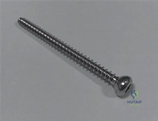
Candidate: I would assess her bone quality, noting she is likely osteoporotic due to steroids. I would consider either internal fixation or a hemiarthroplasty/total hip replacement. If I fix it, I'd use screws, but the bone might be soft, so I'd be worried about pull-out.
The candidate fails to bridge the gap between "poor bone" and the biological healing environment. They treat this as a purely mechanical problem (fixation) rather than a biological one (cellular function, remodeling capacity). They also ignore the specific impact of corticosteroids on the Wnt/beta-catenin pathway and osteoblast apoptosis.
I would structure my response by addressing: 1) Biological Status: Corticosteroid use induces osteoblast apoptosis and inhibits Wnt signaling, resulting in reduced bone mineral density and impaired secondary healing. 2) Bone Quality Assessment: The 'personality' of the fracture indicates poor screw purchase potential, increasing the risk of cut-out or non-union. 3) Surgical Strategy: Given the physiological, cellular, and biomechanical limitations, this patient likely falls out of the criteria for internal fixation. I would favor joint replacement (arthroplasty) to provide immediate weight-bearing, acknowledging that the host's impaired bone-remodeling potential makes biological union of a femoral neck fracture less predictable and potentially prone to complications like AVN or failure of fixation.
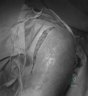
Look at this image. This patient has a pathological fracture through a lytic lesion. Explain the pathophysiology of why this bone failed and how the cellular environment dictates your management.
Candidate: This looks like a metastatic lesion. The cancer has eaten away the bone, making it weak. I would biopsy it and then put a nail in it to fix the fracture.
The candidate describes the lesion superficially. They fail to mention the RANKL-OPG axis, the difference between osteolytic and osteoblastic lesions, and the concept that the bone is biologically incapable of healing, thus requiring load-bearing fixation rather than a biological construct.
This is a metastatic lytic lesion. The pathology is driven by a 'vicious cycle': tumor cells secrete factors (like PTHrP) that upregulate RANKL expression on osteoblasts, leading to excessive osteoclast recruitment and aggressive bone resorption. Because this bone is biologically compromised and unlikely to undergo normal fracture healing, the management goal is internal stabilization (load-sharing), typically with an intramedullary device supplemented with PMMA cement. The cement serves as an immediate structural filler, compensating for the lack of inherent biological regenerative capacity while providing stability to allow early mobilization.
You mentioned Wolff's Law during our discussion. If I apply a rigid compression plate to a diaphyseal fracture, how does this interact with Wolff's Law, and what is the biological consequence of "stress shielding"?
Candidate: Wolff's Law says bone remodels based on stress. If you put a strong metal plate on, the plate takes the stress. So the bone underneath gets weak because it doesn't get enough load. That's stress shielding.
The candidate understands the concept at a high school level but lacks the technical terminology. They don't explain the role of the osteocyte as the mechanosensor or the molecular signaling (Sclerostin/Wnt pathway) that facilitates this atrophy.
Wolff's Law dictates that bone adapts to the mechanical environment. A rigid plate creates a 'stress-shielded' environment where the metal carries the majority of the physiological load. Biologically, the osteocytes sense reduced fluid shear stress within the lacunocanalicular system, leading to an upregulation of Sclerostin. Sclerostin inhibits the Wnt/β-catenin pathway in osteoblasts, resulting in decreased bone formation and increased resorption. This leads to regional osteopenia (bone atrophy) beneath the plate, which can create a stress riser at the ends of the implant and potentially compromise the bone's structural integrity if the implant is removed prematurely.
