Lower Limb Trauma: Mastering This Oral Examination Question
Key Takeaway
Looking for accurate information on Lower Limb Trauma: Mastering This Oral Examination Question? Managing a pilon fracture, a common oral examination question, involves a phased approach. Initial management focuses on stabilizing the injury with a spanning external fixator for soft tissue resuscitation. A CT scan provides detailed fracture mapping. Definitive treatment then involves careful surgical planning for anatomical reduction, fracture stabilization, and early mobilization, considering soft tissue condition and optimal timing.
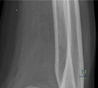
A 60-year-old patient presents with a high-energy peri-prosthetic fracture. Please analyze the provided radiograph. Describe the fracture pattern, classify it, and outline your management plan.
Candidate: "The radiograph shows a transverse diaphyseal fracture occurring just proximal to the femoral component of a total knee arthroplasty. The implant appears well-fixed. Based on the Vancouver classification for peri-prosthetic knee fractures, this is a Type B1 fracture. My management would involve open reduction and internal fixation using a lateral locking compression plate, ensuring anatomical reduction and stable fixation."
Candidates often fail to explicitly assess the stability of the implant. A poor answer would skip the classification or misclassify it (e.g., calling it a simple distal femur fracture). Furthermore, failing to mention the secondary need to ensure the joint is not infected or that the patient is medically fit for a major revision-type construct is a common oversight.
A perfect response begins with a systematic description: "This is a peri-prosthetic distal femur fracture proximal to a TKA." It correctly classifies the fracture using the Vancouver system (Type B1 = stable implant; Type B2 = loose implant; Type B3 = loose implant with poor bone stock). The "Gold Standard" response addresses: 1. Classification: Vancouver B1. 2. Implant stability: Confirmed by radiographic assessment of the cement-bone interface. 3. Surgical Strategy: Choice of a long lateral locking plate (spanning the entire femur if necessary) vs. retrograde nail (if implant geometry permits). 4. Patient Factors: Acknowledging the fragility of the patient and the need for bone grafting if comminution is present.
